1. Introduction to Interrupted Aortic Arch Type A
Interrupted aortic arch distal to the left subclavian artery, designated clinically as Type A, is a critical congenital cardiovascular anomaly. This condition is characterized by a complete physical separation between the aortic arch and the descending aorta. The interruption occurs precisely after the branching of the third major vessel that supplies the upper body.
Because the main conduit for systemic blood flow is completely severed, the lower half of the infant’s body cannot receive oxygenated blood directly from the left ventricle. Survival is entirely dependent on the presence of a patent ductus arteriosus, a fetal blood vessel that connects the pulmonary artery to the descending aorta.
Without rapid medical intervention to maintain this fetal connection, the condition is uniformly fatal shortly after birth. Fortunately, advancements in neonatal intensive care and specialized pediatric cardiac surgery have made it possible to successfully reconstruct the aorta, allowing affected children to survive and thrive.
2. Anatomical Classification and Structure
The aorta is the main artery carrying oxygen-rich blood from the heart to the entire body. As it arcs over the heart, it gives rise to three primary branches in a normal anatomical configuration. These are the innominate artery, the left common carotid artery, and the left subclavian artery.
In the classification system for interrupted aortic arch, the lettering corresponds to the location of the missing segment. Type A indicates that the interruption is located distal to the left subclavian artery. This is the second most common form of interrupted aortic arch, trailing behind Type B.
Because the interruption is distal to the left subclavian artery, the left ventricle pumps blood to the head, right arm, and left arm normally. The descending aorta, however, which supplies the abdomen and legs, is physically detached from this system and relies entirely on alternative pathways established during fetal development.
3. Embryology and Developmental Origins
During normal embryonic development, the cardiovascular system undergoes a complex series of folding, septation, and remodeling phases. The aortic arch system develops from multiple pairs of branchial arches. The left fourth branchial arch typically forms the segment of the definitive aortic arch between the left common carotid and the left subclavian arteries.
The exact embryological failure leading to a Type A interruption is distinct from Type B. In Type A, it is the embryonic dorsal aorta segment located just beyond the left subclavian artery that fails to develop or regresses abnormally.
This developmental error occurs very early in pregnancy, usually within the first two months. While the precise environmental or cellular trigger for this specific failure remains a subject of ongoing scientific investigation, it results in a profound anatomical defect requiring complex reconstructive strategies.
4. Pathophysiology and Fetal Circulation
Before birth, fetal circulation bypasses the fluid-filled lungs. Blood is shunted away from the pulmonary system through the ductus arteriosus directly into the descending aorta. In a fetus with an interrupted aortic arch, this normal fetal adaptation essentially masks the severity of the defect, allowing normal growth and organ development in utero.
Upon birth and the infant’s first breaths, pulmonary vascular resistance drops, and blood is directed into the lungs to be oxygenated. Normally, the ductus arteriosus constricts and closes within the first few days of life as oxygen levels rise.
In a neonate with a Type A interruption, the natural closure of the ductus arteriosus results in an immediate and catastrophic loss of blood flow to the lower body. The abdominal organs, kidneys, and lower extremities are suddenly deprived of perfusion, leading to rapid onset tissue hypoxia and systemic metabolic failure.
5. Genetic Testing and Chromosomal Factors
Unlike Type B interrupted aortic arch, which is very strongly associated with the 22q11.2 microdeletion syndrome, Type A has a much lower association with this specific genetic anomaly. However, genetic factors still play a crucial role in proper cardiac development.
Infants born with Type A interruption may still undergo genetic testing, including chromosomal microarray analysis, to identify any underlying genetic syndromes or distinct microdeletions that could influence overall health and development.
Understanding the genetic profile helps the medical team anticipate other potential congenital anomalies or developmental challenges. It also provides parents with valuable information regarding the risk of recurrence in future pregnancies and the long-term multidisciplinary care needs of their child.
6. Associated Structural Cardiac Anomalies
An isolated interrupted aortic arch is an extreme rarity. The complex morphogenetic processes that form the arch are intricately tied to the septation of the heart chambers. Therefore, structural defects within the heart itself are nearly always present.
A ventricular septal defect is the most common associated anomaly. This opening between the right and left ventricles allows blood to shunt between the chambers, which can stabilize pressure but also leads to mixing of oxygenated and deoxygenated blood.
Additionally, patients with Type A interruption frequently present with left ventricular outflow tract obstruction. This can involve narrowing below the aortic valve or abnormalities of the aortic valve itself, such as a bicuspid aortic valve, which further complicates the initial surgical repair and long-term prognosis.
7. Clinical Symptoms in the First Week of Life
The clinical presentation of interrupted aortic arch Type A hinges entirely on the status of the ductus arteriosus. In the first 24 to 48 hours, while the ductus remains widely patent, the infant may appear relatively well, though subtle signs like rapid breathing might be present.
As the ductus begins its physiological closure, symptoms escalate rapidly. The infant will exhibit profound feeding intolerance, extreme lethargy, and a grayish skin tone. The lack of blood flow to the kidneys significantly decreases urine output, a crucial clinical marker of deteriorating systemic perfusion.
Without immediate identification and treatment, the infant progresses into profound cardiogenic shock. This is characterized by severe metabolic acidosis, absent lower extremity pulses, and respiratory distress as the heart fails to maintain necessary cardiac output.
8. Clinical Examination and Pulse Discrepancy
A meticulous physical examination is the first step in identifying this critical congenital heart defect. In an infant with ductal closure, the most striking finding is the profound difference in pulse strength between the upper and lower extremities.
In Type A interruption, the pulses in both arms and the neck will be strong and bounding, reflecting the unobstructed flow from the left ventricle. In stark contrast, the femoral pulses in the groin and the pedal pulses in the feet will be extremely weak or entirely absent.
Blood pressure measurements confirm these findings. A gradient will be evident, with high blood pressure in both arms and significantly lower, sometimes unmeasurable, blood pressure in the legs. Auscultation often reveals a loud murmur, primarily originating from the associated ventricular septal defect or left ventricular outflow tract obstruction.
9. Diagnostic Imaging Protocols
Echocardiography serves as the definitive diagnostic tool for evaluating congenital heart defects. High-resolution two-dimensional and color Doppler echocardiography provide a comprehensive anatomical map of the heart and the great vessels.
The echocardiogram will specifically demonstrate the discontinuity of the aortic arch. By tracing the arch and identifying the branching vessels, the pediatric cardiologist can confirm that the left subclavian artery arises proximal to the interruption, securing the diagnosis of Type A.
In cases where the anatomy is highly complex or if the exact length of the missing segment needs to be measured precisely for surgical planning, a computed tomography angiography scan may be performed. This imaging modality provides exceptional three-dimensional detail of the vascular structures.
10. Distinguishing Left-Sided Obstructive Lesions
The sudden cardiovascular collapse of a neonate demands rapid differentiation between several critical conditions. The differential diagnosis for an infant presenting with shock and absent femoral pulses includes severe coarctation of the aorta.
While coarctation involves a severe narrowing of the aorta rather than a complete absence of a segment, the clinical manifestation during ductal closure is virtually indistinguishable from an interrupted aortic arch. Immediate echocardiography is required to differentiate the two.
Hypoplastic left heart syndrome is another major differential diagnosis. In this condition, the left side of the heart is profoundly underdeveloped. While it also presents with systemic hypoperfusion, echocardiography will easily identify the severe underdevelopment of the left ventricle and mitral valve, distinguishing it from an interrupted arch.
11. Emergency Medical Management
Immediate medical stabilization is paramount and focuses entirely on restoring systemic blood flow. The administration of continuous intravenous prostaglandin E1 is a life-saving intervention. This medication prevents the ductus arteriosus from closing and can even reopen a closing ductus, thereby restoring perfusion to the lower body and reversing metabolic acidosis.
Simultaneously, the infant requires admission to a neonatal or pediatric cardiac intensive care unit. Mechanical ventilation is often necessary to reduce the workload on the infant’s failing cardiovascular system and to carefully manage oxygenation.
Inotropic support using medications like dopamine or milrinone may be initiated to improve the contractility of the heart muscle. Careful fluid management and correction of electrolyte imbalances and acidosis are critical components of the preoperative stabilization protocol.
12. Comparison of Diagnostic Features
Properly classifying the specific type of interrupted arch dictates the surgical approach. The table below outlines the distinct diagnostic features of Type A compared to general features of arch interruptions.
| Diagnostic Feature | Observation in Type A Interruption |
|---|---|
| Location of Defect | Distal to the left subclavian artery |
| Upper Extremity Pulses | Strong in BOTH right and left arms |
| Lower Extremity Pulses | Weak or absent |
| Vascular Dependence | Ductus arteriosus dependent for lower body perfusion |
| Genetic Link | Lower incidence of 22q11.2 deletion compared to Type B |
13. Surgical Repair Strategies
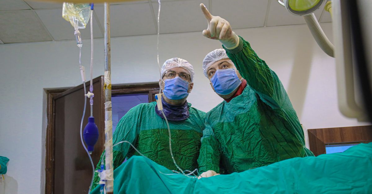
The definitive treatment for interrupted aortic arch Type A is complete surgical reconstruction. This is a highly complex open-heart procedure performed under cardiopulmonary bypass. Deep hypothermic circulatory arrest may be utilized to protect the brain and vital organs while the surgeon operates on the delicate arch structures.
The surgical objective is to re-establish a continuous, unobstructed aortic arch. The surgeon mobilizes the descending aorta and brings it into direct contact with the proximal aortic arch. A primary end-to-end anastomosis is usually performed, directly suturing the two segments together.
During the same operation, the surgical team repairs associated defects. The ventricular septal defect is closed with a patch, and the patent ductus arteriosus is securely ligated and divided. If there is significant left ventricular outflow tract obstruction, it must also be addressed surgically to ensure unimpeded blood flow into the newly reconstructed aorta.
14. Postoperative Intensive Care
The postoperative period is a critical phase of recovery. The infant remains in the pediatric cardiac intensive care unit and requires meticulous monitoring of cardiovascular hemodynamics. The heart must adapt to pumping against a new, continuous systemic resistance without the pressure relief previously provided by the ventricular septal defect.
Controlling blood pressure is a major focus. Systemic hypertension is common after arch reconstruction and can place undue stress on the surgical suture lines. Intravenous medications are used to keep blood pressure within strict, safe parameters.
Monitoring for signs of organ dysfunction, managing pain, and ensuring adequate nutrition are essential components of the recovery process. The intensive care team remains highly vigilant for complications such as bleeding, arrhythmias, and fluid accumulation around the heart or lungs.
15. Long-Term Health and Prognosis
With modern surgical techniques, the survival rate for infants undergoing repair of interrupted aortic arch Type A is excellent. However, these patients require lifelong follow-up with a specialized congenital cardiologist.
The most common long-term complication is the development of a narrowing, or stricture, at the site of the aortic anastomosis as the child grows. Routine echocardiograms are essential to detect this narrowing early. If severe, it can often be treated successfully in the cardiac catheterization laboratory using balloon angioplasty or stent placement.
Patients also need regular evaluation of left ventricular function and surveillance of the aortic valve and subaortic area, as problems in these regions can manifest years after the initial surgery. With appropriate follow-up, the majority of patients lead active, productive lives.
16. When to Consult a Pediatric Cardiologist
For parents of a child who has undergone arch repair, knowing when to seek specialized medical advice is crucial for long-term health. Routine, scheduled appointments with a pediatric cardiologist are mandatory throughout childhood and adolescence.
Immediate consultation is necessary if a child develops unexplained fatigue, shortness of breath during physical exertion, or complaints of chest pain. These symptoms can be indicators of recurrent aortic narrowing or progressive left ventricular outflow tract obstruction.
Additionally, if a pediatrician notes a new heart murmur or a significant difference in blood pressure between the arms and legs during a routine check-up, a prompt evaluation by the pediatric cardiology team is strongly advised to assess the integrity of the aortic repair.
17. Frequently Asked Questions (FAQ)
1. How is an interrupted aortic arch different from aortic coarctation?
While both conditions impede blood flow to the lower body, an interrupted aortic arch is a complete anatomical disconnection of the vessel. Coarctation is a severe narrowing or pinching of the vessel, but the tissue remains physically continuous.
2. What role does the ductus arteriosus play in this condition?
The ductus arteriosus is a temporary fetal blood vessel. In interrupted aortic arch, it acts as a critical detour, allowing blood from the right side of the heart to reach the lower body, bypassing the disconnected aorta. Its patency must be maintained medically until surgery.
3. Is deep hypothermia always used during surgery?
Deep hypothermic circulatory arrest is a common technique that involves cooling the baby’s body to very low temperatures to reduce cellular metabolic demand. This allows the surgeon to stop the bypass machine temporarily to perform delicate work on the arch without causing brain injury. It is frequently, but not always, used depending on the specific surgical approach.
4. Can a patient with a repaired aortic arch live a normal lifespan?
Yes, with successful initial surgery and rigorous, lifelong medical follow-up to manage potential late complications like arch narrowing, patients have an excellent prognosis and can achieve a normal life expectancy.
18. Bibliography
Disclaimer: The content is for informational purposes only and does not replace medical advice. Always consult your doctor for personalized treatment.