1. Introduction to Interrupted Aortic Arch Type B
Interrupted aortic arch between the left subclavian and left common carotid artery, clinically classified as Type B, is a rare and severe congenital heart defect. In this condition, there is a complete anatomic discontinuity between the ascending and descending segments of the aorta at the specific location between these two major branching vessels. This interruption prevents oxygenated blood from flowing directly from the heart to the lower half of the body through the normal aortic pathway.
This specific anatomical configuration requires immediate medical recognition upon birth. The systemic circulation to the lower body relies entirely on a temporary fetal structure known as the ductus arteriosus. As this structure naturally closes shortly after birth, the newborn experiences a critical reduction in systemic blood flow.
Prompt intervention is essential for survival. Modern pediatric cardiology and cardiovascular surgery have developed highly effective stabilization protocols and surgical reconstruction techniques to restore normal blood flow and improve long-term outcomes for affected infants.
2. Understanding the Aortic Arch Anatomy
The normal aorta arises from the left ventricle and forms an arch before descending through the chest into the abdomen. Along this arch, three main blood vessels typically branch off to supply the upper body. These are the innominate artery, the left common carotid artery, and the left subclavian artery.
In an interrupted aortic arch, a segment of this continuous vessel is entirely missing. The location of the missing segment determines the classification of the defect. Type B is the most common form of this rare defect.
In Type B, the interruption occurs exactly between the left common carotid artery, which supplies blood to the left side of the brain, and the left subclavian artery, which supplies blood to the left arm. Consequently, the ascending aorta supplies the head and right arm, while the descending aorta must receive blood from another source to supply the left arm and the lower body.
3. Embryological Development and Malformation
The development of the human heart and its major vessels occurs during the first trimester of pregnancy. The aortic arch forms from a complex remodeling of embryonic branchial arches. The normal left aortic arch develops primarily from the fourth left branchial arch.
An interrupted aortic arch Type B results from an early developmental failure. Specifically, the embryonic left fourth aortic arch fails to develop or involutes prematurely. This failure leaves a physical gap in the developing vascular tree.
The exact cellular mechanisms causing this failure involve neural crest cell migration. These specialized cells travel from the developing neural tube to the heart and are crucial for the proper formation of the aortic arch and the separation of the main pulmonary artery from the aorta.
4. Pathophysiology and Hemodynamic Alterations
The hemodynamic consequences of this defect are profound. Because the aorta is disconnected, the left ventricle can only pump blood to the vessels arising before the interruption. In Type B, this means oxygenated blood reaches the right arm and both sides of the head.
The lower half of the body and the left arm depend entirely on blood crossing from the pulmonary artery through the patent ductus arteriosus into the descending aorta. This blood is typically a mixture of oxygenated and deoxygenated blood, resulting in lower oxygen levels reaching the lower body.
Furthermore, the right ventricle must pump blood not only to the lungs but also to the entire lower body. This places an extraordinary workload on the right side of the heart. As pulmonary vascular resistance drops after birth, blood tends to flood the lungs, reducing systemic flow and leading to cardiac strain.
5. Genetic Associations and Deletion Syndromes
There is a powerful established link between interrupted aortic arch Type B and specific genetic microdeletions. The most prominent of these is the 22q11.2 deletion syndrome, historically known as DiGeorge syndrome.
Research indicates that a significant majority of infants diagnosed with Type B interrupted aortic arch carry this specific genetic microdeletion. This genetic anomaly affects the migration of neural crest cells during embryonic development, leading directly to the arch malformation.
Identifying this genetic factor is a critical step in comprehensive patient care. Patients with this deletion may present with additional clinical challenges, including immune system deficiencies, low blood calcium levels due to underactive parathyroid glands, and developmental delays, all of which require specialized multidisciplinary management.
6. Associated Congenital Heart Defects
Interrupted aortic arch Type B rarely occurs as an isolated anomaly. The embryonic pathways that form the aortic arch are closely linked to the structures that divide the lower chambers of the heart and the main outflow tracts.
A ventricular septal defect is present in nearly all cases of Type B interruption. This hole between the lower pumping chambers allows blood to mix and helps maintain pressure in the right ventricle, which is necessary to drive blood through the ductus arteriosus to the lower body.
Additionally, the aortic valve is frequently abnormal. A bicuspid aortic valve or subaortic narrowing can occur, further complicating the blood flow out of the left ventricle. These associated defects must be addressed during comprehensive surgical planning.
7. Clinical Presentation in Neonates
Infants born with this condition often appear healthy at the exact moment of birth because the ductus arteriosus remains open. This structure maintains circulation to the lower body in the immediate postnatal period.
However, within the first few days of life, the ductus arteriosus begins its natural closure process. As this channel constricts, blood flow to the lower half of the body is drastically reduced. The infant will rapidly develop signs of poor perfusion, including lethargy, poor feeding, and cold lower extremities.
If the closure continues without medical intervention, the infant will progress to cardiogenic shock and severe metabolic acidosis. The lack of oxygen delivery to vital abdominal organs and the kidneys leads to rapid clinical deterioration.
8. Physical Examination Findings
During a clinical assessment, healthcare providers rely on specific physical signs to suspect ductal-dependent lesions. A key finding in interrupted aortic arch Type B is a discrepancy in pulses. The pulses in the right arm and the neck are typically strong, while the pulses in the left arm and both legs are weak or completely absent.
Measuring blood pressure in all four extremities is a standard diagnostic step. In Type B, there is a marked difference in pressure, with higher pressures in the right arm compared to the legs. The left arm blood pressure will also be low because the left subclavian artery arises below the interruption.
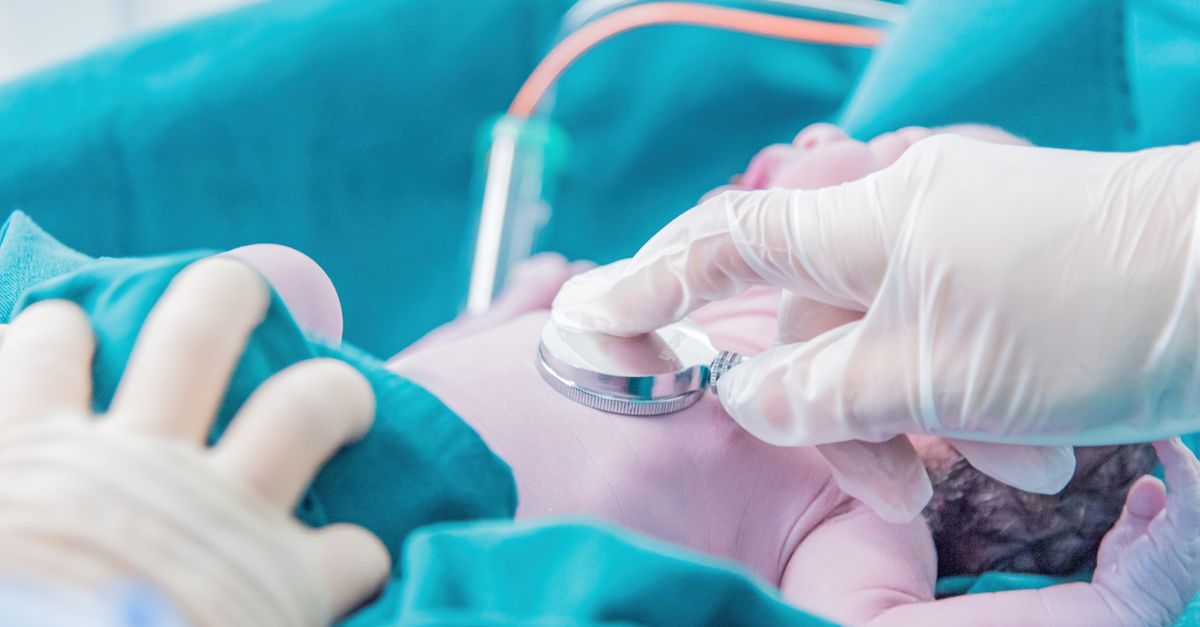
Auscultation of the heart often reveals a prominent heart murmur. This murmur is primarily generated by the associated ventricular septal defect rather than the interrupted arch itself. The infant may also exhibit signs of respiratory distress, such as rapid breathing and retractions, due to excessive blood flow to the lungs.
9. Diagnostic Imaging and Echocardiography
Echocardiography is the gold standard for diagnosing this condition. High-resolution ultrasound waves allow pediatric cardiologists to visualize the exact anatomy of the heart and the aortic arch in real time without radiation exposure.
The echocardiogram will clearly show the lack of continuity between the ascending and descending aorta. It will precisely locate the interruption between the left common carotid and left subclavian arteries, confirming the Type B classification.
Additionally, the echocardiogram evaluates the size and location of the ventricular septal defect, assesses the function of the heart valves, and measures the size of the patent ductus arteriosus. Advanced imaging techniques like cardiac magnetic resonance imaging or computed tomography angiography are occasionally utilized for complex preoperative planning.
10. Differential Diagnosis in Neonatal Shock
When a newborn presents with sudden cardiovascular collapse in the first week of life, several critical conditions must be considered. The clinical picture of interrupted aortic arch is very similar to other left-sided obstructive heart lesions.
Severe coarctation of the aorta is a primary differential diagnosis. While coarctation involves a severe narrowing rather than a complete disconnect, the physiological impact during ductal closure is nearly identical. Hypoplastic left heart syndrome must also be ruled out, as it presents with similar systemic circulatory failure.
Overwhelming neonatal sepsis is another critical condition that mimics cardiogenic shock. Differentiating between severe infection and structural heart disease requires rapid clinical assessment, blood cultures, and immediate bedside echocardiography to establish the correct diagnosis.
11. Initial Medical Stabilization
The absolute priority in medical management is maintaining or reopening the ductus arteriosus. This is achieved through a continuous intravenous infusion of prostaglandin E1. This medication acts directly on the ductal tissue, preventing it from constricting and thereby restoring life-saving blood flow to the lower body.
Alongside prostaglandin therapy, intensive care support is mandatory. Many infants require endotracheal intubation and mechanical ventilation to reduce the metabolic work of breathing and optimize oxygen delivery. Careful management of intravenous fluids and inotropic medications helps support the failing heart muscle.
Oxygen therapy must be administered with extreme caution. High levels of oxygen can cause the blood vessels in the lungs to dilate, stealing blood away from the systemic circulation. Providers balance oxygen and carbon dioxide levels to ensure adequate blood flow reaches both the lungs and the body.
12. Preoperative Clinical Data Summary
Understanding the anatomical and clinical differences is vital for accurate treatment. The following table summarizes key aspects of Interrupted Aortic Arch Type B.
| Anatomical Feature / Clinical Marker | Description in Type B |
|---|---|
| Location of Interruption | Between left common carotid and left subclavian artery |
| Primary Source of Lower Body Blood Flow | Patent ductus arteriosus |
| Most Common Associated Defect | Ventricular septal defect |
| Strongest Genetic Association | 22q11.2 microdeletion syndrome |
| Initial Life-Saving Medication | Prostaglandin E1 continuous infusion |
13. Surgical Reconstruction Techniques
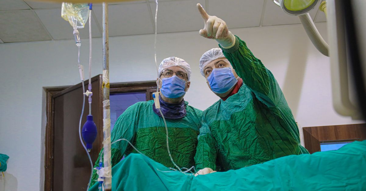
Definitive treatment requires complex open-heart surgery, typically performed within the first week or two of life once the infant is medically stabilized. The surgery is conducted under cardiopulmonary bypass, which takes over the function of the heart and lungs during the procedure.
The primary goal of the operation is to reconstruct a continuous aortic pathway. The surgeon meticulously mobilizes the descending aorta and brings it up to join the ascending aorta. The vessels are sutured directly together to create a permanent, unobstructed conduit for blood to flow to the entire body.
Simultaneously, the surgeon addresses the associated structural defects. The ventricular septal defect is closed using a synthetic patch or a piece of the patient’s own pericardium. The patent ductus arteriosus, no longer needed, is securely tied off and divided.
14. Postoperative Care and Complications
The immediate postoperative period requires intensive monitoring in a dedicated pediatric cardiac intensive care unit. The infant may experience low cardiac output, requiring continued medication support to help the heart muscle pump effectively against the newly reconstructed aorta.
Vigilant monitoring for bleeding, arrhythmias, and respiratory complications is standard protocol. The reconstructed aortic arch tissue can sometimes become narrowed as the healing process occurs. This narrowing at the surgical site requires close observation and potential intervention.
Another specific complication involves the recurrent laryngeal nerve, which controls the vocal cords and is located very close to the aortic arch. Temporary or permanent injury to this nerve during dissection can lead to feeding difficulties and a hoarse cry, requiring specialized speech and swallowing therapy.
15. Long-Term Prognosis and Follow-Up
The long-term outlook for children who undergo successful repair has improved dramatically in recent decades. Most children grow and develop well, participating in normal childhood activities. However, lifelong follow-up with a pediatric or adult congenital cardiologist is mandatory.
A significant number of patients may develop narrowing at the site of the arch reconstruction as they grow. This complication can often be treated in the cardiac catheterization laboratory using balloon dilation or stent placement, avoiding a second open-heart surgery.
Patients with the 22q11.2 deletion syndrome require comprehensive, multidisciplinary care. Endocrine evaluation for calcium regulation, immunological testing, and developmental assessments are integral parts of their long-term health management strategy.
16. When to Seek Immediate Medical Attention
Recognizing the signs of circulatory compromise in a newborn is vital for parents and caregivers. Since newborns are often discharged before the ductus arteriosus fully closes, symptoms can manifest at home.
Immediate emergency medical care is required if an infant exhibits unusual lethargy, difficulty waking up for feedings, or extreme weakness. Changes in skin color, particularly a pale or grayish appearance, or cyanosis around the lips and nail beds, are critical warning signs.
Additionally, if the infant begins breathing very rapidly, sweating heavily during feeding, or if the lower legs feel unusually cold compared to the upper body, parents must contact emergency services without delay. These symptoms indicate an urgent need for medical evaluation and potential prostaglandin therapy.
17. Frequently Asked Questions (FAQ)
1. Can interrupted aortic arch Type B be diagnosed before birth?
Yes, advancements in fetal echocardiography allow for the detection of interrupted aortic arch during the second trimester of pregnancy. A prenatal diagnosis enables medical teams to plan for immediate care and prostaglandin administration right at the time of delivery.
2. Is interrupted aortic arch hereditary?
While the defect itself is not strictly passed from parent to child in a traditional manner, the strong association with the 22q11.2 deletion syndrome means genetics play a major role. Genetic counseling is highly recommended for parents to understand the risks for future pregnancies.
3. Will my child need more surgeries in the future?
While the initial surgery is intended to be a complete repair, some children develop narrowing at the repair site or face issues with the aortic valve over time. These issues might require catheter-based interventions or, occasionally, additional surgeries later in life.
4. Can a child with a repaired interrupted aortic arch play sports?
Most children can participate in recreational sports and normal physical activities. However, competitive or high-intensity sports should be discussed with the treating cardiologist, who will provide individualized recommendations based on regular exercise testing and heart function assessments.
18. Bibliography
Disclaimer: The content is for informational purposes only and does not replace medical advice. Always consult your doctor for personalized treatment.