1. Introduction
Intraductal papillary-mucinous tumours are complex cystic neoplasms arising from the epithelial lining of the pancreatic ducts. When a tumour is histologically classified as exhibiting moderate dysplasia, it signifies a critical transition point in the biological progression of the disease. In contemporary pathological classification, moderate dysplasia is often grouped within the broader spectrum of low-grade lesions, yet clinically, it represents a state where the cellular architecture is becoming increasingly disorganized and genetically unstable.
These tumours are no longer entirely indolent, but they have not yet acquired the severe, tissue-destroying characteristics of high-grade dysplasia or invasive carcinoma. The management of moderate dysplasia requires a highly nuanced and individualized approach. Physicians must carefully balance the significant risks associated with major pancreatic surgery against the increasing risk of malignant transformation. Utilizing high-resolution imaging, endoscopic fluid analysis, and multidisciplinary expertise is paramount for identifying the precise moment when surgical intervention is required to secure a definitive cure.
2. Understanding the Pathophysiology
The fundamental physiological disruption begins within the branching ductal system of the pancreas. The epithelial cells lining these ducts undergo a neoplastic transformation, growing outward into the ductal lumen to form complex, microscopic papillary fronds. Simultaneously, these cells hyper-secrete thick, gelatinous mucin. This continuous secretion creates a physical mechanical obstruction within the narrow ducts, leading to increasing internal pressure and the subsequent formation of dilated, fluid-filled cysts.
The term moderate dysplasia describes the specific microscopic appearance of these abnormal cells. At this stage, the cells exhibit noticeable nuclear enlargement, crowding, and a loss of normal cellular polarity. The architectural pattern of the papillae becomes more complex and disorganized compared to early, low-grade lesions. While the structural integrity of the ductal basement membrane remains intact, preventing true tissue invasion, the increasing cellular disarray indicates a progressive loss of normal biological regulation.
3. Cellular Origins and Mechanisms
The neoplastic process originates from the multipotent stem cells residing within the pancreatic ductal epithelium. The progression to moderate dysplasia is driven by the sequential accumulation of specific somatic genetic mutations. The initial transformation is almost universally driven by mutations in the KRAS and GNAS genes, which act as continuous cellular growth signals instructing the cells to divide and produce mucin.
As the tumour evolves into the moderate dysplasia phase, the cells begin to accumulate secondary genetic alterations. The genomic stability that characterizes earlier lesions begins to degrade. While the cells typically have not yet acquired the catastrophic mutations in the TP53 or SMAD4 tumour suppressor genes that define highly aggressive carcinomas, the increasing genetic burden propels the tumour along the neoplastic continuum. This active molecular evolution is the primary reason these lesions require intense clinical scrutiny.
4. Key Risk Factors
Intraductal papillary-mucinous tumours most frequently affect older demographics, with diagnoses peaking in individuals in their sixties and seventies. The condition arises sporadically in most patients, but several distinct clinical and environmental factors significantly increase the risk of developing these progressively dysplastic lesions.
Chronic tobacco use is the most powerful modifiable risk factor, known to induce severe genetic damage within the pancreatic epithelium and accelerate neoplastic progression. A clinical history of chronic pancreatitis also creates an inflammatory microenvironment conducive to abnormal cellular proliferation. Furthermore, individuals harboring specific inherited genetic mutations, such as BRCA2 mutations, or those with familial adenomatous polyposis, exhibit a higher baseline susceptibility to pancreatic neoplasms and necessitate early, rigorous clinical screening.
5. Clinical Presentation
The clinical presentation of a tumour with moderate dysplasia is heavily dictated by its exact anatomical location and the volume of mucin it produces. Lesions confined to the smaller side branches of the pancreatic duct may remain entirely asymptomatic and evade detection until an abdominal scan is performed for an unrelated issue. However, as the dysplasia progresses and mucin production increases, symptoms become more prevalent.
Patients frequently experience recurrent, dull, aching pain in the upper abdomen that may radiate to the back. This discomfort results directly from the elevated pressure within the obstructed ductal system. If the thick mucin partially or completely blocks the main pancreatic duct, the patient may suffer from repeated episodes of acute pancreatitis, characterized by severe abdominal pain, nausea, and vomiting. Compression of the adjacent bile duct by an expanding cyst can lead to visible jaundice.
6. Early Warning Signs
Identifying the transition from a silent, indolent cyst to a symptomatic, moderately dysplastic tumour is critical for timely intervention. A sudden change in the character or frequency of abdominal pain is a primary warning sign. Patients may notice that previously mild indigestion has escalated into sharp, severe episodes of upper abdominal distress following meals, suggesting increasing ductal obstruction.
Systemic signs of progressive pancreatic damage must be treated with high clinical suspicion. The sudden onset of diabetes mellitus in an older patient, without corresponding changes in body weight or diet, is a profound indicator of exocrine and endocrine pancreatic failure. Unexplained, progressive weight loss combined with pale, floating stools indicates a severe lack of digestive enzymes reaching the intestine, mandating immediate and comprehensive radiological evaluation.
7. Diagnostic Imaging
High-resolution imaging is the most critical tool for evaluating the structural threat posed by the tumour. Magnetic resonance imaging combined with magnetic resonance cholangiopancreatography is the optimal diagnostic modality. This specialized imaging protocol provides exceptional soft-tissue contrast, allowing radiologists to meticulously examine the architecture of the cysts and the entire pancreatic ductal tree.
For a tumour with moderate dysplasia, the radiologist specifically looks for high-risk structural features. These include a main pancreatic duct dilated to more than five millimeters, a rapid increase in the overall size of the cyst, or the presence of thickened cyst walls. Computed tomography with intravenous contrast is also utilized to evaluate the surrounding blood vessels and ensure the tumour remains localized, providing essential data for multidisciplinary surgical planning.
8. Endoscopic Evaluation
When cross-sectional imaging reveals worrisome structural features, an endoscopic ultrasound is performed to obtain a definitive tissue diagnosis and directly assess the cyst’s internal lining. A specialized endoscope is advanced into the stomach and duodenum, placing the ultrasound probe in direct proximity to the pancreas. This allows the gastroenterologist to detect tiny solid nodules within the cyst wall that are invisible on standard magnetic resonance imaging.
During the procedure, fine-needle aspiration is frequently utilized to collect fluid directly from the cyst. This fluid undergoes rigorous cytological and biochemical analysis. The presence of elevated carcinoembryonic antigen confirms the mucinous nature of the lesion. Highly specialized cytologists evaluate the aspirated cells to confirm the degree of nuclear atypia, differentiating moderate dysplasia from highly aggressive carcinoma in situ.
9. Molecular Diagnostics
The integration of molecular pathology into the evaluation of aspirated cyst fluid provides a deeper understanding of the tumour’s biological potential. Identifying the classic KRAS and GNAS mutations confirms the fundamental diagnosis of an intraductal papillary-mucinous tumour.
More importantly, the molecular analysis screens for the presence of advanced, high-risk genetic alterations. The complete absence of mutations in the TP53, PIK3CA, and SMAD4 genes provides significant reassurance that the tumour remains in the moderately dysplastic phase and has not yet crossed the threshold into highly aggressive malignancy. This detailed molecular profile heavily influences the clinical decision between continued strict surveillance and immediate surgical resection.
10. Differential Diagnosis
Accurately distinguishing this moderately dysplastic tumour from other pancreatic lesions is vital for patient safety and appropriate therapeutic planning.
| Pancreatic Lesion Type | Differentiating Characteristics |
|---|---|
| Serous Cystadenoma | A purely benign, non-mucinous cyst that frequently displays a central calcification and poses essentially zero risk of malignancy. |
| Pancreatic Pseudocyst | An inflammatory fluid collection arising after acute pancreatitis, completely lacking the dysplastic epithelial lining seen in neoplasms. |
| Mucinous Cystic Neoplasm | Occurs almost exclusively in women, typically in the tail of the pancreas, and does not physically connect to the main ductal system. |
| High-Grade Dysplasia / Carcinoma | Features severe cellular atypia, solid tissue masses invading the ductal wall, and advanced genetic mutations requiring immediate major surgery. |
11. Surgical Interventions
The decision to proceed with surgical resection for a tumour with moderate dysplasia is highly individualized. Surgery is generally recommended if the lesion involves the main pancreatic duct, if the cyst is larger than three centimeters, if solid nodules are present, or if the patient is suffering from recurrent acute pancreatitis.
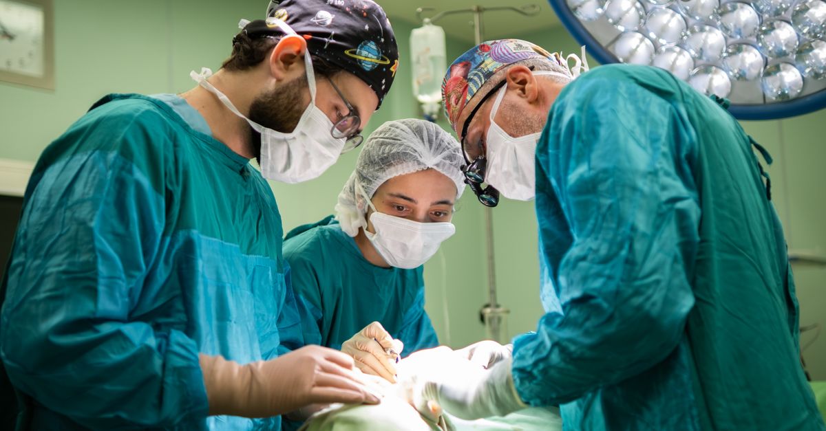
The surgical procedure is dictated by the anatomical location of the lesion. Tumours in the head of the pancreas require a pancreaticoduodenectomy (Whipple procedure), a complex operation involving the removal of the pancreatic head, duodenum, and bile duct. Tumours in the body or tail are managed with a distal pancreatectomy. During the operation, the surgeon relies on intraoperative frozen section pathology to ensure the resection margins are completely free of dysplastic cells, guaranteeing total eradication of the disease.
12. Systemic and Targeted Therapies
Because a tumour with moderate dysplasia remains a non-invasive, localized lesion, systemic oncological therapies such as traditional cytotoxic chemotherapy and radiation therapy are strictly contraindicated. These aggressive treatments are designed to combat rapidly dividing, invasive cancer cells and provide absolutely no clinical benefit for precancerous ductal abnormalities.
The entire therapeutic strategy relies upon timely and precise surgical resection. The goal is to physically remove the dysplastic tissue before the accumulating genetic mutations enable the cells to invade surrounding structures. A successful surgical extraction provides a complete and permanent cure for that specific localized lesion, eliminating the need for any pharmacological oncological interventions.
13. Post-Treatment Monitoring
Survivorship following the surgical resection of a dysplastic pancreatic tumour necessitates a rigorous and enduring surveillance protocol. The genetic factors and environmental exposures that initiated the primary tumour place the entire remaining pancreatic ductal system at a heightened risk for developing new, independent neoplastic lesions over the patient’s lifetime.
The standard surveillance regimen involves alternating high-resolution magnetic resonance imaging and endoscopic ultrasound every six to twelve months. Patients also undergo routine metabolic blood panels to monitor fasting glucose levels and assess overall pancreatic exocrine function. This proactive monitoring ensures that the remaining pancreas functions healthily and that any new dysplastic lesions are detected at an early, highly curable stage.
14. Prognostic Factors
The clinical outlook for an individual diagnosed with moderate dysplasia is exceptionally favorable, provided the tumour is completely resected prior to the onset of high-grade dysplasia or invasive carcinoma. The most significant prognostic indicator is the final pathology report confirming negative surgical margins and the absolute absence of invasive cancer cells.
When the disease is completely removed at this precancerous stage, the disease-specific survival rate approaches one hundred percent. However, the prognosis is heavily reliant on the timing of the intervention. Therefore, adherence to strict surveillance protocols and reliance on expert multidisciplinary surgical teams are the critical elements that ensure a normal life expectancy and a positive long-term outcome.
15. When to Consult a Specialist
The discovery of any complex cystic lesion in the pancreas demands an immediate referral to a specialized gastroenterologist or a dedicated pancreatic surgeon. Determining the exact degree of dysplasia and the associated risk of malignancy is highly complex and requires clinical expertise far beyond the scope of general medical practice.
Patients with increasing abdominal pain, recurrent pancreatitis, or new-onset diabetes must be evaluated at a high-volume tertiary care center. These specialized facilities possess the advanced endoscopic technologies and the integrated multidisciplinary tumor boards necessary to accurately interpret complex molecular and radiological data, ensuring the patient receives the safest, most precise surgical or surveillance strategy available.
16. Frequently Asked Questions (FAQ)
1. What does moderate dysplasia mean in a pancreatic cyst?
It means the cells lining the cyst are showing significant abnormal changes. They are no longer completely benign, but they have not yet turned into a true, invasive cancer. It is a critical warning stage.
2. Will I definitely need surgery for this condition?
Surgery is frequently recommended at this stage, especially if the cyst is large, causing pain, or showing solid areas on a scan. The goal is to remove it before it becomes dangerous.
3. Can this tumour spread to other parts of my body?
At the moderate dysplasia stage, the abnormal cells are entirely confined within the pancreatic ducts and cannot spread to other organs. They only gain the ability to spread if they mutate into invasive cancer.
4. How is the surgery performed?
The type of surgery depends on where the tumour is located. If it is in the head of the pancreas, a complex procedure called a Whipple is performed. If it is in the tail, the surgeon removes just that specific section of the pancreas.
5. Will I need to take digestive enzymes after surgery?
It depends on how much of your pancreas is removed. Some patients need to take oral enzyme supplements with their meals to help digest food properly, which your medical team will carefully monitor and manage.
17. Bibliography
Disclaimer: The content is for informational purposes only and does not replace medical advice. Always consult your doctor for personalized treatment.