1. Introduction
A pancreatic mass refers to any abnormal growth or lesion developing within the tissues of the pancreas. The pancreas is a vital, dual-functioning glandular organ situated deep within the abdominal cavity, nestled behind the stomach and in front of the spine. Due to this hidden, retroperitoneal location, masses growing within the pancreas are notoriously silent in their early stages, evading detection during routine physical examinations.
When a pancreatic mass is discovered, it initiates an urgent and complex diagnostic cascade. The pathology can range from harmless inflammatory fluid collections and slow-growing benign cysts to very aggressive malignant tumors. Pancreatic cancer, specifically pancreatic ductal adenocarcinoma, is one of the most lethal human malignancies because it frequently metastasizes before causing any localized symptoms.
Determining the exact nature of the mass relies on high-resolution cross-sectional imaging and specialized endoscopic biopsy techniques. The cellular origin of the tumor, its precise location within the gland, and its relationship to the surrounding major blood vessels dictate the entire oncological and surgical strategy. This article explores the function of the pancreas, the various types of pancreatic neoplasms, and the clinical pathways used to diagnose and treat them.
2. Anatomy and Function of the Pancreas
To comprehend the impact of a pancreatic mass, one must understand the organ’s anatomy and dual physiological roles. Anatomically, the pancreas is divided into the head, the neck, the body, and the tail. The head of the pancreas sits snugly in the curve of the duodenum (the first part of the small intestine) and is intimately associated with the common bile duct.
Physiologically, the pancreas is composed of two distinct types of tissue. The exocrine tissue constitutes the vast majority of the gland. It produces powerful digestive enzymes that travel through the pancreatic duct into the intestines to break down fats, proteins, and carbohydrates.
The endocrine tissue consists of tiny clusters of cells known as the Islets of Langerhans. These cells produce vital hormones, primarily insulin and glucagon, which are secreted directly into the bloodstream to regulate the body’s blood sugar levels. A mass can originate from either the exocrine ducts or the endocrine cells, presenting entirely different clinical profiles.
3. Pancreatic Ductal Adenocarcinoma
When the general public hears the term “pancreatic cancer,” it almost always refers to pancreatic ductal adenocarcinoma. This very aggressive malignancy accounts for over ninety percent of all pancreatic tumors. It originates from the epithelial cells lining the exocrine ducts.
These tumors are characterized by a dense, fibrous tissue reaction known as a desmoplastic stroma. This fibrous shield surrounds the cancer cells, making the tumor incredibly hard and physically squeezing shut the small blood vessels within it. This poor blood supply makes it notoriously difficult for traditional intravenous chemotherapy drugs to penetrate the tumor and destroy the cancer cells.
Furthermore, ductal adenocarcinomas have a strong propensity to invade the dense network of nerves surrounding the pancreas, causing severe, radiating back pain, and they readily spread to the nearby liver and regional lymph nodes very early in the disease process.
4. Pancreatic Neuroendocrine Tumors
A completely different category of solid pancreatic mass is the Pancreatic Neuroendocrine Tumor. These arise from the hormone-producing endocrine cells. They are much less common than adenocarcinomas and, generally speaking, have a significantly slower growth rate and a better overall prognosis.
Neuroendocrine tumors are classified as either functional or non-functional. Functional tumors overproduce specific hormones, leading to dramatic clinical syndromes. For example, an insulinoma secretes massive amounts of insulin, causing dangerous, recurrent episodes of low blood sugar (hypoglycemia) that can lead to fainting or seizures.
Non-functional neuroendocrine tumors do not secrete excess hormones. Consequently, they remain asymptomatic for long periods and are often only discovered when they grow large enough to press on adjacent organs or when they metastasize to the liver.
5. Cystic Neoplasms of the Pancreas
With the increasing use of abdominal computed tomography scans for unrelated issues, the incidental discovery of cystic pancreatic masses has risen dramatically. These are fluid-filled sacs rather than solid tumors. While some are completely benign, others are precancerous lesions that require careful surveillance or surgical removal.
Intraductal Papillary Mucinous Neoplasms are a common type of precancerous cyst. They grow within the main pancreatic duct or its side branches and produce a thick, sticky mucus. Over time, the cells lining these cysts can undergo dysplastic changes and transform into invasive cancer.
Mucinous Cystic Neoplasms are thick-walled cysts that almost exclusively occur in the body or tail of the pancreas in middle-aged women. Because they carry a significant risk of malignant transformation, surgical resection is usually recommended upon diagnosis.
6. Pseudocysts and Inflammatory Swellings
Not all pancreatic masses are true neoplasms. A pancreatic pseudocyst is a benign collection of inflammatory fluid and tissue debris. They develop as a complication of acute or chronic pancreatitis, which is a severe inflammation of the pancreas usually caused by gallstones or heavy alcohol consumption.
During an attack of pancreatitis, the digestive enzymes leak out of the ducts and damage the surrounding tissue. The body walls off this fluid with a rim of scar tissue, forming a pseudocyst.
Pseudocysts are not cancerous and do not turn into cancer. Many resolve on their own as the inflammation subsides. However, if they grow very large, they can cause pain, compress the stomach causing nausea, or become infected. Symptomatic pseudocysts are treated by draining the fluid, often using an endoscopic procedure through the stomach wall.
7. The Impact of Location: Head versus Tail
The anatomical location of a solid pancreatic mass dictates the patient’s symptoms and the timing of diagnosis. Approximately two-thirds of adenocarcinomas develop in the head of the pancreas.
Because the common bile duct runs directly through the head of the pancreas to reach the intestine, a mass here quickly pinches the duct closed. This mechanical blockage prevents bile from leaving the liver, resulting in painless jaundice. The skin and eyes turn yellow, the urine becomes dark brown, and the stools become pale. This visible sign often leads to an earlier diagnosis.
Conversely, tumors growing in the body or tail of the pancreas have ample room to expand before they compress any vital structures. They do not cause jaundice. Patients often remain completely asymptomatic until the tumor is very large or has metastasized, leading to delayed diagnoses and poorer outcomes.
8. Clinical Warning Signs and Symptoms
Aside from jaundice, pancreatic malignancies produce several subtle, systemic symptoms that patients and physicians must recognize. Unexplained, significant weight loss is a hallmark sign. It results from both the metabolic demands of the tumor and a decrease in digestive enzymes reaching the gut, causing poor nutrient absorption.
Pain is a common presenting symptom, typically described as a dull, gnawing ache in the upper abdomen that radiates straight through to the mid-back. The pain is often worse when lying flat and may improve slightly when sitting forward. This indicates the tumor is irritating the deep celiac nerve plexus behind the pancreas.
New-onset diabetes in an older adult with no significant risk factors can also be an early warning sign. The growing tumor destroys the insulin-producing cells of the pancreas, leading to a sudden inability to regulate blood sugar, months before other symptoms appear.
9. Advanced Radiographic Imaging
When a pancreatic mass is suspected, specialized imaging is required. A standard abdominal ultrasound is often the first test performed to investigate jaundice, but it is insufficient for evaluating the pancreas itself, which is often obscured by intestinal gas.
The gold standard imaging modality is a “Pancreatic Protocol” computed tomography scan. This utilizes specific timing of intravenous contrast dye to highlight the subtle differences in blood flow between normal pancreatic tissue and a tumor. It provides exquisite detail of the mass’s relationship to the critical arteries and veins that supply the liver and intestines.
Magnetic resonance imaging is also utilized, particularly for characterizing cystic lesions of the pancreas. It can clearly visualize the internal dividing walls of a cyst and demonstrate whether the cyst communicates with the main pancreatic duct, which is crucial for determining its risk of malignancy.
10. Endoscopic Ultrasound Evaluation
While cross-sectional imaging provides a roadmap, the pancreas’s deep location requires a specialized approach for a closer look and tissue sampling. Endoscopic Ultrasound is a highly advanced diagnostic procedure performed by a specialized gastroenterologist.
During this procedure, a flexible endoscope equipped with a high-frequency ultrasound probe at its tip is passed through the mouth and into the stomach and duodenum. Because the stomach sits directly on top of the pancreas, the ultrasound probe provides incredibly detailed, millimeter-accurate images of the pancreatic tissue without the interference of body fat or bone.
Endoscopic ultrasound is vastly superior to a standard computed tomography scan for identifying very small tumors and thoroughly evaluating the local lymph nodes for signs of cancer spread.
11. Tissue Biopsy and Pathological Analysis
A definitive diagnosis of cancer requires a tissue biopsy. The safest and most effective method to biopsy a pancreatic mass is via Fine Needle Aspiration during an Endoscopic Ultrasound procedure.
While watching the ultrasound monitor in real-time, the gastroenterologist passes a very thin needle through the wall of the stomach or duodenum directly into the center of the pancreatic mass. The aspirated cells are immediately smeared onto a slide and evaluated by a cytopathologist.
Obtaining a tissue diagnosis is absolutely mandatory before any patient can begin systemic chemotherapy. It confirms whether the mass is a ductal adenocarcinoma, a neuroendocrine tumor, or an unusual metastasis from another organ, ensuring the correct oncological drugs are prescribed.
12. Data Summary: Evaluating a Pancreatic Mass
The following table summarizes the key features differentiating the major types of pancreatic masses.
| Type of Mass | Cellular Origin | Key Clinical Characteristics |
|---|---|---|
| Ductal Adenocarcinoma | Exocrine Ducts | Solid, dense tumor, causes jaundice if in the head, very aggressive. |
| Neuroendocrine Tumor | Islet Cells | Solid, highly vascular, may secrete hormones (e.g., insulin), slower growth. |
| Mucinous Cystic Neoplasm | Epithelial lining | Fluid-filled cyst, precancerous, typically found in the body/tail. |
| Pseudocyst | Inflammatory response | Fluid collection following pancreatitis, totally benign. |
This clinical stratification determines the urgency and the specific modality of the medical intervention.
13. Surgical Resection: The Whipple Procedure
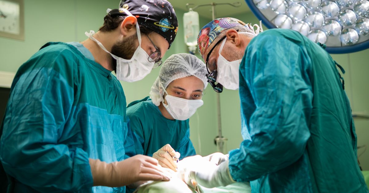
For patients with localized ductal adenocarcinoma or precancerous cysts, surgical resection offers the only potential for a cure. If the mass is located in the head of the pancreas, the standard operation is the pancreaticoduodenectomy, universally known as the Whipple procedure.
The Whipple is one of the most complex operations in general surgery. The surgeon must remove the head of the pancreas, the gallbladder, the duodenum, a portion of the bile duct, and sometimes a part of the stomach. Afterward, the digestive tract must be meticulously reconstructed, sewing the remaining pancreas, bile duct, and stomach back into the small intestine to restore digestive continuity.
If the mass is located in the body or tail of the pancreas, a distal pancreatectomy is performed. This involves removing the left side of the pancreas, usually along with the spleen, as they share a closely intertwined blood supply.
14. Determining Surgical Resectability
Not all pancreatic masses can be surgically removed. The crucial factor determining “resectability” is the tumor’s relationship to the major blood vessels passing directly behind the pancreas, specifically the superior mesenteric artery and the celiac axis.
If the computed tomography scan shows that the tumor is wrapping around or invading these vital arteries, the tumor is deemed unresectable, as removing these arteries would fatally compromise blood flow to the intestines and liver.
If the tumor is just touching the vessels, it is considered borderline resectable. These patients often receive intensive chemotherapy and radiation upfront to shrink the tumor and pull it away from the blood vessels, potentially allowing for a safe surgical resection at a later date.
15. Systemic Oncological Therapies
Because pancreatic adenocarcinoma is a systemic disease that sheds microscopic cancer cells early in its development, surgery alone is rarely sufficient. Systemic chemotherapy is a cornerstone of treatment.
Adjuvant chemotherapy is administered after a successful surgery to eradicate any remaining microscopic disease and significantly improve long-term survival. Neoadjuvant chemotherapy is increasingly used before surgery to shrink borderline tumors and test the tumor’s biological aggressiveness before committing the patient to a massive operation.
For advanced or metastatic disease, palliative chemotherapy is utilized to control tumor growth, alleviate pain, and extend the patient’s quality of life. Modern research is heavily focused on targeted therapies and immunotherapies, aiming to breach the tumor’s protective fibrous shield and improve patient outcomes, employing strategies similar to the multidisciplinary approach used for a complex mediastinal mass.
16. Frequently Asked Questions FAQ
1. Is a cyst on the pancreas the same as pancreatic cancer?
No. A cyst is a fluid-filled sac, whereas cancer is a solid tumor. While some cysts are completely harmless, others are precancerous and can eventually turn into cancer if left alone. A specialist must evaluate the cyst using specialized imaging to determine its risk.
2. Why does a pancreatic tumor cause jaundice?
The tube that carries bile from your liver to your intestines runs straight through the head of the pancreas. If a tumor grows there, it pinches the tube shut. The bile backs up into your blood, turning your skin and eyes yellow.
3. What is a Whipple procedure?
It is a major surgery used to remove tumors in the head of the pancreas. The surgeon removes the tumor, the first part of the intestine, the gallbladder, and part of the bile duct, and then reconnects the remaining organs so you can digest food.
4. Can pancreatic cancer be cured?
If caught very early before it has spread, and if it can be completely removed with surgery, there is a chance for a cure. However, because it rarely causes early symptoms, it is most often found at an advanced stage where treatment focuses on extending life and controlling symptoms.
5. How is a biopsy of the pancreas performed?
Because the pancreas is deep in the body, doctors use a procedure called an Endoscopic Ultrasound. You are put to sleep, and a flexible camera with an ultrasound is passed down your throat into your stomach. A tiny needle is then passed through the stomach wall to safely sample the tumor.
17. Bibliography
Disclaimer: The content is for informational purposes only and does not replace medical advice. Always consult your doctor for personalized treatment.