1. Introduction
A loud grinding noise in the knees during lunges is clinically known as crepitus, resulting from rough articular cartilage rubbing behind the kneecap or the patella tracking poorly within the femoral groove. This mechanical friction, often an early sign of patellofemoral pain syndrome or chondromalacia patellae, amplifies under the substantial compressive load of deep knee flexion. Management requires a careful clinical assessment of muscular imbalances, followed by targeted physical therapy to strengthen the quadriceps and correct the biomechanical alignment of the lower extremity.
2. Anatomy of the Patellofemoral Joint
The knee is a complex hinge joint designed to absorb massive impact forces while providing dynamic mobility. The front portion of this joint, where the grinding typically occurs, is the patellofemoral joint. It consists of the patella (kneecap) resting in the trochlear groove of the femur (thigh bone).
The underside of the patella and the surface of the trochlear groove are coated in a thick layer of hyaline cartilage. This specialized tissue is exceptionally smooth, lubricated by synovial fluid, allowing the kneecap to glide silently and effortlessly up and down the groove as the knee bends and straightens.
3. The Mechanics of a Lunge
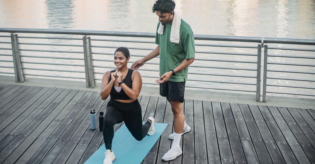
A lunge is a closed kinetic chain exercise that places immense biomechanical demand on the patellofemoral joint. As the knee descends into deep flexion, the quadriceps muscle contracts forcefully to control the descent. This muscular contraction pulls the patella tightly against the femur.
The compressive force between the kneecap and the thigh bone increases exponentially as the angle of knee flexion increases. If the articular cartilage is perfectly smooth and the patella is aligned precisely, this force is distributed evenly. However, any structural roughness or misalignment translates this intense compression into audible, grinding friction.
4. Defining Joint Crepitus
The medical term for the grinding, crunching, or popping noises emanating from a joint is crepitus. It is critical to differentiate between physiological and pathological crepitus. Physiological popping is caused by the sudden release of nitrogen gas bubbles within the synovial fluid, similar to cracking a knuckle. This type of pop is usually a single, sharp sound and is entirely harmless.
Pathological crepitus, which sounds like crushed glass or gravel grinding together, indicates underlying mechanical dysfunction. The continuous crunching noise heard during the descent and ascent of a lunge represents the physical collision of uneven cartilaginous surfaces, alerting the clinician to structural degradation.
5. Chondromalacia Patellae
The most frequent cause of grinding in the younger athletic population is chondromalacia patellae. This condition involves the softening, fraying, and eventual breakdown of the hyaline cartilage on the underside of the kneecap.
Instead of a smooth, frictionless surface, the cartilage becomes fibrillated, resembling the texture of crabmeat. When this rough, degraded surface is dragged across the femur under the intense compressive load of a lunge, it produces a loud, gritty vibration. While the grinding itself may be painless initially, progressive cartilage loss eventually exposes the underlying sensory nerves in the bone, leading to significant discomfort.
6. Patellar Tracking Dysfunction
Proper function of the patellofemoral joint requires the kneecap to stay perfectly centered within its groove. Patellar tracking dysfunction occurs when muscular imbalances pull the kneecap off-center, usually dragging it too far toward the outside (lateral) edge of the knee.
This lateral pull forces the kneecap to ride up on the rigid bony ridge of the femoral groove. The cartilage is not designed to bear weight on this sharp ridge. The resulting mechanical grinding is loud and highly destructive to the delicate articular cartilage. Addressing this misalignment is a fundamental component of treating chronic knee pain.
7. Muscular Imbalances and the VMO
The alignment of the patella is governed by a tug-of-war between the quadriceps muscles. The vastus lateralis on the outside of the thigh is inherently large and powerful. It is counterbalanced by the vastus medialis obliquus on the inside of the thigh, which is smaller and frequently weak or neurologically inhibited.
When an individual performs a lunge with a weak vastus medialis obliquus, the dominant outer muscle easily wins the anatomical tug-of-war, pulling the kneecap laterally. Furthermore, a tight iliotibial band running down the outside of the leg can exacerbate this lateral tethering, guaranteeing a grinding collision with every repetition.
8. The Q-Angle and Biomechanics
The Quadriceps angle, or Q-angle, is a clinical measurement that dictates the line of pull of the quadriceps muscle relative to the kneecap. It is determined by the width of the pelvis in relation to the center of the knee. Women naturally possess a wider pelvis and consequently a larger Q-angle.
A larger Q-angle creates a sharper diagonal pull on the patella, inherently increasing the risk of lateral tracking dysfunction. Individuals with wide hips, flat feet (overpronation), or knock-knees (genu valgum) possess altered biomechanics that significantly magnify the grinding forces during closed chain exercises like lunges.
9. Osteoarthritis of the Knee
In individuals over the age of fifty, loud grinding during weight-bearing flexion is frequently the hallmark of advanced knee osteoarthritis. Osteoarthritis involves the progressive loss of the joint’s cartilage cushion.
As the condition advances, the cartilage may erode completely, leaving exposed bone. Bone grinding on bone produces a remarkably loud, coarse crepitus. The joint attempts to stabilize itself by growing new, irregular bone formations called osteophytes or bone spurs. These bony ridges further destroy the smooth gliding mechanism of the knee, making deep flexion exercises mechanically harsh and painful.
10. Synovial Plica Syndrome
The synovial membrane lining the knee joint sometimes develops thick, fibrotic bands of tissue known as plicae. These are essentially anatomical remnants from fetal development. While usually asymptomatic, a plica can become inflamed and thickened due to repetitive friction or trauma.
During a lunge, as the knee bends, a thickened medial plica can snap abruptly over the medial femoral condyle. This creates a palpable catching sensation and a loud, localized pop or grind. Synovial plica syndrome is uniquely characterized by a dull, aching pain on the inner aspect of the kneecap that flares up specifically during repetitive bending.
11. Clinical Diagnosis and Assessment
A physician or physical therapist diagnoses the specific cause of crepitus through a targeted physical examination. The clinician will place a hand firmly over the patient’s kneecap while instructing them to perform a slow squat or lunge, physically feeling the exact location and quality of the grinding vibrations.
The patellar apprehension test involves the clinician attempting to push the kneecap laterally to assess joint stability and check for tracking dysfunction. The therapist will also analyze the patient’s lunge form, looking for collapsing arches in the feet or inward dropping of the knees, which highlight the proximal weakness in the gluteal muscles driving the poor mechanics.
12. Diagnostic Imaging Technologies
Standard weight-bearing X-rays are the initial imaging modality utilized to evaluate the joint space. A specialized view, called the sunrise view or merchant view, is captured with the knee bent, looking down across the kneecap. This specific angle clearly displays if the patella is tilted or tracking improperly within the groove.
If soft tissue damage, such as a severe cartilage defect or a meniscal tear, is suspected, magnetic resonance imaging provides unparalleled high-resolution imaging. An MRI allows the orthopedic surgeon to grade the exact depth and severity of chondromalacia patellae.
13. Physical Therapy and Neuromuscular Re-education
The gold standard for eliminating painful grinding involves rigorous physical therapy. The primary objective is to correct the muscular imbalances pulling the kneecap off center. Targeted exercises isolate and strengthen the vastus medialis obliquus and the gluteus medius.
Strengthening the gluteal muscles ensures the femur remains externally rotated and stable during a lunge, preventing the knee from collapsing inward. Therapists utilize myofascial release techniques and stretching protocols to lengthen a tight iliotibial band and lateral retinaculum, releasing the excessive outward tension on the patella.
14. Data: Characteristics of Knee Noises
| Type of Sound | Anatomical Cause | Clinical Significance |
|---|---|---|
| Single, sharp pop (Painless) | Nitrogen gas bubble release. | Benign, normal joint physiology. |
| Grinding or crunching (Crepitus) | Cartilage fraying (Chondromalacia). | Indicates friction; requires muscular correction. |
| Snapping or catching | Synovial Plica or Meniscal tear. | Tissue impingement; may require medical intervention. |
| Coarse, bone-on-bone grinding | Advanced Osteoarthritis. | Severe cartilage loss; necessitates activity modification. |
15. Form Modification and Joint Preservation
Until the biomechanical imbalances are corrected, patients must modify their exercise routine to protect the articular cartilage. Performing shallow lunges limits the extreme compressive forces that occur past ninety degrees of flexion.
Utilizing supportive footwear or custom orthotics corrects foot overpronation, establishing a stable foundation for the entire leg. Taping the kneecap with specialized kinesiology tape can manually guide the patella into the center of the groove, providing immediate, albeit temporary, relief from the grinding friction during rehabilitation sessions.
16. Frequently Asked Questions
1. Is it safe to keep doing lunges if my knees grind but don’t hurt?
Pain-free grinding is a warning sign of poor mechanics. While it may not hurt today, continuing to grind the cartilage under heavy loads will eventually wear it down to the sensitive bone. You should modify your form and strengthen your stabilizing muscles to stop the friction.
2. Does taking collagen or glucosamine supplements rebuild the worn cartilage?
Clinical evidence is highly mixed. Cartilage has no direct blood supply, making it exceptionally difficult for oral supplements to reach and repair the tissue. Supplements may reduce overall joint inflammation, but they cannot physically regrow degraded cartilage or fix a tracking issue.
3. Will a knee sleeve stop the grinding noise?
A neoprene knee sleeve provides warmth and mild compression, which feels supportive and can reduce minor swelling. However, a standard sleeve does not exert enough mechanical force to realign a tracking kneecap, so it will not stop the underlying grinding mechanism.
4. Why does the grinding only happen on the way down into the lunge?
The descent phase requires an eccentric contraction, where the quadriceps muscles must lengthen while under extreme tension to act as a brake. This eccentric load pulls the kneecap against the femur with maximum force, making structural imperfections much more audible.
5. When does loud knee grinding require surgery?
Surgery is only considered when extensive physical therapy completely fails, the pain becomes severely debilitating, and imaging confirms major structural defects, such as large pieces of loose cartilage floating in the joint or a severely malaligned patella requiring a lateral release procedure.
17. Bibliography
Disclaimer: The content is for informational purposes only and does not replace medical advice. Always consult your doctor for personalized treatment.