1. Introduction to Knee Region Pain
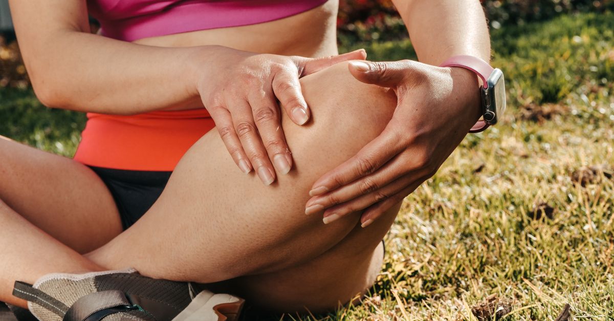
Pain encompassing the broader region of the knee, rather than just the internal joint space, is a highly frequent and debilitating clinical complaint. The knee region is a complex biomechanical transition zone connecting the powerful musculature of the thigh to the structural levers of the lower leg. Because it functions as a primary load-bearing mechanism during dynamic activities such as running, jumping, and climbing, the surrounding soft tissues are subjected to immense mechanical tension and repetitive stress.
The diagnostic evaluation of knee region pain requires a systematic clinical approach to differentiate between true intra-articular joint destruction, such as osteoarthritis, and the multitude of extra-articular or periarticular conditions that affect the surrounding tendons, ligaments, and bursae. Patients typically present with localized aching, a deep burning sensation, or sharp pain that correlates directly with specific phases of physical movement.
Establishing the precise anatomical origin of these symptoms is the critical foundation for formulating an effective therapeutic intervention. Comprehensive medical management ranges from targeted physiotherapy aimed at restoring muscular balance and flexibility to specific pharmacological pain control. Early clinical assessment prevents minor localized inflammation from evolving into chronic, stiff, and severely degenerated tissue states, ultimately preserving long-term ambulatory function.
2. Anatomy of the Periarticular Knee Structures
The knee region is supported and stabilized by a dense network of extra-articular soft tissues. The most prominent structure is the extensor mechanism, comprising the massive quadriceps muscle, the quadriceps tendon, the patella, and the patellar tendon, which anchors the entire complex to the tibial tubercle. This mechanism provides the immense power required to extend the leg against gravity.
On the lateral aspect of the knee courses the iliotibial band, a thick strip of fascia extending from the pelvis down to the lateral tibia. This band provides vital lateral stability during the stance phase of walking and running. On the medial aspect, the pes anserinus represents the conjoined tendons of three separate thigh muscles (sartorius, gracilis, and semitendinosus) that insert onto the upper inner tibia.
To facilitate smooth, frictionless movement between these powerful tendons and the underlying bones, the knee region is equipped with multiple fluid-filled sacs known as bursae. The prepatellar bursa sits directly in front of the kneecap, while the pes anserine bursa cushions the medial tendons. Any disruption in the delicate biomechanical balance of these structures leads to concentrated mechanical stress and subsequent periarticular pain.
3. Patellofemoral Pain Syndrome
Patellofemoral pain syndrome is a highly prevalent musculoskeletal condition characterized by severe, aching pain localized directly behind or surrounding the patella. This condition frequently affects young, active adults and is fundamentally driven by abnormal biomechanical tracking of the kneecap as it glides within the trochlear groove of the femur during movement.
The abnormal tracking is frequently caused by a muscular imbalance, specifically weakness in the vastus medialis obliquus muscle of the inner quadriceps, combined with tightness in the lateral structures such as the iliotibial band. Patients report pain that significantly worsens during activities that heavily load the flexed knee, such as squatting, running, or walking down stairs.
A classic clinical feature is the “movie theater sign,” where the patient experiences deep, aching anterior knee pain after sitting with the knees bent for a prolonged period. Management completely eschews surgical intervention, focusing intensely on a structured physical therapy program designed to stretch tight lateral tissues and dynamically strengthen the core and quadriceps musculature to restore proper patellar alignment.
4. Patellar Tendinopathy
Patellar tendinopathy, clinically recognized as “jumper’s knee,” is an overuse injury characterized by chronic, painful cellular degeneration within the tendon that connects the kneecap to the shinbone. This condition is overwhelmingly associated with sports that require repetitive, explosive jumping and landing, such as basketball or volleyball.
The immense eccentric forces generated during landing lead to microscopic tearing and disorganized collagen formation at the inferior pole of the patella. Patients experience a severe, burning ache localized strictly to the patellar tendon. The pain is typically worse at the beginning of a physical activity, often subsiding as the tendon warms up, only to return with increased severity once the activity ceases.
Direct clinical palpation of the patellar tendon elicits pinpoint tenderness. Management requires strict activity modification to halt repetitive microtrauma. A structured physical therapy program emphasizing eccentric strengthening exercises on a decline board is the gold standard for stimulating collagen remodeling and restoring the structural integrity of the diseased tendon.
5. Iliotibial Band Syndrome
Iliotibial band syndrome is a leading cause of severe pain localized to the lateral aspect of the knee region, predominantly affecting long-distance runners and cyclists. During the continuous, repetitive flexion and extension of the knee, the tight fibrous band forcefully snaps back and forth over the lateral femoral epicondyle, a prominent bony ridge.
This chronic, repetitive friction incites localized inflammation in the underlying tissues and the band itself. Patients report a sharp, burning pain on the outside of the knee that typically begins a few miles into a run and progressively worsens until the athlete is forced to stop. The pain rapidly dissipates with rest but predictably returns during the next physical exertion.
Clinical evaluation involves specific provocative maneuvers, such as the Noble compression test, to reproduce the localized friction pain. Management involves strict activity modification, the application of localized cryotherapy, and a rigorous physical therapy program focused on deep tissue massage, foam rolling, and strengthening the gluteal muscles to control pelvic stability during the gait cycle.
6. Pes Anserine Bursitis
Pain localized to the medial aspect of the knee region, just below the joint line, is frequently attributed to pes anserine bursitis. The pes anserine bursa is a fluid-filled sac that cushions the conjoined tendons of the medial thigh muscles where they insert onto the tibia.
This condition is prevalent among overweight individuals with underlying knee osteoarthritis, as the altered joint mechanics place excessive stretch on the medial tendons. It is also common in runners who abruptly increase their mileage. Patients present with highly localized, aching pain on the inner, lower portion of the knee that frequently radiates down the medial shin.
The pain is characteristically worsened by climbing stairs or rising from a seated position. Direct clinical palpation over the pes anserine insertion elicits significant tenderness. Treatment involves targeted physical therapy to stretch the hamstring muscles, weight management, and localized corticosteroid injections to rapidly reduce the bursal inflammation.
7. Prepatellar Bursitis
The prepatellar bursa is located superficially, directly between the skin and the anterior surface of the patella. Prepatellar bursitis, colloquially known as “housemaid’s knee,” occurs when this sac becomes actively inflamed and accumulates excess synovial fluid. The inflammation is typically triggered by prolonged, repetitive kneeling on hard surfaces, causing chronic mechanical friction.
The knee develops a distinct, highly visible, fluctuant swelling directly over the kneecap. In non-infectious cases, the localized swelling may be relatively painless unless subjected to direct physical pressure. However, if the bursa becomes infected through a microscopic break in the overlying skin, the condition evolves into septic bursitis.
In septic bursitis, the area becomes intensely erythematous, warm, and exquisitely painful. This is a medical urgency requiring immediate clinical aspiration of the fluid for microbiological culture and prompt initiation of targeted systemic antibiotic therapy to prevent the infection from spreading into the true joint space.
8. Osgood-Schlatter Disease
Osgood-Schlatter disease is a frequent cause of anterior knee region pain in growing adolescents, particularly those engaged in running and jumping sports. It is an traction apophysitis, involving painful inflammation at the insertion point of the patellar tendon on the tibial tubercle, the prominent bony bump just below the kneecap.
During periods of rapid skeletal growth, the bones grow faster than the surrounding muscles and tendons. The resulting increased tension, combined with repetitive mechanical pulling from the powerful quadriceps, causes microscopic avulsion fractures at the cartilaginous growth plate. Adolescents present with localized pain, distinct swelling, and a markedly prominent tibial tubercle.
The pain is exacerbated by direct pressure, kneeling, or explosive jumping. Management is entirely conservative, relying on activity modification, localized cryotherapy, and targeted stretching of the quadriceps and hamstrings. The condition universally resolves once the adolescent’s skeletal growth is complete and the growth plate fuses into solid bone.
9. Referred Pain from the Hip
It is a critical clinical imperative to recognize that pain perceived in the knee region may actually originate from severe pathology within the hip joint. Because the femoral nerve supplies sensory branches to both the hip and the anterior knee, the brain frequently misinterprets signals of deep hip distress as originating in the knee.
Conditions such as severe hip osteoarthritis, avascular necrosis, or slipped capital femoral epiphysis in children frequently present with knee pain as the primary, and sometimes only, clinical complaint. Referred pain is characteristically described as a deep, poorly localized ache that is not associated with any localized knee swelling or mechanical symptoms.
Crucially, active or passive movement of the knee joint itself does not typically exacerbate this referred pain. Instead, the pain is reproduced or worsened by specific rotational movements of the hip joint. Recognizing this distinction directs clinical attention and radiographic evaluation toward the hip, preventing profound misdiagnosis.
10. Meniscal and Ligamentous Pathology
While primarily intra-articular issues, injuries to the menisci and the collateral ligaments present with distinct regional pain patterns. The medial and lateral collateral ligaments stabilize the knee against side-to-side forces. A sprain of these ligaments, typically from a direct blow to the side of the knee, causes acute, sharp pain directly over the affected ligament.
Meniscal tears typically occur during dynamic activities that involve sudden, forceful twisting of the knee while the foot is planted. Patients describe a deep, sharp pain localized distinctly to the inner or outer joint line. The clinical hallmark of a torn meniscus is the presence of distinct mechanical symptoms, including sudden catching, painful locking, or a sensation that the knee is physically giving way.
Diagnosis relies on specific provocative maneuvers and is confirmed via magnetic resonance imaging. Symptomatic tears or severe ligamentous ruptures frequently require arthroscopic surgical intervention to restore dynamic joint stability and prevent rapid, early-onset joint degeneration.
11. Regional Degenerative Osteoarthritis
Osteoarthritis is a profound, progressive disease characterized by the gradual deterioration of the protective articular cartilage. As the cartilage wears away over decades of continuous mechanical loading, the underlying subchondral bones grind directly against one another, inciting localized inflammation and affecting the entire knee region.
Patients suffering from regional osteoarthritis typically report a deep, generalized ache that is reliably provoked by physical exertion, particularly prolonged walking or descending stairs. As the disease advances, patients experience profound morning stiffness and a progressive, structural loss of terminal extension and flexion.
To compensate for the loss of cartilage and increasing mechanical instability, the body forms prominent osteophytes, or bone spurs, altering the mechanical axis of the entire lower leg. Management relies heavily on comprehensive weight reduction, targeted physical therapy to strengthen the surrounding musculature, and eventual total joint arthroplasty for definitive pain relief.
12. Differentiating Anterior, Lateral, and Medial Pain
A meticulous clinical analysis of the specific location and mechanical triggers of the pain directs the clinician toward an accurate anatomical diagnosis.
| Regional Location of Pain | Common Associated Diagnosis |
|---|---|
| Anterior pain, directly on the patellar tendon, worse when jumping | Patellar Tendinopathy (Jumper’s Knee) |
| Lateral pain, burning sensation during long-distance running | Iliotibial Band Syndrome |
| Medial pain, localized below the joint line, worse on stairs | Pes Anserine Bursitis |
| Anterior pain with visible, fluctuant swelling directly over the kneecap | Prepatellar Bursitis |
The presence of an acutely hot, red, and swollen knee region accompanied by a systemic fever warrants immediate advanced clinical evaluation to rule out a severe, limb-threatening bacterial infection.
13. Diagnostic Evaluation and Imaging
The clinical examination of the knee region involves precise palpation of bony landmarks, assessing the integrity of specific ligaments and tendons through manual stress testing, and observing the patient’s dynamic gait pattern. Standard weight-bearing plain radiographs are the universally accepted first line of imaging.
When complex soft tissue injury is suspected, such as severe tendinosis, occult meniscal tears, or ligamentous ruptures, magnetic resonance imaging provides unparalleled anatomical resolution. This advanced imaging modality is the absolute gold standard for comprehensively evaluating both the intra-articular and extra-articular structures of the knee region.
Diagnostic ultrasound is increasingly utilized for the dynamic, real-time assessment of superficial tendons, such as the patellar tendon, and for accurately confirming the presence and extent of localized bursitis. Furthermore, clinical joint aspiration is mandatory to extract and analyze fluid from a swollen bursa or joint capsule to rule out active infection.
14. Physical Therapy and Biomechanics
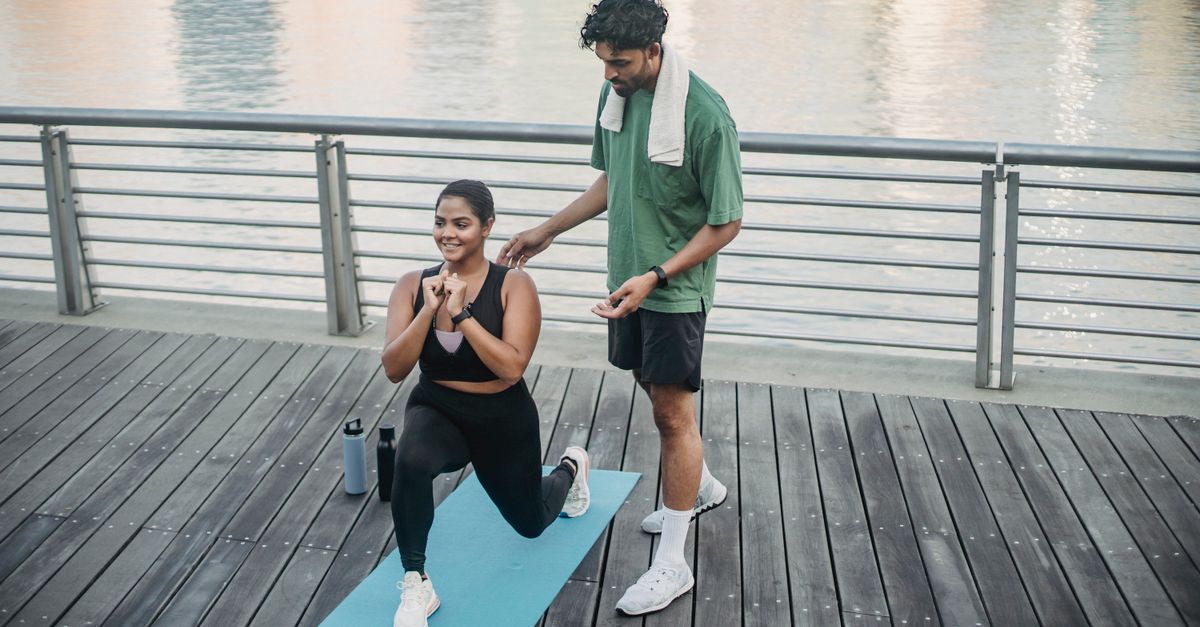
The initial management of pain in the knee region relies heavily on conservative, non-pharmacological interventions. The absolute cornerstone of long-term management is comprehensive physical therapy. A structured, progressive exercise program prevents severe muscular deconditioning and enhances the dynamic stability provided by the quadriceps and hamstring muscles.
Biomechanical correction is a fundamental component of conservative care. For conditions like patellofemoral pain syndrome and iliotibial band syndrome, therapy focuses intensely on strengthening the core and gluteal muscles to prevent the thigh bone from rotating internally during the gait cycle.
Utilizing specialized, custom-molded orthotics supports collapsing foot arches and alters the mechanical axis of the lower limb, significantly redistributing pressure away from damaged structures. Comprehensive weight loss is also considered a primary, highly effective therapeutic intervention to directly unload the knee region.
15. Medical and Surgical Interventions
Pharmacological intervention utilizes oral nonsteroidal anti-inflammatory drugs to mitigate acute mechanical pain and reduce soft tissue edema. For localized, severe pain that does not respond to oral medications, targeted corticosteroid injections provide rapid, potent suppression of the inflammatory response in conditions like pes anserine or prepatellar bursitis.
When conservative measures fail to control intractable pain, or when structural tendon or joint destruction severely impairs ambulatory function, surgical intervention is required. Modern knee surgery heavily utilizes arthroscopic techniques, allowing surgeons to utilize minimally invasive instruments to thoroughly evaluate and repair the regional structures.
Through small incisions, the surgeon can debride degenerative tendon tissue, repair torn meniscal fragments, or reconstruct completely ruptured ligaments. For severe, end-stage osteoarthritis affecting the entire knee region, total knee arthroplasty provides comprehensive joint replacement, offering profound pain relief and the full restoration of mechanical function.
16. Frequently Asked Questions FAQ
1. Why does my knee hurt when I sit at my desk for a long time?
This is a classic presentation of patellofemoral pain syndrome, often called the “movie theater sign.” When your knee is bent for prolonged periods, the kneecap is compressed tightly against the underlying femur. This sustained mechanical pressure reduces blood flow and irritates the underlying cartilage, causing a deep, aching pain.
2. Should I run through the pain on the outside of my knee?
Absolutely not. Sharp, lateral knee pain during running is typical of iliotibial band syndrome. Attempting to run through this specific friction pain will rapidly increase the localized inflammation, leading to a much longer recovery period. Immediate rest and targeted physical therapy are necessary.
3. Can tight hamstrings cause knee pain?
Yes. The hamstring muscles cross the back of the knee region. If they are chronically tight, they alter the normal biomechanics of the pelvis and the knee joint, increasing the compressive forces placed on the kneecap and predisposing the individual to conditions like pes anserine bursitis.
4. Is a prominent bump below my teenager’s knee normal?
A prominent, painful bump precisely on the tibial tubercle in an active adolescent is the hallmark sign of Osgood-Schlatter disease. It is a very common, benign inflammation of the growth plate caused by rapid bone growth and tight thigh muscles. It resolves completely once skeletal growth finishes.
5. How do I know if my knee pain is coming from my hip?
Referred pain from the hip usually presents as a deep, poorly localized ache in the front of the knee without any actual swelling or tenderness in the knee tissues themselves. Furthermore, moving the knee does not worsen the pain, but rotating the hip joint will reliably reproduce the severe discomfort.
17. Bibliography
Disclaimer: The content is for informational purposes only and does not replace medical advice. Always consult your doctor for personalized treatment.