
1. Introduction
Kyphoscoliosis is a complex spinal deformity characterized by the abnormal curvature of the vertebral column in two anatomical planes. It involves both an excessive outward curvature of the thoracic spine (kyphosis) and a lateral, twisting curvature (scoliosis). This dual-plane distortion alters the body’s center of gravity and can significantly affect the function of internal organs, particularly the lungs and the heart, depending on the severity of the spinal angulation.
The progression of this structural anomaly varies considerably among patients. Mild forms may remain asymptomatic and require only routine monitoring, while more severe curvatures can cause significant respiratory restriction, chronic back discomfort, and neurological deficits. Timely identification and intervention are essential to prevent structural deterioration and maintain physical function.
Modern orthopedic management offers a range of therapeutic options. Depending on the patient’s age, skeletal maturity, and the degree of curvature, treatment may involve specialized physical therapy, custom orthotic bracing, or surgical spinal fusion to stabilize the vertebral column and restore anatomical alignment.
2. Anatomy and Biomechanics of the Spine
The human spine is a column of interlocking bones, called vertebrae, separated by shock-absorbing intervertebral discs. In a typical spine, the cervical (neck) and lumbar (lower back) regions curve gently inward, while the thoracic (upper back) region curves gently outward. This natural S-shape provides balance and mechanical strength, allowing for upright posture and shock absorption during movement.
In kyphoscoliosis, this biomechanical harmony is disrupted. The lateral curve of the scoliosis component introduces a twisting or rotational force to the vertebral bodies, causing the ribs to shift and form a prominence on one side of the back. Simultaneously, the kyphotic component exaggerates the forward rounding of the upper back.
These combined forces place uneven pressure on the intervertebral discs and the facet joints that connect the vertebrae. Over time, this asymmetrical loading accelerates degenerative changes, contributing to chronic discomfort and reduced spinal flexibility.
3. Pathophysiology and Curvature Progression
The exact mechanism that drives the progression of kyphoscoliosis depends on its underlying etiology. During periods of rapid skeletal growth, such as the adolescent growth spurt, the spine is particularly vulnerable to structural deformation. If a curvature develops, the asymmetrical forces applied to the growing bone can alter normal bone development according to the Hueter-Volkmann law.
This law states that increased pressure on one side of a growth plate slows bone growth, while reduced pressure accelerates it. Consequently, the vertebrae begin to grow in a wedge shape, which structurally locks the deformity in place and facilitates further curvature progression if left unchecked.
In adult-onset or degenerative cases, the pathophysiology involves the progressive failure of the intervertebral discs and supporting ligaments. As the discs lose height and hydration, the spine loses its structural integrity, allowing the vertebrae to tilt and rotate out of alignment under the force of gravity.
4. Congenital and Idiopathic Etiologies
Congenital kyphoscoliosis is present at birth and results from a failure of the vertebrae to form or segment properly during embryonic development. Infants may be born with hemivertebrae (partially formed vertebrae) or unsegmented bars (vertebrae fused together). These structural anomalies dictate an asymmetric growth pattern as the child matures.
Idiopathic kyphoscoliosis is the most common presentation, emerging most frequently during adolescence. The term “idiopathic” indicates that the exact cause is unknown, although genetic factors are strongly suspected to play a role. Adolescents with this condition often experience rapid progression of the curvature during their peak growth years.
Identifying the etiology is crucial, as congenital curves tend to be more rigid and progressive, often requiring earlier surgical intervention compared to idiopathic curves, which may respond to conservative bracing.
5. Neuromuscular and Syndromic Causes
Neuromuscular kyphoscoliosis occurs as a secondary complication of central or peripheral nervous system disorders. Conditions such as cerebral palsy, muscular dystrophy, and spinal muscular atrophy compromise the muscle strength and coordination required to support the spinal column. The resulting muscle imbalance pulls the flexible spine into complex, often severe curvatures.
Syndromic kyphoscoliosis is associated with underlying connective tissue disorders, such as Marfan syndrome or Ehlers-Danlos syndrome. In these conditions, the ligaments and supportive structures of the spine are inherently weak and hypermobile, failing to maintain adequate spinal alignment under normal loads.
These forms of the deformity are often challenging to manage, as the underlying systemic conditions complicate both conservative therapies and surgical interventions, requiring a coordinated, multidisciplinary medical approach.
6. Respiratory and Cardiovascular Implications
One of the most concerning complications of severe kyphoscoliosis is its impact on pulmonary function. The thoracic cage, composed of the ribs and sternum, attaches directly to the thoracic spine. As the spine twists and curves forward, the volume of the chest cavity is physically reduced, and the ribs lose their normal capacity to expand during inhalation.
This structural restriction leads to restrictive lung disease. Patients may experience diminished vital capacity, which is the maximum amount of air a person can expel from the lungs after a maximum inhalation. Chronic hypoventilation can result in reduced oxygen levels in the blood, leading to fatigue and diminished exercise tolerance.
If the curvature becomes profound, the distorted chest wall can compress the heart and major blood vessels. This places an increased workload on the right ventricle of the heart to pump blood through the compressed pulmonary vasculature, potentially leading to cor pulmonale, a serious form of right-sided heart failure.
7. Clinical Symptoms and Patient Presentation
The presentation of kyphoscoliosis varies widely. In mild cases, patients may only notice a cosmetic asymmetry, such as uneven shoulders, a prominent shoulder blade, or a noticeable hump on one side of the back when bending forward.
As the curvature progresses, pain becomes a more prominent symptom. Muscular fatigue is common, as the paraspinal muscles work continuously to counteract the abnormal biomechanical forces and maintain balance. Patients frequently report a deep, aching discomfort in the upper and mid-back that worsens after prolonged sitting or standing.
In advanced or degenerative cases, patients may exhibit neurological symptoms. If the misaligned vertebrae narrow the spinal canal or compress the nerve roots exiting the spine, the patient may experience radiating pain, numbness, tingling, or weakness in the legs.
8. Diagnostic Physical Examination
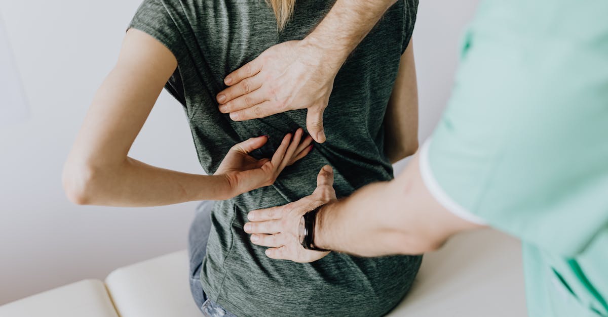
The clinical evaluation begins with a detailed physical examination. The physician assesses the patient’s overall posture, observing the alignment of the head, shoulders, and pelvis. A crucial component of this evaluation is the Adam’s Forward Bend Test, where the patient bends forward at the waist with arms hanging down.
This position highlights the rotational component of the scoliosis, making any rib prominence clearly visible. A scoliometer may be used during this test to measure the angle of trunk rotation, providing a quantifiable metric to track progression.
The physician will also perform a thorough neurological assessment, checking reflexes, muscle strength, and sensation in the extremities to rule out spinal cord or nerve root compression.
9. Radiological Evaluation and Measurement
Definitive diagnosis and treatment planning rely on precise radiological imaging. Full-length, weight-bearing spinal radiographs (X-rays) taken from the front (coronal plane) and the side (sagittal plane) are the standard diagnostic tools.
On these radiographs, the physician measures the Cobb angle, which quantifies the severity of both the scoliosis and the kyphosis. A Cobb angle greater than 10 degrees defines the presence of a curvature, while angles exceeding 40 to 50 degrees often indicate a need for surgical intervention.
If neurological symptoms are present, or if surgery is being planned, a magnetic resonance imaging (MRI) scan is typically ordered. An MRI provides detailed visualization of the spinal cord, nerve roots, and intervertebral discs, allowing the surgical team to identify any points of neural compression or structural anomalies within the spinal canal.
10. Data Summary: Curvature Classifications
Understanding the degree of curvature helps dictate the appropriate clinical management. The table below outlines the general correlation between Cobb angle measurements and typical treatment strategies.
| Degree of Curvature (Cobb Angle) | Clinical Classification | Standard Treatment Approach |
|---|---|---|
| 10 to 25 degrees | Mild Deformity | Observation, physical therapy, regular radiographic monitoring |
| 25 to 40 degrees | Moderate Deformity | Custom orthotic bracing (if still growing), specialized exercises |
| 40 to 50+ degrees | Severe Deformity | Surgical evaluation for spinal fusion and instrumentation |
11. Conservative Management and Physical Therapy
For mild to moderate curvatures, particularly in adults or adolescents nearing skeletal maturity, conservative management is the first line of treatment. The primary goal is not to reverse the curvature, which is generally not possible without surgery, but to manage symptoms, prevent further progression, and maintain spinal mobility.
Physical therapy is a cornerstone of this approach. Therapists prescribe specific, targeted exercises to strengthen the core musculature and improve posture. Techniques such as the Schroth method, which involves specific breathing patterns and asymmetric strengthening exercises, can help patients improve their trunk symmetry and respiratory mechanics.
Pain management may involve the use of nonsteroidal anti-inflammatory drugs to reduce muscle inflammation and discomfort. Patients are also educated on proper body mechanics and ergonomics to reduce daily mechanical stress on the spine.
12. Orthotic Bracing for Adolescents
If an adolescent with idiopathic kyphoscoliosis presents with a moderate curvature and has significant skeletal growth remaining, custom orthotic bracing is frequently recommended. The brace applies external corrective pressure to the spine and ribs, steering the growth of the vertebrae in a straighter alignment.
To be effective, the brace typically needs to be worn for a significant portion of the day, often up to 18 hours or more. The rigid plastic brace is custom-molded to the patient’s specific body shape and the exact pattern of their curvature.
While bracing can be socially and physically challenging for young patients, strict adherence to the wearing schedule is the most effective non-surgical method for halting curve progression and avoiding the need for spinal fusion surgery.
13. Surgical Indications and Goals
Surgical intervention is considered when the curvature exceeds 40 to 50 degrees, continues to progress despite bracing, causes severe pain that cannot be managed conservatively, or begins to compromise cardiopulmonary function.
The primary goals of surgery are to halt the progression of the deformity, achieve a reasonable anatomical correction, stabilize the spine, and relieve any pressure on the spinal cord or nerve roots. Surgeons carefully evaluate the flexibility of the curve before the procedure, often using special bending X-rays to determine how much of the deformity can be safely corrected.
For related information on nerve compression issues that may arise from spinal deformities, reading about sciatica can provide additional clinical context regarding neurological symptoms radiating into the lower extremities.
14. Spinal Fusion and Instrumentation
The standard surgical procedure for severe kyphoscoliosis is posterior spinal fusion with instrumentation. The surgeon makes an incision along the back to access the affected vertebrae. Specialized hardware, including pedicle screws, hooks, and titanium or cobalt-chrome rods, are attached to the bone.
The surgeon uses these rods to gently derotate and realign the spine, correcting both the kyphosis and the scoliosis. Once the spine is in its improved position, bone graft material is placed between the vertebrae. Over the following months, the bone graft stimulates the vertebrae to heal and fuse together into a single, solid mass of bone.
This fusion prevents any further movement in that specific section of the spine, permanently stabilizing the correction. Advanced neuromonitoring techniques are used throughout the surgery to continuously assess the function of the spinal cord and ensure neurological safety.
15. Postoperative Care and Rehabilitation
Recovery from spinal fusion surgery requires a dedicated rehabilitation period. Patients typically remain in the hospital for several days to manage post-surgical pain and begin early mobilization with physical therapists. Early walking is strongly encouraged to promote circulation and aid healing.
For the first few months, patients are restricted from bending, lifting, and twisting to allow the bone graft to fuse successfully. Full fusion typically takes six to twelve months, during which radiographic monitoring continues.
Once the fusion is solid, patients undergo physical therapy to strengthen the surrounding musculature and adapt to the new biomechanics of their spine. While a fused section of the spine will not bend, most patients retain sufficient mobility in their neck and lower back to perform daily activities comfortably.
16. Long-Term Prognosis
The long-term outlook for individuals treated for kyphoscoliosis is generally favorable, especially when diagnosed and managed appropriately. Patients who undergo successful spinal fusion typically experience significant relief from pain, stabilization of their cardiopulmonary function, and a notable improvement in physical appearance.
However, a spinal fusion does place increased mechanical stress on the unfused vertebrae located immediately above and below the fused section. This can lead to adjacent segment disease over a period of years, potentially requiring further intervention later in life.
Routine medical follow-up remains essential to monitor spinal health, ensure the integrity of surgical hardware, and address any late-onset degenerative changes.
17. Frequently Asked Questions (FAQ)
1. Can kyphoscoliosis be corrected without surgery?
While physical therapy and bracing can halt the progression of a curve and manage symptoms, they cannot permanently straighten a spine that has already developed a structural curvature. Surgery is the only method to physically correct a severe structural deformity.
2. Is it safe to exercise with this spinal condition?
Yes, maintaining core strength and flexibility through low-impact exercises like swimming, walking, and targeted physical therapy is highly beneficial for supporting the spine and reducing discomfort.
3. Will my child’s curve definitely get worse?
Progression is most likely during periods of rapid growth. Once a patient reaches skeletal maturity, mild to moderate curves often stabilize and do not worsen significantly, though severe curves can slowly progress in adulthood due to gravity and disc degeneration.
4. How long does the pain last after spinal fusion surgery?
Acute surgical pain typically subsides over the first few weeks and is managed with medication. Most patients return to school or light work within four to six weeks, though the complete bone fusion process takes several months.
5. Does kyphoscoliosis affect life expectancy?
Mild to moderate curvatures do not affect life expectancy. However, profound, untreated deformities that severely restrict lung and heart function can potentially reduce lifespan due to cardiopulmonary complications.
18. Bibliography
Disclaimer: The content is for informational purposes only and does not replace medical advice. Always consult your doctor for personalized treatment.

