1. Introduction
A lump on the face is a localized swelling or mass that can arise from various dermal or subcutaneous structures, most commonly indicating benign conditions such as epidermoid cysts, lipomas, or localized infections. The bottom line is that while the vast majority of facial lumps are harmless and aesthetically manageable, any persistent, rapidly growing, hard, or fixed facial mass requires prompt medical evaluation to definitively rule out infectious etiologies, salivary gland pathology, or malignant neoplastic processes. Proper clinical management relies on a thorough anatomical assessment to direct targeted diagnostic testing and appropriate surgical or pharmacological interventions.
The human face is a highly complex anatomical region comprising multiple tissue layers, including the epidermis, dermis, subcutaneous fat, intricate musculature, and specialized glandular structures. A mass can originate from any of these specific tissues. Because the face is the center of human interaction and identity, the appearance of a new lump frequently causes significant psychological distress and anxiety for the patient, regardless of the underlying pathology.
Differentiating a benign blemish from a clinically significant mass is the primary goal of the initial evaluation. Clinicians must meticulously assess the size, consistency, mobility, and growth rate of the lesion, combining these physical findings with the patient’s medical history to formulate an accurate differential diagnosis and establish a safe, effective treatment protocol that prioritizes both oncological safety and cosmetic outcomes.
2. Anatomical Considerations of the Facial Tissues
To accurately diagnose a facial lump, one must understand the layered anatomy of the face. The outermost layer is the epidermis, supported by the underlying dermis, which houses hair follicles, sebaceous (oil) glands, and sweat glands. Pathology originating here, such as blocked glands or follicular infections, typically presents as superficial, easily palpable lumps closely tied to the overlying skin.
Beneath the dermis lies the subcutaneous tissue, primarily composed of adipose (fat) cells and connective tissue fascia. Benign tumors of fat cells, known as lipomas, are common in this layer. This layer also contains a vast network of blood vessels and superficial nerves, meaning vascular malformations or nerve sheath tumors can present as subcutaneous facial masses.
Deeper still are the muscles of facial expression and the major salivary glands, most notably the parotid gland, which sits anterior to and just below the ear. Masses originating in the parotid gland or the deep musculature present as firm, deep-seated swellings that are less mobile and require advanced imaging to evaluate fully. Furthermore, the extensive lymphatic network of the face drains into specific regional lymph nodes, which can enlarge in response to infection or malignancy.
3. Epidermoid and Sebaceous Cysts
Epidermoid cysts, frequently mislabeled by the general public as sebaceous cysts, are among the most common benign lumps found on the face and neck. These cysts form when epidermal cells, which normally shed from the surface of the skin, move deeper into the dermis and multiply, creating a walled sac. This sac slowly fills with keratin, a thick, cheese-like protein produced by the skin cells.
Clinically, an epidermoid cyst presents as a smooth, round, slow-growing, and painless subcutaneous nodule. A defining characteristic is often the presence of a central punctum, a small, dark comedone (blackhead) opening on the surface of the skin directly overlying the mass. The cyst is typically freely mobile beneath the skin but tethered to the overlying epidermis at the site of the punctum.
True sebaceous cysts, which originate from the sebaceous glands and contain sebum rather than keratin, are actually quite rare and are more properly termed steatocystomas. While epidermoid cysts are benign, they can occasionally rupture, releasing the keratin into the surrounding tissue. This triggers an intense inflammatory response, causing the cyst to become rapidly swollen, red, hot, and exquisitely painful, mimicking an acute bacterial infection.
4. Lipomas: Benign Adipose Tumors
A lipoma is a benign, slow-growing tumor composed entirely of mature adipose (fat) tissue enclosed in a thin, fibrous capsule. They are incredibly common, though they appear less frequently on the face compared to the trunk or extremities. On the face, they most commonly present on the forehead, the cheeks, or along the jawline.
Upon physical examination, a lipoma is classically described as a soft, doughy, or rubbery mass. It is typically asymptomatic, painless, and highly mobile, slipping easily beneath the examining fingers. Because they originate in the subcutaneous fat layer, the overlying skin remains completely normal in color and texture and moves independently of the tumor.
Lipomas grow very slowly over months or years and almost never undergo malignant transformation. The primary clinical indication for treating a facial lipoma is cosmetic concern or if the tumor grows large enough to cause physical discomfort or compress adjacent facial nerves, leading to localized numbness or muscle weakness.
5. Infectious Causes: Abscesses and Furuncles
Acute localized infections are a frequent cause of rapidly developing, painful facial lumps. A furuncle, commonly known as a boil, is a deep infection of a hair follicle, usually caused by *Staphylococcus aureus* bacteria. It presents as a tender, red, inflammatory nodule that may eventually develop a central pustule.
If an infection spreads deeper into the subcutaneous tissues or if an epidermoid cyst becomes secondarily infected, a skin abscess can form. An abscess is a walled-off collection of purulent material (pus). Clinically, an abscess is characterized by the classic signs of acute inflammation: significant localized swelling, intense erythema (redness), heat, and severe pain. The mass is typically fluctuant, meaning it feels like a fluid-filled balloon when pressed.
Severe infections on the face, particularly those located in the “danger triangle” (the area from the corners of the mouth to the bridge of the nose), require urgent medical attention. The venous drainage of this specific anatomical region communicates directly with the cavernous sinus in the brain. A superficial infection left untreated in this area can theoretically spread internally, leading to a catastrophic central nervous system thrombosis.
6. Enlarged Lymph Nodes (Lymphadenopathy)
The face and scalp possess a rich network of lymphatic vessels that drain into specific groups of regional lymph nodes, primarily the preauricular nodes (in front of the ear), the submandibular nodes (under the jawline), and the submental nodes (under the chin). When the immune system responds to an inflammatory stimulus, these nodes multiply their lymphocyte populations, causing them to physically enlarge—a condition known as lymphadenopathy.
The most common cause of a swollen facial or submandibular lymph node is a reactive inflammatory response to a localized infection. Dental abscesses, severe acne, sinus infections, or viral upper respiratory tract infections frequently cause these nodes to become enlarged, tender, and rubbery. This reactive swelling is a normal physiological defense mechanism and typically resolves completely once the underlying infection is cleared. This is similar to evaluating generalized swollen lymph nodes throughout the body.
However, a lymph node that is progressively enlarging, painless, firm, and fixed to the underlying deep tissues raises high clinical suspicion for malignancy. This could represent a primary lymphoma or, more commonly in the head and neck region, metastatic spread from a squamous cell carcinoma originating in the oral cavity, throat, or skin.
7. Salivary Gland Pathology
The parotid gland is the largest of the major salivary glands, situated on the side of the face, wrapping around the angle of the mandible just anterior to the earlobe. A lump appearing in this specific anatomical region requires specialized evaluation, as it strongly suggests parotid pathology.
The most common cause of a mass in the parotid gland is a pleomorphic adenoma, a benign, slow-growing tumor comprising both epithelial and connective tissue components. It typically presents as a firm, painless, and mobile mass at the angle of the jaw. Another common benign parotid tumor is a Warthin tumor, which often appears in older males and can occasionally be bilateral.
While the majority of parotid masses are benign, a significant percentage (approximately twenty percent) are malignant. Warning signs of a malignant salivary gland tumor include rapid growth, severe localized pain, fixation to the overlying skin or deep muscle, and crucially, any signs of facial nerve paralysis (weakness in the facial muscles on the affected side), as the facial nerve courses directly through the substance of the parotid gland.
8. Dermatofibromas and Benign Skin Growths
The dermal layer of the facial skin frequently develops small, benign fibrous growths. A dermatofibroma is a common, firm, pea-sized nodule that arises within the dermis. It typically feels like a hard little button embedded in the skin. When the skin surrounding a dermatofibroma is pinched, the nodule characteristically dimples inward, a classic clinical sign aiding in diagnosis.
Seborrheic keratoses are extremely common benign epidermal growths that appear frequently on the face, particularly in older adults. While they are often flat, they can become raised and verrucous (wart-like), presenting as a waxy, “stuck-on” appearing lump ranging in color from tan to dark black. Though benign, their dark appearance often prompts patients to seek medical evaluation to rule out melanoma.
Actinic keratoses are rough, scaly patches that can sometimes become hyperkeratotic and present as small, raised bumps. They are considered precancerous lesions caused by chronic sun exposure and require localized treatment to prevent progression to squamous cell carcinoma.
9. Warning Signs of Malignancy
While the vast majority of facial lumps are benign, clinicians maintain a high index of suspicion for primary skin cancers and soft tissue sarcomas. Basal cell carcinoma is the most common skin cancer globally and frequently occurs on the sun-exposed areas of the face. It typically presents as a slow-growing, pearly or translucent nodule that may develop a central ulceration and bleed easily with minor trauma.
Squamous cell carcinoma presents as a firm, red nodule or a rough, scaly lesion that may crust or bleed. It grows more rapidly than basal cell carcinoma and carries a higher risk of localized invasion and metastasis to the regional lymph nodes.
Clinical red flags that mandate immediate biopsy of a facial mass include rapid and progressive enlargement over a period of weeks, a mass that is stony hard upon palpation, an irregular or indistinct border, fixation to deep tissues rendering the mass immobile, overlying skin ulceration, associated numbness or motor weakness (indicating nerve involvement), and the simultaneous presence of hard, enlarged regional lymph nodes.
10. Clinical Evaluation and Patient History
The diagnostic process begins with a meticulous clinical history. The clinician will establish the exact timeline of the lump’s appearance. A mass that appears suddenly overnight and is exquisitely tender strongly points toward an acute infectious process like an abscess or an inflamed cyst. A mass that has been present for years without changing size is almost certainly a benign entity like a lipoma.
The physician will inquire about associated symptoms. The presence of fever, chills, or generalized malaise suggests systemic infection. A history of recent dental pain or oral sores directs the investigation toward reactive lymphadenopathy from an odontogenic source. The patient’s occupational and environmental history, particularly regarding cumulative lifetime sun exposure, is crucial for assessing skin cancer risk.
Any history of previous skin cancers or a strong family history of neoplastic syndromes drastically alters the clinical risk profile, prompting a lower threshold for aggressive diagnostic testing and surgical biopsy.
11. Physical Examination Techniques
The physical examination requires precise inspection and careful palpation of the mass and the surrounding anatomical structures. The clinician inspects the overlying skin for erythema, scaling, ulceration, or the presence of a central punctum.
Palpation determines the primary characteristics of the mass. The clinician assesses the consistency: is it soft and doughy (lipoma), firm and rubbery (cyst or lymph node), fluctuant (abscess), or rock-hard (malignancy or calcification)? The mobility is tested in multiple planes; a lesion that moves freely within the subcutaneous tissue is reassuring, whereas a lesion anchored firmly to the underlying bone or muscle raises significant oncological concern.
A comprehensive examination of the head and neck regional lymph nodes is mandatory for any unidentified facial mass. The physician will palpate the submental, submandibular, preauricular, and cervical lymph node chains to assess for any concurrent lymphadenopathy, providing critical context regarding potential infectious spread or malignant metastasis.
12. Data Summary: Common Facial Masses and Characteristics
The following table summarizes the key clinical characteristics used to differentiate the most common types of lumps found on the face.
| Type of Mass | Typical Characteristics | Common Anatomical Location |
|---|---|---|
| Epidermoid Cyst | Firm, round, mobile, central punctum possible | Anywhere on the facial skin |
| Lipoma | Soft, doughy, slow-growing, asymptomatic | Subcutaneous; forehead, cheeks |
| Abscess / Furuncle | Rapid onset, red, hot, very painful, fluctuant | Hair-bearing areas, areas of trauma |
| Reactive Lymph Node | Tender, rubbery, enlarged but mobile | Jawline, in front of the ear, neck |
| Parotid Tumor | Firm, deep-seated, slow-growing | In front of and immediately below the ear |
13. Diagnostic Imaging for Deep Facial Lumps
While superficial dermal lesions like cysts and lipomas are typically diagnosed clinically without the need for imaging, deep-seated facial masses, particularly those in the parotid gland or deep musculature, require advanced radiological assessment prior to any surgical intervention.
Ultrasound is the highly preferred initial imaging modality. It is non-invasive, does not use ionizing radiation, and is exceptionally proficient at distinguishing between solid tumors and fluid-filled cysts. Ultrasound can also evaluate the vascularity of a mass using color Doppler, helping to differentiate benign growths from highly vascularized malignant tumors.
If a mass is large, fixed, or suspected to be malignant, a computed tomography scan or magnetic resonance imaging is required. These advanced modalities provide unparalleled anatomical detail, delineating the exact margins of the tumor and its precise relationship to critical structures such as the facial nerve, the major blood vessels, and the underlying facial bones.
14. Biopsy Protocols and Histopathological Analysis
The definitive diagnosis of any suspicious solid facial mass relies entirely on histopathological analysis obtained through a biopsy. For large or deep-seated masses, fine-needle aspiration is frequently the first step. A thin needle is guided into the mass, often under ultrasound guidance, to extract a sample of cells. The cytologist examines these cells under a microscope to determine if they are benign, inflammatory, or malignant.
While fine-needle aspiration is excellent for confirming benign cysts or diagnosing specific salivary gland tumors, it sometimes yields insufficient tissue architecture for a definitive diagnosis of complex malignancies. In these cases, a core needle biopsy or an incisional biopsy is required to obtain a larger piece of intact tissue.
For small, suspicious skin lesions (such as a suspected basal cell carcinoma), an excisional biopsy is the standard of care. The clinician surgically removes the entire lump along with a small margin of normal surrounding skin. The entire specimen is sent to pathology, providing both a definitive diagnosis and, frequently, a complete curative treatment in a single procedure.
15. Medical and Pharmacological Treatment
The treatment of a facial lump is entirely dictated by the underlying diagnosis. Acute infectious processes, such as abscesses or severely inflamed cysts, require prompt medical management. The primary treatment for an abscess is incision and drainage, a minor surgical procedure where the physician opens the cavity to release the trapped purulent material, immediately relieving the pressure and pain.
Oral antibiotics are frequently prescribed in conjunction with drainage, particularly if there is a significant area of surrounding cellulitis (skin infection) or if the patient exhibits systemic signs of illness. Pathogen-directed antibiotic therapy, covering common skin flora like *Staphylococcus aureus*, is critical for preventing the dangerous internal spread of facial infections.
In cases where an epidermoid cyst is severely inflamed but not infected, an intra-lesional injection of a corticosteroid (such as triamcinolone) can rapidly reduce the swelling and inflammation, flattening the cyst and providing symptomatic relief without the need for immediate surgical excision.
16. Surgical Excision and Cosmetic Considerations
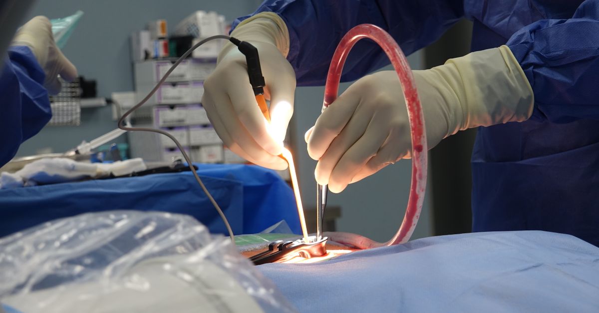
Surgical excision is the definitive treatment for bothersome benign lesions such as large epidermoid cysts, lipomas, or benign salivary gland tumors, as well as the mandatory treatment for confirmed primary skin malignancies.
Because the procedures occur on the face, the surgical approach must prioritize both complete eradication of the pathology and optimal cosmetic outcomes. Surgeons meticulously plan their incisions to fall within the natural relaxed skin tension lines, or Langer’s lines, of the face. Aligning the incision with these natural creases minimizes post-operative tension on the wound, resulting in a significantly thinner, less noticeable scar.
For the removal of epidermoid cysts, it is absolutely critical that the surgeon completely excises the entire fibrous sac intact. If fragments of the sac are left behind in the dermis, the cyst has a highly elevated probability of recurring in the exact same location. For complex resections, such as parotidectomies, highly specialized head and neck surgeons employ intraoperative nerve monitoring to ensure the delicate branches of the facial nerve are preserved.
17. Frequently Asked Questions (FAQ)
1. How can I tell if a lump on my face is a cyst or a tumor?
Clinically, cysts are typically smooth, round, move easily under the skin, and sometimes have a visible central pore. Benign tumors like lipomas are soft and doughy. A mass that is rock-hard, rapidly growing, bleeding, or fixed firmly to the bone is highly suspicious for a malignant tumor and requires immediate medical evaluation.
2. Should I try to pop a cyst on my face at home?
No. Attempting to squeeze or pop a cyst forces the keratin and bacteria deep into the surrounding facial tissues, causing massive inflammation, increasing the risk of a dangerous infection, and frequently leading to severe, permanent scarring.
3. Will removing a lump on my face leave a large scar?
Dermatologists and plastic surgeons use highly specialized techniques to minimize scarring, often hiding incisions in natural wrinkles or skin folds. While any incision leaves a mark, a professionally excised lump usually heals with a very fine, cosmetically acceptable line.
4. Why did a lump suddenly appear near my jawline after I was sick?
This is almost certainly a reactive lymph node. When you have a cold, sinus infection, or dental issue, the lymph nodes in your neck and jawline swell as they multiply white blood cells to fight the infection. They usually shrink back to normal size within a few weeks after the illness resolves.
5. What is the danger triangle of the face?
The danger triangle is the anatomical area covering the nose and the corners of the mouth. The veins here connect directly to blood vessels inside the skull. A severe, untreated infection (like an abscess) in this area carries a theoretical risk of spreading to the brain, making prompt medical treatment essential.
18. Bibliography
Disclaimer: The content is for informational purposes only and does not replace medical advice. Always consult your doctor for personalized treatment.