1. Introduction
A soft tissue mass refers to any abnormal growth or lump that develops within the non-epithelial, extraskeletal tissues of the body. These tissues include fat, muscle, fibrous connective tissue, blood vessels, and peripheral nerves. Because soft tissue is ubiquitous, providing the supportive framework for the entire body, these masses can arise in virtually any anatomical location, from the extremities and the trunk to the deep abdominal spaces.
The discovery of a new lump prompts a critical diagnostic journey. The clinician must accurately differentiate a benign, harmless overgrowth of tissue from a soft tissue sarcoma, which is a rare and aggressive malignancy. Soft tissue encompasses a diverse array of cellular origins, meaning a mass can present with a wide variety of textures, growth rates, and symptomatic profiles.
While the overwhelming majority of superficial lumps are benign lipomas or simple cysts, a mass situated deep within the musculature carries a substantially different risk profile. The diagnostic protocol relies heavily on advanced magnetic resonance imaging and meticulously planned tissue biopsies. This article details the anatomy of soft tissue compartments, the distinct types of benign and malignant masses, and the specialized oncological strategies used to manage them.
2. The Anatomy of Soft Tissue Compartments
Soft tissue is organized into distinct anatomical compartments, separated by thick, unyielding layers of connective tissue known as deep fascia. In the arms and legs, these fascial boundaries separate the muscles into functional groups. These compartments dictate the biological behavior and growth patterns of a soft tissue mass.
When a tumor begins to grow, it generally encounters less resistance expanding longitudinally up and down within its native compartment. The tough fascial walls act as a natural barrier, initially preventing the tumor from growing horizontally into adjacent muscle groups. Consequently, a deep mass often presents as a diffuse firmness or generalized swelling of a specific limb segment rather than a perfectly round, distinct lump.
Understanding compartmental anatomy is absolutely vital for surgical planning. When a malignant tumor is diagnosed, the surgeon must comprehend its precise relationship to these fascial boundaries, as well as the major blood vessels and nerves running between them, to achieve complete tumor resection without leaving microscopic disease behind.
3. Benign Adipocytic Tumors: Lipomas
Lipomas are overwhelmingly the most common soft tissue masses encountered in clinical practice. They are benign neoplasms composed entirely of mature, normal-appearing adipocytes (fat cells). Lipomas almost always develop in the superficial subcutaneous layer, situated just beneath the skin but above the investing muscle fascia.
Clinically, a lipoma is easily identifiable. It feels soft, doughy, and rubbery. It is highly mobile, slipping easily beneath the examiner’s fingers independently of the underlying muscle. Lipomas are completely painless and grow at an exceptionally slow rate, often remaining unchanged for decades.
Unless a lipoma grows large enough to cause cosmetic distress, or compresses a superficial sensory nerve, no medical intervention is required. True lipomas possess zero biological potential to transform into malignant sarcomas.
4. Distinguishing Lipoma from Liposarcoma
While a lipoma is harmless, a liposarcoma is a malignant tumor of fat tissue that requires aggressive oncological treatment. Distinguishing between the two relies on specific clinical and radiological features. Liposarcomas most frequently develop in the deep muscle tissues of the thigh or in the deep retroperitoneal space of the abdomen.
Unlike the soft, doughy texture of a superficial lipoma, a liposarcoma often feels firmer and is fixed deeply within the muscle belly, making it immobile. Furthermore, liposarcomas tend to grow steadily and attain significant sizes.
On magnetic resonance imaging, a benign lipoma appears uniformly composed of bright fat signals. A liposarcoma displays heterogeneous features, showing thick dividing walls, solid non-fatty tissue components, and areas of necrosis. Any “fatty” tumor that is deep, larger than five centimeters, or growing rapidly mandates an expert biopsy to rule out a liposarcoma.
5. Fibrous and Connective Tissue Masses
Fibrous tumors arise from the cells that produce collagen and connective tissue. A common benign fibrous mass is a fibroma, which can present as a firm, smooth nodule near a joint or within a tendon sheath.
Desmoid tumors, also known as aggressive fibromatosis, represent a unique clinical entity. They are technically benign because they do not possess the capacity to metastasize to distant organs like the lungs. However, they are locally aggressive. Desmoid tumors infiltrate deeply into surrounding muscles, tendons, and blood vessels, acting much like a malignancy on a local level.
Managing a desmoid tumor is complex. Surgical removal is difficult due to their infiltrative nature and high rate of local recurrence. Modern treatment strategies often involve a prolonged period of active surveillance, or the use of targeted medical therapies and anti-inflammatory drugs to halt their growth before attempting destructive surgeries.
6. Vascular and Peripheral Nerve Sheath Tumors
Vascular anomalies represent abnormal proliferations of blood vessels or lymphatic channels. Hemangiomas are benign tangles of blood vessels that can form deep within a muscle belly, presenting as a soft, compressible mass that may swell after exercise. Because they are filled with blood, attempting to biopsy a vascular mass without proper imaging can lead to severe hemorrhage.
Peripheral nerve sheath tumors develop from the protective layers surrounding nerve fibers. Schwannomas and neurofibromas are the most common benign variants. They often present as well-defined, firm, oval masses growing directly along the path of a major nerve.
Patients with a nerve sheath tumor may report a “zap” of electricity or a radiating tingling sensation down their arm or leg when the physician presses directly on the mass, known as a positive Tinel’s sign.
7. Soft Tissue Sarcomas
Soft tissue sarcomas are rare, aggressive malignancies encompassing over fifty distinct histological subtypes, including leiomyosarcoma (smooth muscle origin) and undifferentiated pleomorphic sarcoma. They can develop anywhere in the body, though the extremities account for over half of all cases.
These malignancies typically present as a firm, progressively enlarging mass situated deep beneath the investing muscle fascia. A critical and often deceptive characteristic of soft tissue sarcomas is that they are frequently painless in their early stages. The absence of pain often lulls the patient into delaying medical evaluation until the mass has reached a substantial size.
Unlike many carcinomas that spread through the lymphatic system, soft tissue sarcomas favor hematogenous spread, shedding cancer cells directly into the bloodstream. The lungs are overwhelmingly the most common site for distant metastasis, making a staging chest computed tomography scan mandatory upon diagnosis.
8. Identifying Clinical Red Flags
The clinical assessment aims to rapidly identify features that mandate immediate advanced imaging and referral to a specialized oncology center. Size is the most paramount indicator: any soft tissue mass that is larger than five centimeters (roughly the size of a golf ball) is considered suspicious for malignancy until proven otherwise.
Depth is another critical metric. Superficial masses confined to the fat layer are mostly benign. Conversely, any mass located deep to the muscle fascia carries a significantly higher risk of being a sarcoma. Rapid, noticeable growth over a period of weeks to a few months is a classic sign of an aggressive neoplastic process.
Furthermore, a mass that returns after a previous attempt at surgical removal by a general surgeon is a major red flag for a misdiagnosed soft tissue sarcoma. Recurrent deep masses require immediate evaluation by a multidisciplinary sarcoma team.
9. Initial Imaging and Ultrasound
When a soft tissue mass is identified, imaging is required before any surgical intervention. For small, superficial masses, an ultrasound is an excellent, cost-effective initial modality. It flawlessly differentiates a fluid-filled cyst or a hematoma from a solid tissue mass.
Ultrasound provides real-time information regarding the mass’s vascularity using color Doppler techniques. A benign fluid cyst will show no internal blood flow, while a solid tumor will demonstrate internal vascularity.
However, ultrasound cannot penetrate deep through bone or thick muscle layers effectively, nor can it definitively distinguish a benign solid tumor from a malignant sarcoma. If the ultrasound confirms a solid mass, or if the mass is clinically deep or large, advanced cross-sectional imaging is required.
10. MRI: The Gold Standard Diagnostic Tool
Magnetic Resonance Imaging is the absolute gold standard diagnostic tool for evaluating soft tissue masses. MRI provides unparalleled contrast resolution, allowing radiologists to clearly distinguish between fat, muscle, fluid, necrotic tissue, and active tumor tissue.
An MRI with and without intravenous contrast perfectly delineates the exact anatomical boundaries of the mass. It maps the tumor’s relationship to the fascial compartments and measures its precise distance from critical structures like major arteries and major nerves.
This detailed anatomical mapping is the foundation of surgical oncology. The MRI dictates the exact trajectory that must be used for the subsequent tissue biopsy and determines whether the limb or organ can be safely preserved during the final surgical resection.
11. Core Needle Biopsy Principles
Definitive diagnosis relies entirely on a tissue biopsy evaluated by a specialized musculoskeletal pathologist. The biopsy of a suspected sarcoma is a highly specialized procedure. It must be meticulously planned by the oncologist who will eventually perform the definitive surgery.
A poorly placed biopsy tract can contaminate surrounding healthy tissue compartments with cancer cells, compromising the ability to save the patient’s limb. Therefore, core needle biopsies, performed under ultrasound or CT guidance, are the preferred method. They are minimally invasive and minimize tissue disruption compared to an open surgical biopsy.
The needle tract must be placed directly in line with the planned surgical incision. This ensures that during the final operation, the entire path of the biopsy needle is excised along with the main tumor, preventing any local recurrence of the cancer along the biopsy track.
12. Data Summary: Deep vs. Superficial Masses
The following table summarizes the typical characteristics differentiating deep and superficial soft tissue masses.
| Characteristic | Superficial Mass (e.g., Lipoma) | Deep Mass (e.g., Sarcoma) |
|---|---|---|
| Location | Above the muscle fascia (subcutaneous fat). | Below the muscle fascia (intramuscular). |
| Mobility | Highly mobile, slips under the skin. | Fixed, immobile, attached to deep tissue. |
| Growth Rate | Very slow, often unchanged for years. | Progressive, noticeable growth over months. |
| Primary Imaging | Ultrasound is usually sufficient. | MRI with contrast is strictly mandatory. |
This stratification dictates the urgency of the referral and the required imaging protocols.
13. Surgical Resection: Wide Local Excision
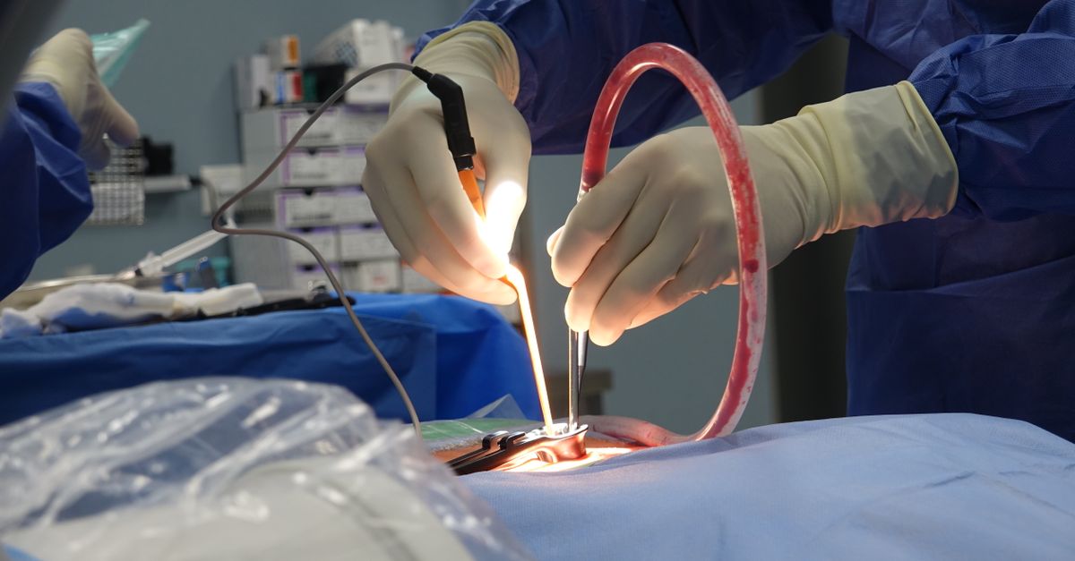
For localized soft tissue sarcomas, the standard of care is surgical resection. The oncological principle dictating the surgery is a “wide local excision.” This means the surgeon must remove the tumor entirely, surrounded by a thick, continuous cuff of normal, healthy tissue in all directions.
This healthy tissue acts as a protective margin, ensuring that any microscopic cancer cells extending slightly beyond the visible tumor are removed. If the tumor is resting against a major blood vessel or a bone, the surgeon must either remove that structure or carefully strip the outer protective layer of the vessel to achieve a negative margin.
Cutting directly into the tumor capsule during surgery significantly increases the risk of leaving microscopic cancer cells behind, which invariably leads to a high rate of local recurrence. This highlights why thorough preoperative MRI planning is essential, similar to the precision required when managing a complex thoracic structure mass.
14. Radiation Therapy and Local Control
Surgery alone is often insufficient for large or high-grade soft tissue sarcomas due to their propensity for microscopic local invasion. Radiation therapy is a cornerstone of treatment to ensure long-term local control of the disease.
Radiation is frequently administered preoperatively (neoadjuvant). Neoadjuvant radiation aims to sterilize the outer edges of the tumor and create a thicker, tougher fibrous capsule around the mass, making the subsequent surgical resection cleaner and safer.
Postoperative radiation may be used if the final pathology report shows that the surgical margins were very close to the tumor cells. The careful sequencing of surgery and radiation requires a coordinated effort by a multidisciplinary tumor board.
15. Systemic Chemotherapy and Surveillance
The role of systemic chemotherapy in soft tissue sarcomas is complex and highly dependent on the specific histological subtype and the tumor’s grade. High-grade sarcomas have cells that divide rapidly and appear very abnormal, indicating a high risk of metastasizing to the lungs.
In these high-risk cases, chemotherapy may be given before surgery to eradicate circulating cancer cells and assess the tumor’s biological sensitivity to the drugs.
Following the completion of all treatments, patients enter a rigorous surveillance program. Because soft tissue sarcomas can recur locally or metastasize to the lungs years later, scheduled chest CT scans and physical examinations are mandated for up to a decade, ensuring any recurrence is caught at the earliest possible stage.
16. Frequently Asked Questions FAQ
1. Is a squishy lump on my arm likely to be a sarcoma?
No. Soft, squishy, painless lumps that you can easily move around just under the skin are almost always benign lipomas. Sarcomas typically feel much firmer and are rooted deep within the muscle tissue.
2. Why does my doctor want an MRI if the lump doesn’t hurt?
Pain is not a reliable indicator of cancer in soft tissues. Many aggressive sarcomas grow silently without causing any pain. An MRI is needed to look at the internal structure of the mass and determine its exact depth and borders.
3. What is a core needle biopsy?
Instead of cutting you open with a scalpel, a doctor uses a specialized hollow needle, guided by a scanner, to extract tiny cylinders of tissue from the mass. This provides the laboratory with the tissue architecture they need without spreading cancer cells into healthy areas.
4. Can a benign lipoma turn into a liposarcoma?
A true, benign lipoma does not possess the genetic ability to transform into a malignant liposarcoma. They are completely separate disease processes from the very beginning.
5. Why do they need to remove healthy muscle around the tumor?
Sarcomas send out microscopic, invisible tentacles of cancer cells into the surrounding normal tissue. To ensure the cancer does not grow back, the surgeon must remove the main tumor along with a safety buffer of healthy tissue surrounding it.
17. Bibliography
Disclaimer: The content is for informational purposes only and does not replace medical advice. Always consult your doctor for personalized treatment.