1. Introduction to Neonatal Birth Trauma Hemorrhage
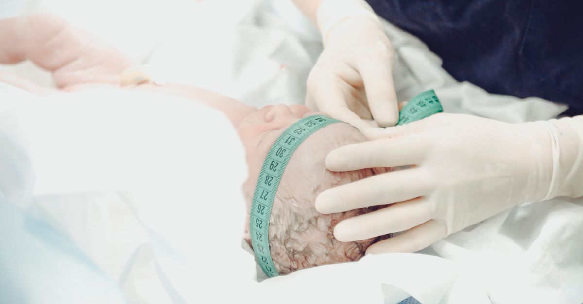
An intracranial hemorrhage resulting from birth trauma is a critical neonatal condition characterized by bleeding within the cranial vault of a newborn, occurring as a direct consequence of mechanical forces endured during the labor and delivery process. While modern obstetric practices have significantly reduced the incidence of severe birth trauma, the mechanical passage of the fetal head through the maternal pelvis inherently subjects the delicate neonatal cranium and its vascular structures to immense compressive and shearing forces.
When these mechanical forces exceed the physiological tolerance of the fetal tissues, the result is the tearing of cerebral blood vessels. Depending on the exact nature of the forces applied—whether rotational, compressive, or tractional—the bleeding can manifest in various compartments, most notably as a subdural hemorrhage between the protective membranes, or as an intraparenchymal cerebral hemorrhage directly within the brain tissue itself.
The clinical presentation of these traumatic hemorrhages can range from entirely asymptomatic, incidental findings to devastating, life-threatening neurological collapse within hours of birth. Prompt recognition of the clinical signs of elevated intracranial pressure and seizure activity in the neonate is paramount. Rapid diagnostic imaging, meticulous supportive neuro-intensive care, and occasionally, emergency neurosurgical intervention are necessary to stabilize the infant and optimize long-term neurodevelopmental outcomes.
2. Anatomy of the Fetal Cranium
To understand why the newborn brain is susceptible to mechanical trauma during delivery, one must appreciate the unique, adaptive anatomy of the fetal skull. Unlike an adult skull, which is a rigid, fused bone box, the fetal cranial vault is composed of distinct, pliable bony plates separated by wide, fibrous gaps known as sutures and larger membranous spaces called fontanelles.
This unfused structure serves a vital physiological purpose. During vaginal delivery, the extreme compressive forces of the maternal pelvis cause the fetal skull bones to overlap and shift—a process known as molding. Molding significantly reduces the diameter of the fetal head, facilitating its passage through the birth canal.
However, this necessary deformability has a structural cost. The brain and its protective meningeal layers are attached to the inner surface of these shifting skull bones. The rapid overlapping and extreme shifting of the bony plates place immense, acute traction on the underlying venous sinuses and the delicate bridging veins that drain blood from the brain into those sinuses, rendering them highly vulnerable to mechanical tearing.
3. Biomechanics of Birth Trauma
Birth trauma resulting in intracranial hemorrhage is fundamentally a mechanical injury driven by disproportionate forces. In a normal, uncomplicated vaginal delivery, the gradual pressure of uterine contractions allows the fetal head to mold slowly and safely.
Trauma occurs when these forces become excessive, abrupt, or directionally abnormal. A precipitous (extremely rapid) labor deprives the fetal skull of the necessary time to mold gradually, leading to sudden, violent compression and decompression of the cranial vault. Conversely, prolonged, obstructed labor causes the fetal head to become repeatedly wedged against the rigid maternal pelvic bones, leading to localized cranial trauma and ischemic stress.
Rotational forces are particularly destructive. If the fetal head is turned sharply while the body remains stationary, or if extreme traction is applied during the delivery of the shoulders, the shearing forces generated inside the skull can easily tear the bridging veins and the internal folds of the dura mater (such as the tentorium cerebelli), resulting directly in a subdural hemorrhage.
4. Subdural Hemorrhage in the Neonate
A subdural hemorrhage is the most common clinically significant type of intracranial hemorrhage caused by mechanical birth trauma. This bleeding occurs in the potential space between the tough outer covering of the brain (dura mater) and the delicate middle layer (arachnoid mater).
In the neonatal period, subdural bleeding is almost exclusively venous in origin. It typically results from the mechanical tearing of the bridging veins that span the subdural space, or from lacerations of the major dural folds, specifically the tentorium cerebelli (which separates the cerebrum from the cerebellum) or the falx cerebri.
A small, localized subdural hemorrhage is surprisingly common after vaginal delivery and often resolves spontaneously without causing symptoms. However, a significant laceration of the tentorium can lead to a rapidly expanding hematoma in the posterior fossa of the skull. This tight compartment leaves no room for expansion; the accumulating blood rapidly compresses the infant’s brainstem, leading to acute respiratory failure and profound neurological collapse shortly after birth.
5. Intraparenchymal Cerebral Hemorrhage
Intraparenchymal hemorrhage, or true cerebral hemorrhage, involves bleeding directly into the functional tissue (parenchyma) of the neonatal brain. While less common than a subdural hemorrhage in the context of pure mechanical trauma, it represents a severe and highly destructive injury.
This type of bleeding is typically caused by extreme mechanical deformation of the skull that crushes the underlying cortical tissue, or from severe, prolonged hypoxia (lack of oxygen) during a difficult delivery that causes the microscopic capillary beds within the brain to rupture.
An intraparenchymal hemorrhage directly destroys the neurons and glial cells in the affected area. As the blood breaks down, it induces profound localized inflammation and cerebral edema (swelling). This dual insult of direct tissue destruction and secondary inflammatory swelling frequently leads to significant, long-term focal neurological deficits, the exact nature of which depends on the specific lobe of the brain damaged.
6. Obstetric Risk Factors for Trauma
Specific maternal, fetal, and intrapartum factors significantly elevate the statistical risk of mechanical birth trauma and subsequent intracranial hemorrhage. Identifying these high-risk scenarios allows the obstetric and neonatal teams to prepare for immediate intervention upon delivery.
Fetal macrosomia (a significantly larger than average birth weight) is a primary risk factor, as the large fetal head is inherently subjected to much greater compressive forces while navigating the maternal pelvis. Cephalopelvic disproportion, where the maternal pelvis is anatomically too small for the fetal head, similarly guarantees a highly traumatic passage.
Operative vaginal delivery is strongly associated with an increased risk of traumatic hemorrhage. The application of obstetric forceps places direct, firm compressive force on the lateral aspects of the fetal skull. Vacuum extraction utilizes strong suction on the fetal scalp, which can generate profound tractional forces that stretch the internal cranial structures. While both interventions are often necessary to expedite a compromised delivery, they fundamentally amplify the mechanical stress placed on the infant’s neurovasculature.
7. Clinical Signs in the Newborn
The clinical presentation of a traumatic intracranial hemorrhage in a neonate is highly variable. Symptoms may be apparent immediately in the delivery room, or they may evolve gradually over the first 24 to 48 hours of life as the internal bleeding slowly accumulates.
A classic, immediate sign of a significant intracranial bleed is a depressed level of consciousness. The infant may appear profoundly lethargic, exhibit a weak or absent cry, and demonstrate poor muscle tone (hypotonia, or “floppiness”). The infant often struggles to establish or maintain normal, spontaneous respirations, requiring immediate breathing support.
As the hematoma expands and intracranial pressure rises, specific physical signs emerge. The anterior fontanelle (the “soft spot” on top of the head) may feel distinctly full, tense, or bulging. The infant may develop an abnormal, high-pitched cry indicative of neurological pain. They may also exhibit abnormal eye movements, such as a fixed, downward gaze, which is a classic sign of severe pressure on the brainstem.
8. Seizures and Neurological Deficits
Seizures are a prominent, alarming, and highly specific clinical manifestation of an intracranial hemorrhage in a newborn. The physical presence of the blood clot, combined with the resulting tissue swelling and hypoxia, chemically and mechanically irritates the delicate neonatal cerebral cortex.
Neonatal seizures often do not look like the dramatic, full-body convulsions seen in older children. They can be incredibly subtle. Caregivers and clinicians must watch for repetitive, involuntary movements such as lip-smacking, chewing motions, rhythmic bicycling movements of the legs, or brief, repetitive stiffening of a single arm or leg. Unexplained episodes of apnea (pauses in breathing) accompanied by heart rate drops are also a frequent presentation of subtle seizure activity.
Focal neurological deficits may also become apparent. If the hemorrhage damages the motor cortex on one side of the brain, the infant may demonstrate a lack of spontaneous movement or significant weakness in the opposite arm and leg (hemiparesis), though this can be difficult to definitively assess in a hypotonic newborn.
9. Diagnostic Imaging Modalities
When a traumatic intracranial hemorrhage is suspected based on the delivery history or the infant’s clinical presentation, rapid and accurate neuroimaging is mandatory to confirm the diagnosis, locate the bleeding, and guide intervention.
Cranial ultrasonography is the standard, first-line imaging modality utilized in the neonatal intensive care unit. It is highly advantageous because it is portable, can be performed directly at the bedside without moving a critically ill infant, and utilizes no ionizing radiation. The ultrasound probe is placed over the anterior fontanelle, providing excellent visualization of the brain’s ventricles and deep structures to quickly rule out massive bleeding.
However, ultrasound is less sensitive for detecting subtle subdural hemorrhages located near the surface of the skull or in the posterior fossa. If a significant traumatic injury is suspected but the ultrasound is inconclusive, a non-contrast Computed Tomography (CT) scan is required. A CT scan rapidly and definitively identifies all types of acute intracranial blood and bone fractures. Magnetic Resonance Imaging (MRI) provides the most detailed assessment of the actual brain tissue injury, but it is typically performed days later once the infant is stable enough for a prolonged scan.
10. Comparing Neonatal Hemorrhage Types
Differentiating the cause and location of neonatal bleeding guides the medical team’s expectations for recovery and potential complications. The table below outlines key differences.
| Hemorrhage Type | Primary Mechanism | Typical Clinical Outcome |
|---|---|---|
| Subdural Hemorrhage (SDH) | Mechanical trauma, tearing of bridging veins or dural folds. | Variable. Small SDHs resolve; large posterior fossa SDHs are life-threatening emergencies. |
| Intraparenchymal Hemorrhage | Severe trauma or severe hypoxic-ischemic injury. | High risk of focal neurological deficits, cerebral palsy, or post-traumatic epilepsy. |
| Cephalohematoma (Extracranial) | Bleeding under the scalp periosteum, distinct from brain bleeding. | Benign. Causes temporary jaundice; resolves spontaneously over weeks. |
| Intraventricular Hemorrhage (IVH) | Primarily caused by extreme prematurity, not usually mechanical birth trauma. | Graded I-IV; high grades carry severe risks of hydrocephalus and developmental delay. |
11. Emergency Neonatal Resuscitation
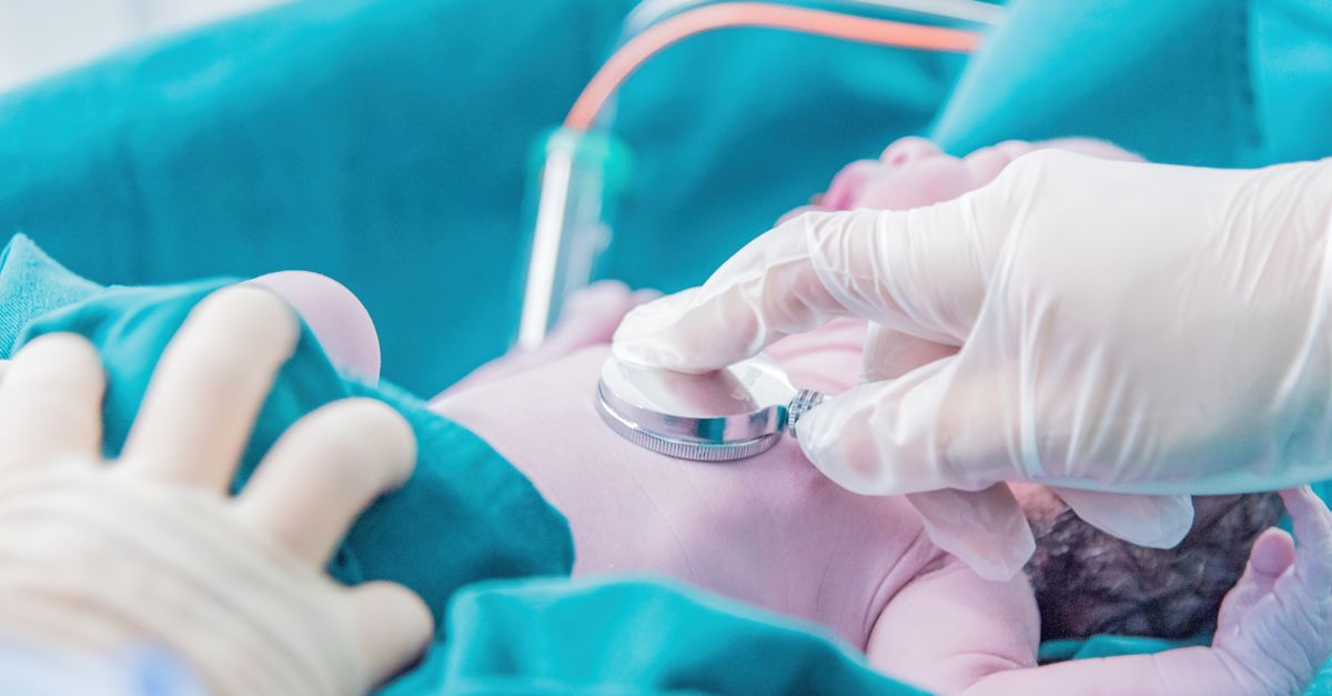
The immediate management of a neonate with a suspected traumatic intracranial hemorrhage adheres strictly to the principles of neonatal resuscitation. The primary goal is to ensure adequate oxygen delivery to the injured brain to prevent secondary hypoxic-ischemic damage.
If the infant is apneic or struggling to breathe, the airway is secured, and positive pressure ventilation is initiated. In severe cases, endotracheal intubation is required. The medical team rigorously monitors oxygen saturations and carbon dioxide levels, as elevated carbon dioxide can cause the cerebral blood vessels to dilate, dangerously exacerbating any internal brain swelling.
Intravenous access is established immediately to support cardiovascular function. If the internal bleeding is substantial, the infant can rapidly develop hypovolemic shock, requiring emergency blood transfusions or fluid boluses to maintain systemic blood pressure and ensure the remaining healthy brain tissue is adequately perfused. If the infant’s blood clotting factors are deficient, specific blood products are administered to halt the ongoing hemorrhage.
12. Medical Management and Seizure Control
Once the infant is stabilized and admitted to the neonatal intensive care unit (NICU), the focus shifts to minimizing secondary brain injury and controlling complications. Continuous cardiovascular and neurological monitoring is essential.
Controlling neonatal seizures is a primary objective, as prolonged seizure activity rapidly depletes the brain’s energy reserves and exacerbates the underlying tissue damage. First-line anticonvulsant medications, typically phenobarbital or levetiracetam, are administered intravenously. Continuous EEG monitoring is frequently employed to detect “silent” electrical seizures that do not produce visible physical movements.
Strict metabolic control is also maintained. The infant’s blood glucose levels are monitored closely, as hypoglycemia is detrimental to the injured brain. The core body temperature is strictly regulated, as fevers drastically increase the brain’s metabolic demand and worsen cellular swelling, leading to further neural destruction.
13. Neurosurgical Considerations
The vast majority of traumatic intracranial hemorrhages in neonates do not require surgical intervention. Small to moderate subdural hemorrhages and most intraparenchymal bleeds are managed conservatively with supportive medical care; the infant’s body will slowly resorb the clotted blood over a period of weeks to months.
However, emergency neurosurgery is absolutely indicated in specific, life-threatening scenarios. If an infant develops a massive subdural hematoma, particularly in the posterior fossa, that is actively compressing the brainstem and causing rapid neurological deterioration or intractable respiratory failure, a neurosurgeon must perform a craniotomy to evacuate the blood clot and relieve the lethal pressure.
Furthermore, a significant complication of any substantial intracranial bleeding is the development of post-hemorrhagic hydrocephalus. The blood products can block the normal drainage pathways of the cerebrospinal fluid, causing the ventricles in the brain to expand dangerously. If this occurs, a neurosurgeon may need to place a temporary reservoir or a permanent ventriculoperitoneal shunt to drain the excess fluid and protect the brain from pressure damage.
14. Long-Term Neurodevelopmental Prognosis
The long-term prognosis for an infant who has suffered a traumatic intracranial hemorrhage is highly variable and depends entirely on the size, location, and severity of the initial bleeding, as well as the presence of associated hypoxic-ischemic injury.
Infants with small, isolated subdural hemorrhages often experience a complete clinical recovery with no long-term neurological deficits. However, extensive hemorrhages, particularly large intraparenchymal bleeds that destroy cortical tissue, carry a substantial risk of permanent disability.
These infants are at a significantly elevated risk for developing cerebral palsy, characterized by permanent movement and posture disorders. They face higher rates of intellectual disabilities, profound learning deficits, and severe visual or hearing impairments. Additionally, the scarring left behind by the hemorrhage frequently results in post-traumatic epilepsy, requiring long-term treatment with anticonvulsant medications.
15. The Importance of Early Intervention
Because the neonatal brain is highly plastic, capable of significant reorganization and adaptation, early identification of developmental delays is crucial. Infants discharged from the NICU following a traumatic intracranial hemorrhage require enrollment in a rigorous, specialized high-risk infant follow-up program.
These programs involve frequent, multidisciplinary evaluations by pediatric neurologists, developmental pediatricians, physical therapists, and occupational therapists. The goal is to aggressively monitor the child’s achievement of developmental milestones over the first several years of life.
If delays or abnormal motor patterns are identified, early intervention therapies are initiated immediately. Prompt initiation of physical and occupational therapy has been definitively shown to optimize neuroplasticity, significantly improving motor function, maximizing the child’s independence, and mitigating the severity of the long-term neurological impairments.
16. When to Seek Urgent Pediatric Care
For parents of an infant who experienced a difficult delivery or was diagnosed with a minor hemorrhage that did not require NICU admission, vigilance regarding neurological warning signs at home is essential during the first weeks of life.
Immediate emergency pediatric evaluation is required if the newborn exhibits any abnormal, repetitive twitching movements, bicycling motions of the legs, or sudden, unprovoked staring spells, as these are strong indicators of neonatal seizures.
Furthermore, if the infant becomes exceptionally difficult to wake up for feedings, completely refuses to feed, develops a high-pitched, inconsolable cry, or if the soft spot on top of their head (the fontanelle) appears distinctly swollen, bulging, and firm to the touch when the baby is resting quietly, parents must seek immediate medical attention at a pediatric emergency department.
17. Frequently Asked Questions (FAQ)
1. Was this hemorrhage caused by something the doctor did wrong?
A traumatic hemorrhage is a known, inherent risk of childbirth, particularly in difficult, obstructed, or instrument-assisted (forceps/vacuum) deliveries. While the mechanical forces caused the injury, these interventions are usually deployed in emergency situations to save the life of the mother or baby when natural progress halts.
2. Will a small amount of blood on the brain permanently damage my baby?
Not necessarily. Small, asymptomatic subdural hemorrhages are actually quite common after vaginal deliveries and frequently resolve completely on their own without causing any long-term brain damage or developmental issues.
3. Can you tell right away if my baby will have cerebral palsy?
No. Cerebral palsy is a clinical diagnosis based on the child’s failure to meet motor milestones and the development of abnormal muscle tone over time. While a severe hemorrhage increases the risk, the definitive diagnosis of cerebral palsy is usually not made until the child is between 12 and 24 months of age.
4. If my baby had a seizure in the NICU, will they have epilepsy forever?
Neonatal seizures are a symptom of the acute irritation from the bleeding. While some infants who seize in the newborn period go on to develop chronic epilepsy later in childhood, many others never have another seizure once the initial hemorrhage heals and the acute swelling subsides.
18. Bibliography
Disclaimer: The content is for informational purposes only and does not replace medical advice. Always consult your doctor for personalized treatment.