1. Introduction
Pelvic pain is a complex, multifaceted clinical symptom signaling dysfunction within the intricate anatomical basin that houses the urological, gastrointestinal, reproductive, and musculoskeletal systems. This discomfort can present as a sharp, localized stabbing sensation, a dull, pervasive ache, or a cyclical cramping pattern. Prompt clinical evaluation is essential to differentiate benign, self-limiting muscular strains from severe inflammatory, infectious, or structural pathologies that require targeted medical intervention.
The human pelvis serves as a structural foundation for the trunk and a protective enclosure for vital visceral organs. Because these organ systems share overlapping neurological pathways originating from the same spinal nerve roots, pain generated in one specific organ is frequently referred to adjacent structures. This phenomenon often obscures the primary source of the distress. A methodical, multidisciplinary diagnostic approach is necessary to isolate the underlying etiology, allowing clinicians to formulate effective treatment plans that restore functional harmony and alleviate chronic discomfort.
2. Anatomy of the Pelvic Basin
The pelvis is a robust, ring-shaped bony structure composed of the sacrum, the coccyx, and the paired innominate bones. This osseous ring is divided into the greater pelvis, which supports the lower abdominal organs, and the lesser true pelvis, which contains the bladder, rectum, and reproductive organs.
Supporting these visceral structures from below is the pelvic floor, a hammock-like network of striated musculature, primarily the levator ani and coccygeus muscles, reinforced by dense connective tissue fascia. The entire region is richly innervated by the somatic pudendal nerve and extensive autonomic nerve plexuses. Pathology affecting the bone, the supportive musculature, the neurovascular bundles, or the enclosed organs will all manifest as varied presentations of pelvic pain.
3. Urological Etiologies: Cystitis and Calculi
The urinary system is a frequent source of acute and chronic pelvic discomfort. Interstitial cystitis, also known as painful bladder syndrome, is a chronic inflammatory condition of the bladder wall. Patients typically experience significant suprapubic pelvic pain that intensifies as the bladder fills and is only temporarily relieved by urination, leading to profound urinary frequency and urgency.
Acute bacterial cystitis presents similarly but is driven by an active bacterial infection, often accompanied by burning during urination and cloudy urine. Furthermore, urinary calculi located in the distal ureter or at the ureterovesical junction can generate severe, unilateral pelvic pain that radiates toward the groin. The passage of these stones creates intense smooth muscle spasms within the ureter, resulting in characteristic colicky pain.
4. Gastrointestinal Pathologies: IBS and Diverticulitis
Due to the anatomical proximity of the distal colon and rectum, gastrointestinal disorders frequently masquerade as primary pelvic pain. Irritable bowel syndrome is a functional gastrointestinal disorder characterized by visceral hypersensitivity. Patients experience lower abdominal and pelvic cramping that is distinctly associated with changes in bowel habits, such as constipation or diarrhea, and is often relieved following defecation.
Diverticulitis represents an acute inflammatory condition where small outpouchings in the colonic wall become infected. This typically generates localized, constant pain in the left lower quadrant of the pelvis, often accompanied by fever and a change in bowel habits. Inflammatory bowel diseases, including Crohn disease and ulcerative colitis, also present with chronic pelvic aching secondary to persistent mucosal inflammation and potential stricture formation.
5. Musculoskeletal Dysfunction: Pelvic Floor Hypertonicity
A highly prevalent but frequently overlooked cause of chronic pelvic pain is pelvic floor muscle dysfunction. Chronic stress, postural abnormalities, or prolonged guarding against pain from an initial visceral insult can cause the levator ani muscles to enter a state of continuous, involuntary tension.
This chronic hypertonicity leads to localized tissue ischemia and the formation of myofascial trigger points within the pelvic floor. The patient experiences a deep, heavy, aching pain in the pelvis that typically worsens with prolonged sitting or standing. This muscular dysfunction complicates the clinical picture, as the pelvic floor pain often persists long after the initial trigger has resolved.
6. Gynecological Factors in Pelvic Pain
In female patients, the reproductive organs are central considerations in the differential diagnosis. Endometriosis is a primary driver of severe, cyclical pelvic pain. It involves the presence of endometrial-like tissue outside the uterine cavity, which responds to hormonal fluctuations by swelling and bleeding internally. This generates severe inflammation and the formation of fibrotic adhesions that tether pelvic organs together.
Uterine leiomyomas, or fibroids, can cause a persistent, heavy pelvic pressure due to their physical bulk. Adenomyosis, where endometrial tissue infiltrates the uterine muscle wall, causes the uterus to become enlarged and boggy, resulting in severe menstrual cramping. Acute conditions such as an ectopic pregnancy or ovarian torsion present with sudden, agonizing unilateral pain and represent immediate surgical emergencies.
7. Urological Factors in Male Pelvic Pain
In male patients, the prostate gland is a significant anatomical consideration. Acute bacterial prostatitis presents with sudden, severe perineal and pelvic pain, accompanied by fever and urinary retention. This requires rapid antimicrobial therapy.
However, the vast majority of chronic pelvic pain in men is attributed to chronic pelvic pain syndrome. This condition involves persistent pelvic discomfort in the absence of a demonstrable bacterial infection. The pathophysiology is largely believed to be driven by pelvic floor muscle spasms and localized neurological sensitization, often resulting in a continuous, aching pressure behind the scrotum that worsens with prolonged sitting.
8. Neuropathic Pelvic Pain: Pudendal Neuralgia
Pudendal neuralgia is a debilitating neuropathic condition caused by the compression, irritation, or entrapment of the pudendal nerve as it courses through the pelvis. This nerve supplies sensation to the perineum, external genitalia, and the lower rectum.
Patients report sharp, burning, or electric-shock-like pain in the distribution of the nerve. A hallmark clinical sign is that the pain is remarkably exacerbated by sitting and is typically relieved by standing or sitting on a toilet seat, which mechanically offloads pressure from the nerve path. This condition often stems from pelvic trauma, severe childbirth injury, or chronic repetitive strain.
9. Differentiating the Sources of Pelvic Pain
Accurate clinical differentiation relies on a detailed history regarding the onset, triggers, and character of the pain.
| Systemic Origin | Common Condition | Key Pain Characteristics |
|---|---|---|
| Urological | Interstitial Cystitis | Suprapubic pain worsening with bladder filling, severe frequency. |
| Gastrointestinal | Irritable Bowel Syndrome | Cramping pain associated with bowel movements, altered stool form. |
| Musculoskeletal | Pelvic Floor Hypertonicity | Deep, continuous aching pressure, worse with sitting, trigger points present. |
| Neurological | Pudendal Neuralgia | Burning, electrical pain entirely provoked by sitting on firm surfaces. |
10. Clinical Evaluation and Patient History
The clinical assessment of pelvic pain demands a comprehensive and sensitive approach. The clinician takes a detailed history, mapping the pain’s relationship to urination, defecation, sexual activity, and hormonal cycles. Identifying specific exacerbating and relieving factors provides crucial diagnostic clues.
Inquiring about prior abdominal or pelvic surgeries is necessary, as post-surgical adhesions are a prominent cause of chronic dragging or pulling pelvic pain. The clinician must also assess for systemic symptoms such as unexplained weight loss, fever, or hematochezia, which prompt a more urgent investigational pathway.
11. Physical Examination Techniques
The physical examination is tailored to the patient’s anatomical presentation. Palpation of the abdomen evaluates for masses, organomegaly, or localized peritonitis. In female patients, a bimanual pelvic examination assesses the uterus and adnexa for size, mobility, and tenderness, specifically checking for the nodularity associated with endometriosis.
In male patients, a digital rectal examination assesses the prostate gland for size, symmetry, and tenderness. Crucially, across all demographics, the clinician performs a targeted evaluation of the pelvic floor musculature, identifying spastic bands or localized trigger points that indicate primary musculoskeletal dysfunction.
12. Laboratory and Imaging Diagnostics
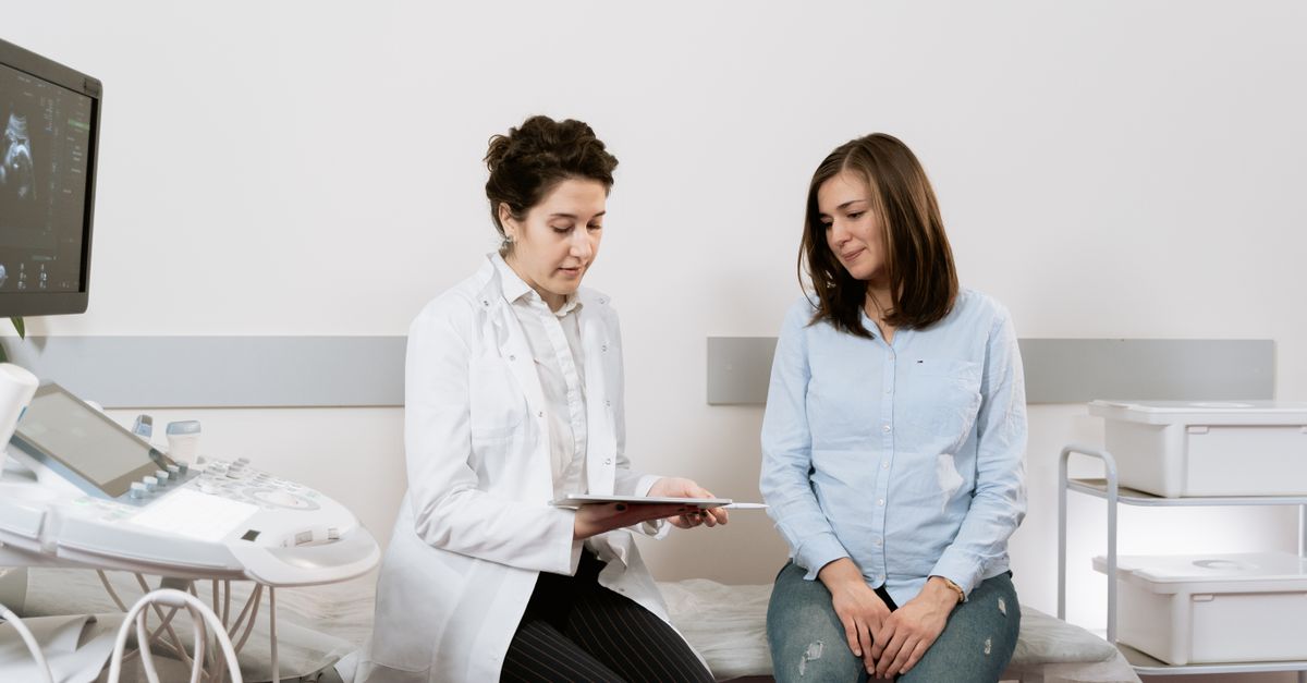
Diagnostic testing begins with a urinalysis and urine culture to rule out infectious cystitis. Basic blood panels evaluate for systemic inflammation or hidden infections. Transvaginal or transabdominal ultrasound serves as the first-line imaging modality to identify structural anomalies such as ovarian cysts, uterine fibroids, or bladder retention.
For complex cases, pelvic magnetic resonance imaging provides superior soft-tissue contrast, allowing clinicians to map deeply infiltrating endometriosis, complex perianal fistulas, or subtle neurological compressions. Diagnostic laparoscopy remains the gold standard for directly visualizing and simultaneously treating peritoneal adhesions and superficial endometriosis.
13. Pharmacological Management
Pharmacological therapy targets the specific physiological pain generators. For pain driven by hormonal fluctuations, oral contraceptives or specialized hormonal modulators are utilized to suppress the cyclical inflammatory response. Nonsteroidal anti-inflammatory drugs provide baseline analgesia for mechanical and inflammatory conditions.
For chronic pelvic pain involving central or peripheral nerve sensitization, neuromodulators such as amitriptyline or gabapentin are highly effective. These medications dampen the hyperactive pain signals traveling from the pelvis to the central nervous system. Muscle relaxants may be prescribed for short-term relief of acute pelvic floor spasms.
14. Pelvic Floor Physical Therapy
When the pelvic pain is driven by or complicated by muscular hypertonicity, specialized pelvic floor physical therapy is the absolute gold standard of care. A certified therapist utilizes manual trigger point release techniques to relax the spastic musculature.
Patients are instructed in biofeedback and diaphragmatic breathing to gain conscious control over their pelvic floor tension. The therapy focuses on paradoxical relaxation, teaching the patient to down-train the nervous system and release the chronic guarding patterns that perpetuate the pain cycle.
15. Interventional Pain Procedures
If conservative therapy fails, interventional pain procedures are considered. Ultrasound-guided nerve blocks targeting the pudendal nerve or the ilioinguinal nerve can provide significant diagnostic confirmation and prolonged therapeutic pain relief by reducing local nerve inflammation.
For refractory muscular pain, trigger point injections using local anesthetics directly into the spastic pelvic floor muscles can break the pain cycle. In highly specific cases, neuromodulation devices can be surgically implanted to stimulate the sacral nerves, effectively altering the perception of pain at the spinal level.
16. When to Seek Emergency Medical Attention
While chronic pelvic pain requires systematic long-term management, certain acute presentations represent life-threatening emergencies. Any sudden, agonizing onset of unilateral pelvic pain, especially if accompanied by nausea, vomiting, or fainting, requires immediate evaluation to rule out an organ torsion or a ruptured viscus.
Furthermore, patients requiring an evaluation for severe abdominal pain accompanied by a rigid abdomen, high fever, and inability to pass gas or stool must seek emergency surgical assessment. Prompt clinical intervention is vital to prevent irreversible organ damage and systemic sepsis.
17. Frequently Asked Questions
1. Why does my pelvic pain worsen as the day goes on?
Pelvic pain that worsens throughout the day is often related to pelvic floor muscle fatigue or venous pooling. Prolonged standing or sitting places continuous mechanical demand on the supportive pelvic muscles, leading to spasms and an aching, heavy sensation by evening.
2. Can digestive issues really cause pain in my lower pelvis?
Yes. The lower intestines and rectum sit directly within the pelvic basin. Conditions like irritable bowel syndrome or severe constipation cause the bowel to dilate and spasm, which is perceived by the brain as deep, generalized pelvic cramping.
3. What is the difference between pelvic pain and regular stomach cramps?
Pelvic pain is located significantly lower, below the level of the belly button and between the hip bones. It often feels deeper and more structurally involved than superficial stomach cramps, frequently radiating to the lower back, groin, or upper thighs.
4. How does physical therapy help with internal pelvic pain?
Just like muscles in your back can form painful knots, the muscles supporting your pelvic organs can become chronically tight and spastic. A specialized physical therapist uses targeted exercises and manual release techniques to relax these hidden muscles, alleviating the deep internal pressure.
5. When should I be worried about sudden pelvic pain?
You should seek emergency medical care if your pelvic pain appears suddenly and severely out of nowhere, is accompanied by a high fever, severe nausea, dizziness, or if you are pregnant and experiencing any new pelvic pain or bleeding.
18. Bibliography
Disclaimer: The content is for informational purposes only and does not replace medical advice. Always consult your doctor for personalized treatment.