1. Introduction
Experiencing a sudden, sharp pain in the lower back is a highly common musculoskeletal complaint, but its intensity indicates that an acute tissue injury or mechanical failure has occurred within the lumbar spine. While sudden back pain is statistically normal due to the immense biomechanical stress placed on the human spine, it demands careful attention. In the majority of cases, this sharp pain is the result of a severe muscular spasm or a ligamentous sprain that, while excruciating, is self-limiting and resolves with conservative care.
However, the lower back is a complex architectural structure housing the spinal cord, exiting nerve roots, intervertebral discs, and multiple articulating joints. A sharp, localized, or radiating pain can sometimes signal structural damage, such as a herniated disc pressing against a nerve, or acute inflammation within the facet joints. Differentiating between a benign muscular issue and a neurological impingement is the primary focus of clinical evaluation.
Management emphasizes calming the acute inflammatory response and preventing the transition from acute injury to chronic pain syndrome. By understanding the specific triggers, radiation patterns, and mechanical limitations associated with the pain, patients can apply appropriate at-home therapies and recognize the vital clinical red flags that necessitate immediate medical imaging and intervention.
2. Anatomy of the Lumbar Spine
The lumbar spine, constituting the lower back, is engineered to support the bulk of the body’s weight while providing exceptional flexibility. It consists of five massive vertebral bones, designated L1 through L5. Between these bones lie the intervertebral discs, acting as hydraulic shock absorbers to dissipate vertical impact forces.
Posterior to the disc space runs the spinal canal, which houses the lower portion of the spinal cord and the cauda equina—a bundle of nerve roots that exit the spine to innervate the pelvis and legs. The vertebrae connect to one another via the facet joints, which guide and limit the rotational and bending movements of the torso.
Enveloping this bony framework is a dense matrix of paraspinal muscles, fascia, and robust ligaments. A failure or acute strain in any single component of this intricate system initiates a cascade of localized inflammation and secondary muscle spasms, culminating in the sharp, debilitating pain that forces a patient to seek relief.
3. Acute Muscular Spasms and Strains
A lumbar muscle strain or ligament sprain is the most frequent cause of sudden, sharp lower back pain. These injuries typically occur during heavy lifting with improper mechanics, abrupt twisting motions, or unexpected loads applied to a cold muscle. The physical tearing of microscopic muscle fibers or ligament tissues immediately triggers localized bleeding and intense inflammation.
In response to this trauma, the body initiates a protective reflex known as a muscle spasm. The massive paraspinal muscles involuntarily contract and lock down to splint the injured area and prevent further movement. This sustained, tetanic contraction is intensely painful and often feels like a sharp knife or a tight band across the lower back.
Patients with acute muscular spasms typically find that the pain is localized to the muscles alongside the spine rather than the spine itself. The pain worsens significantly with movement, particularly bending forward, but does not radiate down the leg past the knee, a key factor distinguishing it from nerve root compression.
4. Lumbar Disc Herniation
The intervertebral discs consist of a tough, fibrous outer ring known as the annulus fibrosus, and a gel-like inner core called the nucleus pulposus. Over time, due to age-related desiccation or acute mechanical overload, the outer ring can tear. A lumbar disc herniation occurs when the inner gel extrudes through this tear into the spinal canal.
Because the spinal canal has limited anatomical space, the extruded disc material frequently compresses the adjacent exiting nerve roots. Furthermore, the chemical composition of the inner disc gel is highly inflammatory to neural tissue. This combination of mechanical pressure and chemical irritation produces a severely sharp, electrical pain.
Unlike a muscle strain, the pain of a herniated disc is often worse when sitting, as sitting increases intradiscal pressure. Coughing, sneezing, or straining heavily exacerbates the pain. If the herniation compresses the sciatic nerve root, the patient will experience sciatica.
5. Sciatica and Nerve Root Compression
Sciatica is not a distinct medical diagnosis; rather, it is a set of symptoms caused by the irritation or compression of the sciatic nerve, the longest and thickest nerve in the body. The nerve originates from the L4 to S3 nerve roots in the lower spine and travels deep through the buttock and down the back of the leg.
When a lumbar nerve root is pinched by a herniated disc or a bone spur, it generates a sharp, shooting, or burning pain that radiates from the lower back directly down the posterior thigh, often extending into the calf and foot. This specific radiation pattern indicates a neuropathic origin.
Alongside the sharp pain, patients routinely experience paresthesia, which manifests as numbness or a pins-and-needles sensation in specific areas of the leg or foot. Clinical evaluation utilizes specialized neural tension tests, such as the straight leg raise, to confirm the presence and severity of root entrapment.
6. Facet Joint Dysfunction
The facet joints are the small, cartilage-lined joints located on the back of the spinal column that link the vertebrae together. Like the knee or hip joints, facet joints are enclosed in a capsule and lubricated by synovial fluid. They are highly innervated and susceptible to acute injury and arthritic degeneration.
A sudden, awkward twisting motion can pinch the highly sensitive joint capsule, causing an acute facet joint sprain. This results in a sharp, localized, catching pain in the lower back. The pain is distinctly different from a disc injury because it typically worsens when the patient leans backward (extension) and rotates, rather than when bending forward.
Facet joint pain can sometimes radiate into the buttocks or the back of the upper thighs, but it rarely travels below the knee. Because the pain is mechanical, patients often find relief by leaning slightly forward or sitting, which opens up the posterior joints and relieves pressure on the inflamed capsule.
7. Sacroiliac Joint Inflammation
The sacroiliac joints are the strong, rigid joints connecting the sacrum at the base of the spine to the large iliac bones of the pelvis. These joints are designed to transfer the entire weight of the upper body into the lower extremities. They are supported by a massive network of ligaments and have very little natural motion.
Sacroiliac joint dysfunction occurs when these joints become inflamed due to trauma, pregnancy-related hormonal laxity, or an underlying inflammatory arthritis like ankylosing spondylitis. The pain is typically felt as a sharp, stabbing sensation unilaterally near the dimple of the lower back.
The pain frequently intensifies during transitions, such as standing up from a low chair, climbing stairs, or rolling over in bed. Restoring proper pelvic biomechanics through targeted physical therapy and utilizing localized anti-inflammatory injections are the mainstays of treating stubborn sacroiliac pain.
8. Spinal Stenosis and Claudication
Spinal stenosis is the gradual narrowing of the spinal canal or the neural foramina (the bony openings where nerve roots exit). This narrowing is most commonly caused by osteoarthritis, where thickened ligaments and bony overgrowths (osteophytes) encroach upon the neural space. While usually a chronic, aching condition, it can provoke sharp pain during specific activities.
The hallmark symptom of lumbar spinal stenosis is neurogenic claudication. Patients experience sharp, cramping pain, heaviness, and weakness in the lower back and legs when walking or standing straight for prolonged periods. The pain reliably subsides when the patient sits down or leans forward, a posture that mechanically widens the spinal canal.
This condition predominantly affects older adults. Unlike the sudden onset of a muscle tear, spinal stenosis develops insidiously over years. Management focuses on core strengthening, epidural steroid injections to reduce nerve inflammation, and, in severe cases, surgical decompression.
9. Renal Causes: Kidney Stones and Infections
It is crucial to recognize that sharp pain in the lower back area is not always musculoskeletal in origin. The kidneys sit against the posterior abdominal wall, situated perfectly in the mid-to-lower back region (the flank). Pathologies within the kidneys can refer excruciating pain to the lumbar area.
Nephrolithiasis, or kidney stones, causes incredibly severe, sharp, colicky pain when a crystalline stone obstructs the ureter. The pain typically originates in the flank and radiates wrapping around the side into the groin. It is frequently accompanied by nausea, vomiting, and blood in the urine, and it fluctuates in intensity.
Pyelonephritis, a severe bacterial infection of the kidney, also causes sharp lower back pain. However, it is invariably accompanied by systemic signs of infection, including a high fever, severe chills, and painful urination. These renal conditions require immediate medical diagnosis utilizing urinalysis and imaging.
10. Biomechanical Imbalances and Posture
Acute lower back pain rarely occurs in a vacuum; it is often the final breaking point of long-standing biomechanical dysfunction. Chronic poor posture, particularly the prolonged sitting associated with modern sedentary lifestyles, physically alters the length and tension of the pelvic musculature.
Prolonged sitting leads to the adaptive shortening of the hip flexors and the weakening of the gluteal muscles and deep core stabilizers. This imbalance, known as lower crossed syndrome, tilts the pelvis forward, exaggerating the natural curve of the lumbar spine (hyperlordosis). This continuous mechanical stress overloads the facet joints and the posterior aspect of the intervertebral discs.
When an individual with these hidden imbalances attempts a physically demanding task, the deconditioned muscles fail to stabilize the spine, resulting in a sudden, sharp injury. Resolving recurrent back pain fundamentally requires correcting these systemic postural faults through dedicated rehabilitation.
11. Immediate Conservative Management Strategies
When a sharp stab of lower back pain strikes, immediate action should focus on minimizing the inflammatory cascade. The classic recommendation of strict bed rest is now obsolete and actively discouraged. Prolonged immobility rapidly weakens the supporting muscles, stiffens the joints, and delays healing.
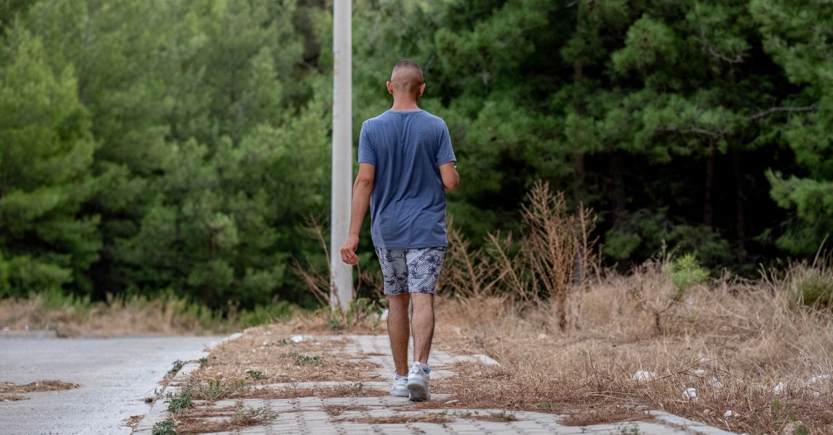
Instead, the concept of relative rest is applied. Patients should avoid the specific movements that trigger the sharp pain, such as deep bending or heavy lifting, but must maintain gentle mobility. Frequent, short walks on flat surfaces promote blood circulation, which flushes inflammatory toxins from the injured tissues and delivers oxygen for repair.
Finding a position of comfort is essential for offloading the spine. The psoas position—lying flat on the floor with the lower legs resting on a chair or couch so the hips and knees are bent at ninety degrees—dramatically reduces the mechanical pressure on the lumbar discs and allows severe muscle spasms to release.
12. Pain Characterization and Diagnosis
Understanding the specific triggers of the pain guides clinical triage. The table below outlines how specific movements correlate with potential anatomical injuries.
| Pain Trigger or Characteristic | Anatomical Implication | Typical Diagnosis |
|---|---|---|
| Worse when bending forward or sitting. Pain radiates below the knee. | Increased pressure on the anterior spine and intervertebral discs. | Lumbar Disc Herniation |
| Worse when leaning backward or twisting. Localized catching pain. | Compression of the posterior spinal joints. | Facet Joint Sprain / Arthropathy |
| Pain wrapping around to the groin, accompanied by nausea. | Distension or obstruction of the renal system. | Kidney Stone (Ureteral Colic) |
13. Physical Therapy and Core Stabilization
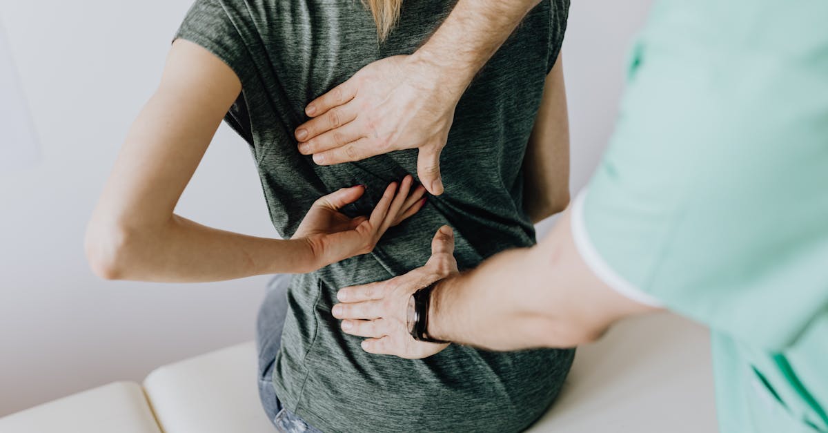
The definitive treatment for resolving lower back pain and preventing future recurrences is a highly structured physical therapy program. A clinical physical therapist will evaluate the patient’s specific movement deficits and design an exercise protocol tailored to their unique pathology.
For disc-related pain, therapists frequently utilize the McKenzie method. This approach employs specific extension exercises that centralize the pain—moving it out of the leg and back toward the spine—facilitating the mechanical retraction of the herniated disc material.
Core stabilization is universally emphasized. The core encompasses much more than the superficial abdominal muscles; it includes the deep transversus abdominis and multifidus muscles that act as a biological corset around the spine. Strengthening these deep stabilizers significantly enhances the load-bearing capacity of the lower back.
14. Pharmacological Pain Management
Medications are utilized adjunctively to break the cycle of acute pain and allow the patient to participate in physical rehabilitation. Nonsteroidal anti-inflammatory drugs (NSAIDs) like ibuprofen or naproxen are the first-line defense for reducing localized tissue swelling and chemical irritation around the joints or nerve roots.
In cases of severe, acute muscular spasms, physicians may prescribe a short course of skeletal muscle relaxants. These medications act on the central nervous system to dampen the neurological signals causing the muscles to lock down. They are highly sedating and are usually reserved for nighttime use during the first few days of injury.
For severe radicular pain caused by nerve compression, medications specifically designed to calm hyperactive nerves, such as gabapentin, or a brief tapering dose of oral corticosteroids may be utilized to rapidly suppress nerve root inflammation.
15. Diagnostic Imaging Guidelines
Patients experiencing sharp lower back pain often immediately request an MRI. However, strict medical guidelines strongly recommend against imaging during the first four to six weeks of acute back pain unless specific red flag symptoms are present. The reason is that most acute injuries resolve naturally within this timeframe, rendering early imaging unnecessary.
Furthermore, spinal imaging is notoriously sensitive and frequently reveals bulging discs or degenerative changes that are completely asymptomatic and unrelated to the current pain. Over-imaging often leads to unnecessary anxiety, misdiagnosis, and invasive treatments.
Imaging is indicated if the pain is the result of severe high-velocity trauma, if the patient has a history of cancer, or if the pain fails to improve after six weeks of rigorous conservative management. In these cases, an MRI provides the necessary detailed visualization of the neural structures and soft tissues.
16. When to Seek Emergency Medical Attention
Certain clinical presentations, known as red flags, indicate that the sharp back pain is a symptom of a dangerous neurological compromise or systemic disease requiring immediate emergency intervention. The most critical of these is Cauda Equina Syndrome.
Cauda Equina Syndrome occurs when a massive central disc herniation completely compresses the bundle of nerve roots at the base of the spine. Symptoms include sudden and severe numbness in the saddle area (groin, buttocks, and inner thighs), profound weakness in both legs making standing impossible, and new-onset loss of bowel or bladder control (incontinence or retention). This is a surgical emergency.
Other red flags include sharp back pain accompanied by an unexplained high fever, which may indicate a spinal epidural abscess, or pain that is unrelenting at night and unaffected by positional changes, which requires investigation for potential spinal malignancy or infection.
17. Frequently Asked Questions (FAQ)
1. Should I use ice or heat for sharp lower back pain?
During the first forty-eight hours of a sudden, sharp injury, applying ice for twenty minutes at a time is recommended to constrict blood vessels and numb the acute inflammatory pain. After the initial acute phase, heat therapy helps relax severe muscle spasms and promotes healing blood flow.
2. Is a firm or soft mattress better for back pain?
A medium-firm mattress is clinically considered optimal for lumbar support. A mattress that is too soft allows the heavy pelvis to sink, forcing the spine into misalignment, while a mattress that is completely rigid creates pressure points and fails to support the natural curvature of the lower back.
3. Can stress cause sharp back pain?
Yes, severe psychological stress heavily activates the sympathetic nervous system, causing generalized muscle tension. The massive paraspinal muscles in the lower back are highly reactive to stress, and chronic tension can precipitate acute, painful muscle spasms without any physical trauma.
4. Will walking make my herniated disc worse?
In most cases, gentle, short-duration walking on flat surfaces is beneficial for a herniated disc. It promotes circulation and helps maintain the mobility of the spinal joints without placing heavy compressive loads on the discs. If walking causes the pain to shoot further down the leg, however, it should be paused.
5. Does cracking my back help relieve the pain?
While gentle spinal manipulation by a qualified professional can temporarily relieve mechanical stiffness, forcefully twisting to “crack” your own back during an acute injury is dangerous. It can severely aggravate a sprained facet joint or push a vulnerable herniated disc further onto a nerve root.
18. Bibliography
Disclaimer: The content is for informational purposes only and does not replace medical advice. Always consult your doctor for personalized treatment.