1. Introduction and Initial Management
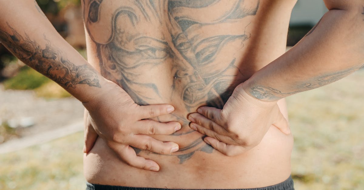
To relieve sudden shooting pain in the lower back, immediately cease any activities that exacerbate the discomfort, assume a resting posture that reduces spinal pressure, and apply ice therapy to the lumbar region to diminish acute inflammation. Short-term use of nonsteroidal anti-inflammatory drugs can mitigate chemical pain signals, but if the shooting sensation travels down the leg or is accompanied by neurological deficits, urgent medical evaluation is necessary to rule out severe nerve root compression.
Acute shooting pain in the lumbar region represents a significant clinical event, often signaling mechanical irritation or compression of the spinal neural structures. Unlike a dull muscular ache, a sudden, sharp, radiating sensation typically points to pathology involving the intervertebral discs, facet joints, or the exiting nerve roots.
Immediate clinical focus centers on controlling the acute inflammatory cascade, stabilizing the lumbar spine, and preventing further neurological insult. Proper diagnostic assessment is mandatory to distinguish between a self-limiting musculoskeletal strain and a structural lesion requiring advanced intervention.
2. Anatomical Structures of the Lumbar Spine
The lumbar spine is constructed of five robust vertebrae, labeled L1 through L5, designed to bear the majority of the body weight and facilitate truncal mobility. These bony structures are separated by fibrocartilaginous intervertebral discs, which act as dynamic shock absorbers during axial loading.
Posteriorly, the vertebrae articulate via facet joints, which guide spinal movement and prevent excessive rotation. A complex network of ligaments and paraspinal muscles encapsulates this bony architecture, providing vital dynamic stability.
The spinal cord terminates near the first lumbar vertebra, transitioning into a bundle of nerve roots known as the cauda equina. These nerves exit the spinal canal through the neural foramina, traveling down into the lower extremities to provide motor function and sensory feedback.
3. The Mechanism of Radicular Pain
Radicular pain, commonly referred to as sciatica when it involves the sciatic nerve, occurs when a spinal nerve root is subjected to mechanical compression or chemical irritation. This irritation generates ectopic neural discharges, which the brain perceives as a sharp, electric, or shooting pain traveling along the specific dermatome supplied by the affected nerve.
The pain is rarely confined to the back; it classically radiates into the buttock, posterior thigh, and sometimes down to the calf and foot. This distinct distribution pattern allows clinicians to pinpoint the exact level of spinal pathology based on the patient symptoms.
In addition to mechanical pressure, an inflammatory response generated by disc material interacting with the immune system releases cytokines, further sensitizing the nerve root and intensifying the perceived pain.
4. Herniated Nucleus Pulposus Explained
An intervertebral disc consists of a tough outer annulus fibrosus and a gel-like inner core called the nucleus pulposus. Disc herniation occurs when a tear in the outer annulus allows the inner nucleus to extrude into the spinal canal or neural foramen.
This displaced nuclear material occupies space intended for the nerve root, causing direct mechanical compression. Furthermore, the nucleus pulposus contains inflammatory proteins that, upon exposure to the systemic circulation, incite a localized neuroinflammatory reaction.
Herniations are most common at the L4-L5 and L5-S1 levels due to the high mechanical stress exerted on the lower lumbar segments during bending and lifting activities.
5. Degenerative Disc Disease and Spinal Stenosis
As the human body ages, intervertebral discs lose their hydration and elasticity, a process classified as degenerative disc disease. This desiccation leads to a loss of disc height, which inherently narrows the neural foramina and increases stress on the facet joints.
Spinal stenosis refers to the pathological narrowing of the central spinal canal or the lateral recesses. This narrowing is often a cumulative result of disc bulging, facet joint hypertrophy, and thickening of the ligamentum flavum, all of which compromise the space available for neural tissues.
Patients with spinal stenosis frequently experience neurogenic claudication, where shooting pain and leg heaviness worsen with standing or walking and are relieved by lumbar flexion, such as sitting or leaning over a shopping cart.
6. Muscle Spasms and Myofascial Triggers
While structural neural compression is a primary cause of shooting pain, severe muscular spasms in the lumbar or gluteal region can also generate sharp, acute symptoms. A sudden, uncoordinated movement can cause microscopic tearing in the paraspinal musculature.
In response to this injury, the muscle contracts forcefully to splint the area and prevent further damage. These protective spasms can be intensely painful and may occasionally compress peripheral nerves, such as the piriformis muscle compressing the sciatic nerve, known as piriformis syndrome.
Myofascial trigger points are localized, hyperirritable nodules within a taut band of skeletal muscle. Activation of these points can send referred shooting pain patterns that mimic structural radiculopathy.
7. Immediate Rest and Activity Modification
The traditional medical advice of prolonged bed rest for back pain has been universally discarded due to its detrimental effects on muscle conditioning and joint stiffness. However, a brief period of modified rest, spanning one to two days, is appropriate for acute, severe shooting pain.
During this brief rest phase, patients should adopt positions that minimize intradiscal pressure. Lying supine with pillows under the knees, or in a lateral decubitus position with a pillow between the legs, often provides substantial mechanical relief.
Following the initial acute phase, early mobilization is strictly encouraged. Patients should resume gentle, low-impact activities like short walks, strictly avoiding heavy lifting, forward bending, or twisting motions that place torsional strain on the lumbar discs.
8. The Role of Thermal Therapy
Thermal modalities are a cornerstone of conservative symptom management. In the first forty-eight hours following a sudden onset of shooting pain, cryotherapy, or cold application, is indicated. Ice causes local vasoconstriction, reducing blood flow and limiting the accumulation of inflammatory mediators around the sensitized nerve root.
After the acute inflammatory window has passed, transitioning to thermotherapy, or heat application, becomes beneficial. Heat promotes vasodilation, increasing local tissue perfusion, accelerating the clearance of metabolic waste, and reducing muscular tension.
Neither ice nor heat will reverse a structural nerve compression, but they modulate sensory input at the spinal cord level, effectively raising the pain threshold and providing temporary comfort.
9. Pharmacological Interventions for Acute Discomfort
Pharmacological management aims to disrupt the pain cycle and reduce neuroinflammation. Nonsteroidal anti-inflammatory drugs, such as ibuprofen or naproxen, inhibit the cyclooxygenase enzymes, thereby decreasing the synthesis of prostaglandins that fuel the inflammatory cascade.
Acetaminophen provides analgesia through central nervous system mechanisms but lacks anti-inflammatory properties, making it less effective for true radicular pain. Muscle relaxants may be prescribed short-term to alleviate severe secondary paraspinal spasms, though they carry a risk of sedation.
For intense neuropathic pain that is refractory to standard analgesics, medications specifically targeting nerve activity, such as gabapentin or pregabalin, may be initiated. These agents stabilize neuronal membranes and decrease the release of excitatory neurotransmitters.
10. Physical Therapy and Targeted Stretching
Once acute symptoms are adequately controlled, a structured physical therapy regimen is essential for long-term recovery and recurrence prevention. A trained physical therapist will design a specific exercise protocol based on the patient directional preference, such as the McKenzie method.
Exercises focused on lumbar extension are often beneficial for discogenic pain, as they encourage the centralized repositioning of herniated disc material away from the nerve roots. Conversely, flexion-based exercises are typically utilized for patients with spinal stenosis.
Stretching the hamstrings, hip flexors, and piriformis muscles helps correct pelvic tilt anomalies and reduces biomechanical stress on the lumbar spine. Strengthening the deep abdominal and multifidus muscles establishes a dynamic muscular corset, providing vital stabilization. Similar stabilization principles are used in managing joint stress, as detailed in our guide on knee joint arthritis.
11. Postural Ergonomics and Core Strengthening
Poor static posture places asymmetrical loads on the intervertebral discs and supporting ligaments. Prolonged sitting with a flexed lumbar spine increases intradiscal pressure significantly compared to standing or lying down.
Ergonomic modifications are mandatory for individuals with desk-bound occupations. Utilizing lumbar support rolls, adjusting monitor height to maintain a neutral cervical spine, and employing sit-to-stand desks can drastically reduce daily spinal stress.
Core strengthening goes beyond simple abdominal exercises. It involves training the entire lumbopelvic complex to fire synchronously, ensuring the spine remains dynamically stabilized during transitional movements and heavy lifting.
12. Epidural Steroid Injections and Advanced Care
When conservative therapies fail to provide relief after several weeks, interlaminar or transforaminal epidural steroid injections may be recommended. This procedure involves the fluoroscopically guided administration of a potent corticosteroid directly into the epidural space surrounding the inflamed nerve root.
The localized delivery of steroids forcefully suppresses the regional inflammatory response, rapidly reducing nerve root edema and pain. While not a definitive cure for structural abnormalities, injections often provide a crucial window of pain relief, allowing the patient to participate fully in physical therapy.
Radiofrequency ablation may be considered if the pain originates from the facet joints rather than a compressed nerve root. This procedure uses thermal energy to sever the medial branch nerves, preventing pain signals from reaching the brain.
13. Red Flag Symptoms Requiring Urgent Evaluation
While most episodes of shooting back pain resolve with conservative care, specific clinical markers, known as red flags, mandate immediate emergency assessment to rule out severe pathologies such as cauda equina syndrome, spinal infection, or occult malignancy.
| Red Flag Symptom | Potential Underlying Pathology |
|---|---|
| Loss of bowel or bladder control | Cauda Equina Syndrome |
| Saddle anesthesia (numbness in groin/perineum) | Cauda Equina Syndrome |
| Progressive motor weakness in the legs | Severe Nerve Root Compression |
| Unexplained fever and chills | Spinal Osteomyelitis or Epidural Abscess |
| History of cancer with new onset severe pain | Spinal Metastasis |
14. Frequently Asked Questions (FAQ)
1. Should I push through the pain to keep my back mobile?
No. Pushing through sharp, shooting pain can worsen nerve damage or increase disc herniation. Gentle movement is good, but any activity that reproduces the sharp, radiating symptoms must be avoided.
2. Does a slipped disc always require surgery?
The vast majority of disc herniations heal through natural physiological processes and conservative treatment over several months. Surgery is generally reserved for progressive neurological deficits or intractable pain failing non-operative care.
3. How do I know if my pain is a muscle spasm or nerve compression?
Muscle spasms usually cause localized, severe tightness and aching in the back itself. Nerve compression causes a sharp, electric pain that travels in a distinct line down the leg, often accompanied by numbness or tingling.
4. Is an MRI necessary for acute back pain?
An MRI is not routinely indicated for acute back pain without red flag symptoms, as imaging often reveals age-related changes that do not correlate with the actual source of the pain. Imaging is reserved for prolonged symptoms or progressive neurological issues.
5. Why is my pain worse when I cough or sneeze?
Coughing and sneezing cause a rapid increase in intra-abdominal and intrathecal pressure, which directly presses on the spinal nerve roots. If a nerve is already sensitized by a herniated disc, this pressure spike will cause a sharp surge of pain.
15. Bibliography
- Read relevant clinical studies on PubMed
- Search for academic articles on Google Scholar
- Read relevant clinical studies on PubMed
- Search for academic articles on Google Scholar
Disclaimer: The content is for informational purposes only and does not replace medical advice. Always consult your doctor for personalized treatment.