1. Introduction
A sudden, sharp clicking sound in the knee during walking is typically caused by a structural mechanical disruption within the joint, most notably a meniscal tear, abnormal tracking of the kneecap, or loose fragments of cartilage catching between the articulating bones. This auditory and physical sensation indicates that smooth, frictionless motion has been compromised, forcing the joint components to snap or rub against one another under weight-bearing loads. When the clicking is accompanied by a distinct, sharp pain, it elevates the symptom from a benign physiological anomaly to a discrete structural injury requiring clinical attention.
The knee is a complex, load-bearing hinge joint that relies on a precise arrangement of bones, ligaments, and fibrocartilage to absorb the impact of every step. Healthy cartilage provides a slick surface, while synovial fluid lubricates the internal compartment. Any irregularity in this gliding mechanism alters the distribution of force. An abrupt onset of painful clicking serves as a distinct biological signal that a specific piece of tissue is being repeatedly caught, pinched, or dragged during the flexion and extension cycle of the leg.
Determining the exact source of the sound requires localizing the pain and identifying the specific phase of the gait cycle where the click occurs. While many patients fear that clicking indicates permanent joint destruction, numerous cases resolve with targeted physical therapy to correct muscular imbalances. However, recognizing the difference between a harmless tendon snap and a degenerative cartilaginous tear is the foundational step in preserving long-term joint function and preventing premature osteoarthritis.
2. Anatomy and Biomechanics of the Knee
To comprehend the mechanical origins of a clicking knee, one must understand its fundamental anatomy. The knee joint forms at the intersection of three bones: the femur in the thigh, the tibia in the lower leg, and the patella acting as the kneecap. Two C-shaped wedges of tough fibrocartilage, the medial and lateral menisci, sit between the femur and tibia to distribute weight and stabilize the joint.
The patella rests within a shallow groove on the front of the femur called the trochlear groove. As the knee bends and straightens, the patella glides smoothly up and down this track. This entire structural unit is wrapped in a joint capsule lined with a synovial membrane, which secretes a viscous fluid to minimize friction.
Smooth movement requires absolute synchrony between the passive ligaments that guide the bones and the active muscles that generate force. The quadriceps muscles pull the patella upward, while the hamstrings bend the knee backward. If any single muscle pulls with disproportionate tension, or if a piece of cartilage loses its smooth contour, the synchronous movement is broken, resulting in a palpable and audible click.
3. Meniscal Tears and Joint Catching
The most prevalent structural cause of a sharp, painful click in the adult knee is a tear in the meniscus. The menisci act as shock absorbers, enduring profound compressive and rotational forces. A sudden twisting motion while the foot is firmly planted on the ground can easily shear this dense fibrocartilage. Furthermore, as individuals age, the meniscal tissue naturally dehydrates and becomes brittle, making it susceptible to degenerative tears from simple daily activities.
When a portion of the meniscus tears, a fragment of the cartilage can fold over or flap loosely within the joint space. As the patient walks and the knee extends, the femur rolls over this loose fragment. The fragment catches between the bones, creating a distinct, sharp click or popping sensation.
Patients with a meniscal tear often describe a pain that is pinpointed to the joint line on either the inner or outer side of the knee. The clicking is frequently accompanied by a feeling of instability, as if the knee might unexpectedly give way, and periods of localized swelling following extended physical activity.
4. Patellofemoral Tracking Syndrome
If the clicking is perceived near the front of the knee, particularly when walking up or down stairs, patellofemoral tracking syndrome is a primary clinical suspicion. This condition occurs when the kneecap fails to glide centrally within the trochlear groove of the femur.
This tracking error is typically driven by a muscular imbalance. If the outer quadriceps muscles are excessively tight and the inner quadriceps muscles are weak, the patella is physically pulled toward the outside of the leg during movement. As the knee bends, the misaligned patella forcefully snaps into or out of its proper groove, generating a loud click or clunk.
Over time, this abnormal tracking damages the protective articular cartilage on the underside of the kneecap. This secondary damage, known as chondromalacia patellae, transforms a painless clicking sensation into a deep, sharp ache that worsens after prolonged periods of sitting with the knees bent.
5. Iliotibial Band Friction Syndrome
The iliotibial band is a thick, fibrous band of connective tissue that runs down the outside of the thigh, from the pelvis to just below the knee. As the knee bends and straightens, this band slides back and forth over the lateral epicondyle, a bony prominence on the outside of the femur.
If the iliotibial band becomes excessively tight due to overuse, poor running biomechanics, or sudden increases in walking distance, it creates significant friction against the bone. Instead of gliding smoothly, the tight band snaps abruptly over the bony ridge.
This snapping produces a sharp, audible click on the lateral side of the knee. The accompanying pain is typically sharp and stinging, localized strictly to the outside of the joint. It is distinctly exacerbated by walking downhill or utilizing stairs, which places maximum tension on the iliotibial band.
6. Osteoarthritis and Cartilage Degeneration
In older adults, a sudden increase in joint noise frequently correlates with the progression of osteoarthritis. Osteoarthritis involves the gradual, structural degradation of the smooth articular cartilage covering the ends of the bones. As this protective layer wears away, the underlying bone becomes exposed and reactive.
The normally slick joint surfaces become rough, pitted, and uneven. When these degraded surfaces rub against one another during walking, they produce a grinding, crunching, or clicking sound clinically referred to as crepitus.
While crepitus itself can be painless, a sudden, sharp pain accompanying the sound suggests that a small fragment of degraded cartilage has broken off or that exposed bone has sustained acute mechanical stress. Osteoarthritic clicking is often accompanied by significant morning stiffness that slowly improves with light movement.
7. Loose Bodies in the Joint Space
Occasionally, small fragments of bone or cartilage detach completely from their original structure and float freely within the synovial fluid of the joint capsule. These fragments are clinically termed loose bodies, or “joint mice.”
As the patient walks and the synovial fluid circulates, these loose bodies migrate throughout the joint. If a fragment drifts between the articulating surfaces of the femur and tibia, it acts like a pebble caught in a door hinge. The joint abruptly catches on the fragment, producing a sharp click and a sudden, intense spike of pain.
The classic clinical feature of a loose body is the unpredictable nature of the symptom. The sharp click may occur agonizingly on one step and then completely disappear for several days as the fragment floats away into a benign recess of the joint capsule.
8. Synovial Plica Syndrome
The synovial plica is a fold or band of tissue within the joint capsule, representing a remnant of fetal development. In most individuals, these folds are thin, elastic, and entirely asymptomatic. However, following a direct blunt trauma to the knee or repeated overuse, a plica can become inflamed, thickened, and fibrotic.
A thickened plica loses its elasticity. During walking, as the knee bends, the thickened tissue snaps forcefully over the medial femoral condyle, the rounded end of the thigh bone. This snapping produces a palpable click on the inner aspect of the kneecap.
Synovial plica syndrome is often confused with a meniscal tear due to the similar location of the pain. However, plica pain frequently feels like a dull ache that sharpens distinctively during the specific snapping event, often accompanied by a sensation of localized catching.
9. Ligamentous Instability
The major ligaments of the knee, including the anterior cruciate ligament and the collateral ligaments, provide essential passive stability. If a ligament is partially torn or chronically stretched from a previous injury, the joint loses its tight structural integrity.
This micro-instability allows the femur and tibia to shift abnormally against one another during the weight-bearing phase of the gait cycle. The bones may subluxate slightly and then snap back into alignment, generating a deep, structural click.
Clicking related to ligamentous laxity is a concerning symptom, as the abnormal shearing forces dramatically accelerate the wear and tear on the meniscus and articular cartilage, predisposing the patient to early-onset osteoarthritis. Patients experiencing joint instability should also refer to our guide on shooting leg pain to differentiate local knee mechanics from referred spinal nerve issues.
10. Table: Differentiating Causes of Knee Clicking
| Underlying Cause | Location of the Click | Distinguishing Clinical Signs |
|---|---|---|
| Meniscal Tear | Inner or outer joint line | Painful catching, swelling after walking, feeling of locking |
| Patellofemoral Tracking | Front of the knee, under kneecap | Worsens on stairs, deep ache after sitting |
| Iliotibial Band Friction | Strictly outside of the knee | Snapping sensation over bone, stinging lateral pain |
| Loose Body | Migratory, unpredictable | Sudden, sharp pain that randomly disappears |
11. Diagnostic Physical Examination
Evaluating a clicking knee begins with a meticulous physical examination to isolate the exact source of the mechanical disruption. The clinician observes the patient’s gait, checking for subtle limping, overpronation of the foot, or valgus collapse, where the knee caves inward during a step.
Palpation of the joint line is crucial. Tenderness specifically along the space between the femur and tibia strongly suggests meniscal pathology. The clinician will gently guide the patella through its groove, feeling for grinding or sudden snapping that indicates tracking issues or chondromalacia.
Specific provocative maneuvers are employed. The McMurray test involves rotating the lower leg while bending and straightening the knee; a palpable clunk accompanied by pain during this maneuver is a classic indicator of a meniscal tear.
12. Advanced Radiographic Imaging
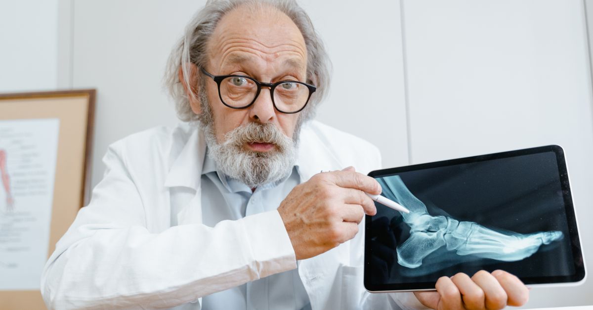
While physical examination is foundational, radiographic imaging provides definitive structural clarity. Standard weight-bearing X-rays are the initial imaging modality. They visualize the bony architecture, identifying narrowed joint spaces characteristic of osteoarthritis or detecting large, calcified loose bodies floating in the joint.
However, X-rays cannot visualize the meniscus, ligaments, or articular cartilage. If a soft tissue injury is suspected based on the clicking and sharp pain, Magnetic Resonance Imaging is the gold standard diagnostic tool.
Magnetic Resonance Imaging provides exquisite, three-dimensional detail of the intra-articular structures. It definitively identifies the precise location and severity of a meniscal tear, visualizes bone marrow edema signifying acute stress, and detects thickened synovial plicae that standard X-rays miss entirely.
13. Conservative Physical Rehabilitation
The overwhelming majority of clicking knees, particularly those driven by patellofemoral tracking or minor degenerative meniscal tears, respond robustly to conservative physical therapy. The objective is to restore perfect biomechanical balance across the joint, relieving the abnormal pressure causing the snap.
Therapy protocols heavily emphasize strengthening the vastus medialis obliquus, the inner teardrop-shaped muscle of the quadriceps, to help pull the kneecap back into a central alignment. Simultaneously, targeted stretching of the iliotibial band and the hamstrings relieves external compressive forces.
Proprioceptive training is also introduced to improve the neurological coordination of the leg muscles during walking. By correcting underlying hip and ankle weaknesses, the knee is no longer forced to compensate for poor mechanics, significantly reducing the occurrence of painful clicking.
14. Medical Management and Injections
To facilitate participation in physical therapy, localized inflammation and pain must be managed. Nonsteroidal anti-inflammatory drugs provide systemic relief from the swelling associated with joint friction.
For severe, localized pain preventing rehabilitation, intra-articular corticosteroid injections deliver a potent anti-inflammatory dose directly into the joint capsule. This rapidly shrinks swollen synovial tissue and inflamed plicae, often eliminating the mechanical click entirely for several months.
Viscosupplementation involves injecting hyaluronic acid, a thick, lubricating gel, into the knee. This therapy aims to artificially restore the shock-absorbing and gliding properties of the synovial fluid, reducing the friction and grinding sounds associated with early osteoarthritis.
15. Surgical Interventions and Arthroscopy
When conservative measures fail and a structural defect consistently causes sharp pain and joint locking, surgical intervention is required. Knee arthroscopy is a minimally invasive surgical procedure where a tiny camera and specialized instruments are inserted into the joint through small portals.
If a meniscal tear is identified, the surgeon will either repair the tear using microscopic sutures or perform a partial meniscectomy, meticulously trimming away the loose, catching flap of cartilage to leave a smooth, stable edge.
For loose bodies, the surgeon simply flushes the joint and removes the floating fragments. If severe patellofemoral tracking is permanently damaging the cartilage, a lateral release procedure can be performed to cut the tight ligaments pulling the kneecap outward, restoring a frictionless, central glide.
16. Frequently Asked Questions (FAQ)
1. Is it normal for my knees to click when I squat, even if it does not hurt?
Yes. Painless clicking or popping during deep bending is very common and usually benign. It is often caused by gas bubbles shifting in the joint fluid or a healthy tendon sliding smoothly over a bone. Only clicking accompanied by pain or swelling requires medical evaluation.
2. Will a torn meniscus heal on its own without surgery?
The outer third of the meniscus has a blood supply and can sometimes heal with rest and physical therapy. The inner two-thirds completely lack a blood supply and cannot heal naturally. If an inner tear is causing a sharp, catching pain, it may require surgical trimming.
3. Can wearing different shoes stop the clicking in my knee?
Yes, absolutely. Shoes with poor arch support allow your foot to roll inward excessively when you walk. This forces your entire leg to rotate inward, placing abnormal twisting stress on the knee joint and kneecap, which frequently leads to painful clicking.
4. Should I stop walking and exercising if my knee is clicking?
You should stop any activity that produces sharp pain. However, complete rest is detrimental to joint health. Transition to low-impact exercises like swimming or stationary cycling, which keep the joint lubricated and the muscles strong without the heavy pounding of walking or running.
5. How do I know if the clicking is just arthritis?
Arthritic clicking usually feels like a grinding, sandy crunch beneath the kneecap and is accompanied by stiffness that is worst in the morning. A sudden, single, sharp pop that catches your joint unpredictably is more likely a mechanical tear or a loose body.
17. Bibliography
Disclaimer: The content is for informational purposes only and does not replace medical advice. Always consult your doctor for personalized treatment.