1. Introduction
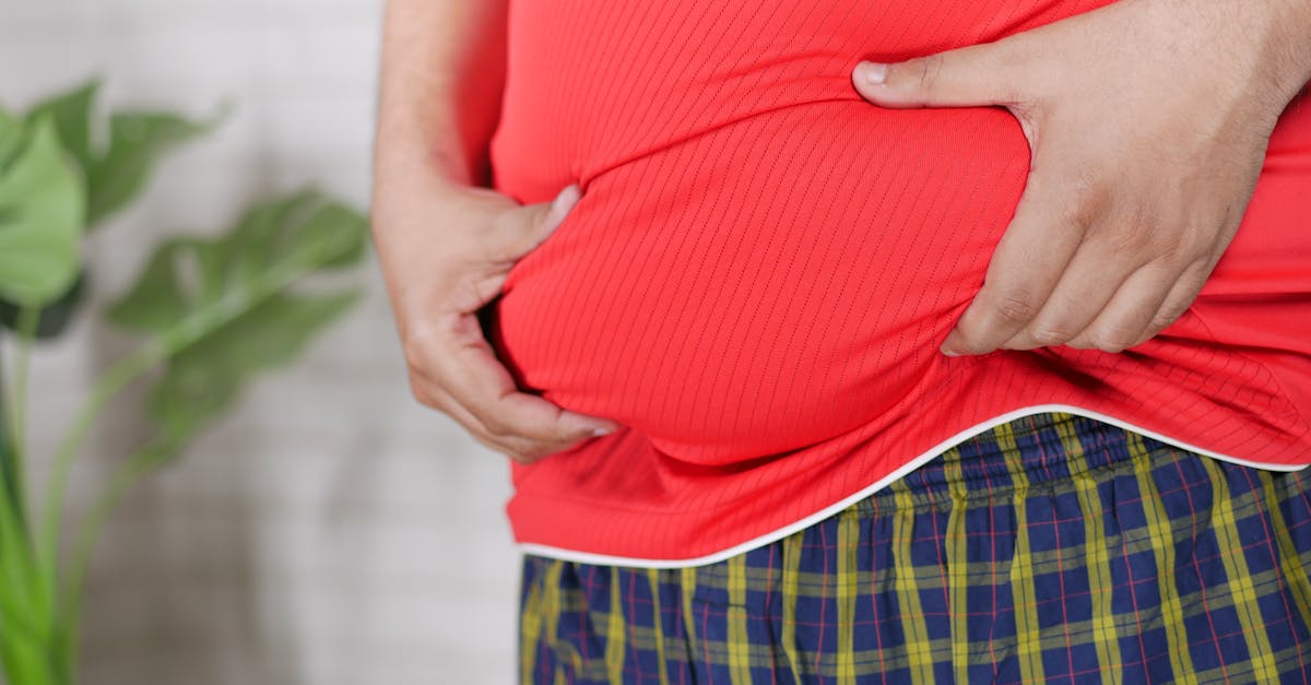
Yes, an umbilical hernia can absolutely cause a sharp, localized pain directly in or around the belly button. This pain occurs when a portion of the intestine or abdominal fat forces its way through a weakness in the fascial ring surrounding the umbilicus, causing physical stretching and nerve compression. When the trapped tissue is subjected to sudden increases in intra-abdominal pressure from coughing or lifting, it generates a sharp, pinching sensation that demands clinical assessment.
The umbilicus is a natural point of structural weakness in the human abdominal wall. While many umbilical hernias are small and entirely asymptomatic, the presence of sudden, sharp pain signifies that the mechanical defect is actively compromising internal tissues. This localized discomfort acts as an early warning system, alerting the patient that the protruding fat or bowel is experiencing significant friction or compression.
Understanding the biomechanics of an umbilical hernia is critical for preventing severe complications. Recognizing the specific physical triggers that elicit the sharp pain helps patients manage their symptoms conservatively while awaiting definitive surgical repair to close the fascial defect and restore abdominal integrity.
2. The Anatomy of the Umbilical Ring
The umbilicus, commonly known as the belly button, is the central scar remaining from the detachment of the fetal umbilical cord. During fetal development, blood vessels pass through an opening in the abdominal muscles called the umbilical ring.
Shortly after birth, this ring typically closes tightly, sealing the abdominal cavity. However, in many individuals, the fascial closure remains incomplete or weakens significantly over time due to mechanical stress. This leaves a small, vulnerable gap precisely in the center of the abdomen.
The abdominal wall consists of tough layers of fascia and muscle designed to contain the internal organs under high pressure. When the umbilical ring fails to maintain its structural integrity, it provides a direct pathway for intra-abdominal contents to escape outward, directly beneath the thin skin of the navel.
3. Pathophysiology of an Umbilical Hernia
An umbilical hernia develops when the constant outward pressure of the abdominal cavity overcomes the tensile strength of the weakened umbilical ring. The inner lining of the abdomen, the peritoneum, pushes through the defect, creating a small sac.
Subsequently, preperitoneal fat or a loop of the small intestine slides into this peritoneal sac. This displacement creates a visible, palpable bulge at the belly button. In adults, these hernias tend to enlarge progressively as the fascial defect stretches over years of continuous mechanical load.
While the defect itself is simply a hole in the fascia, the clinical symptoms arise entirely from the tissues traversing that hole. The interaction between the unyielding fascial edges of the ring and the soft, sensitive tissues trapped within it dictates the severity of the patient’s discomfort.
4. Mechanisms of Sharp Pain Generation
The sharp pain associated with an umbilical hernia is generated through multiple distinct mechanisms. The primary cause is mechanical nerve compression. As tissue is forced through the narrow fascial ring, the somatic nerves innervating the abdominal wall are stretched and pinched.
This compression generates a sudden, acute, stinging pain precisely at the site of the defect. Secondly, if a portion of the highly innervated mesentery or intestine is pulled into the hernia sac, the physical traction on these visceral structures triggers intense, sharp nociceptive signals.
Patients frequently describe the sensation as a tearing or pinching feeling that occurs instantly when they exert themselves. The pain typically subsides quickly once the exertion ceases and the intra-abdominal pressure normalizes, allowing the trapped tissue to relax back into the abdominal cavity.
5. Triggers Increasing Intra-Abdominal Pressure
The intensity of umbilical hernia pain is directly proportional to intra-abdominal pressure. Any physiological action that forces the diaphragm downward and contracts the abdominal wall drastically spikes the internal pressure, driving tissue forcefully into the hernia defect.
Heavy lifting is the most common mechanical trigger. Squatting to pick up a heavy object engages the core musculature intensely, squeezing the abdominal contents against the weakened umbilicus.
Chronic coughing, forceful sneezing, and straining during bowel movements are equally potent triggers. Even prolonged standing can exacerbate the pain, as gravity constantly pulls the abdominal contents downward and outward against the anterior abdominal wall.
6. The Danger of Incarceration
A reducible hernia is one where the protruding bulge can be gently pushed back into the abdomen. While a reducible hernia can cause intermittent sharp pain during exertion, it is not an immediate emergency.
However, if the protruding tissue becomes swollen and firmly stuck within the fascial ring, it transforms into an incarcerated hernia. Incarceration is a severe clinical complication. The narrow fascial ring acts as a rigid tourniquet around the trapped fat or bowel.
An incarcerated hernia presents as a hard, continuous, and exquisitely painful lump that will not flatten when the patient lies down. The sharp, intermittent pain evolves into a constant, severe ache, signaling that the trapped tissue is becoming engorged with fluid.
7. Strangulation and Tissue Ischemia
If an incarcerated hernia is not promptly reduced, it can progress to strangulation. Strangulation occurs when the swelling within the trapped tissue becomes so severe that it completely occludes the arterial blood supply.
Without oxygenated blood, the trapped segment of intestine or fat begins to undergo ischemic necrosis, or tissue death. This is a catastrophic surgical emergency. The pain becomes unbearable and is typically accompanied by overlying skin redness, systemic fever, and signs of a bowel obstruction, such as intractable vomiting.
If a strangulated bowel ruptures, it releases highly toxic intestinal contents directly into the sterile abdominal cavity, leading to life-threatening peritonitis and sepsis.
8. Distinguishing Hernia Pain from Gastrointestinal Distress
Patients experiencing central abdominal pain must distinguish between musculoskeletal hernia pain and true visceral gastrointestinal distress. Hernia pain is remarkably localized; the patient can point with one finger precisely to the belly button.
This pain is overwhelmingly mechanical, meaning it worsens significantly with core movement, coughing, or sitting up from a lying position.
Conversely, gastrointestinal pain, such as that caused by gastroenteritis or early appendicitis, is usually diffuse, crampy, and unrelated to movement of the abdominal wall. Gastrointestinal distress is also consistently accompanied by systemic symptoms like nausea, diarrhea, or a loss of appetite, which are absent in an uncomplicated hernia.
9. Physical Examination Findings
A clinical diagnosis of an umbilical hernia is primarily established through a thorough physical examination. The physician will inspect the umbilicus while the patient is standing, looking for asymmetry or a distinct outward bulge.
The clinician will then perform the Valsalva maneuver, asking the patient to bear down or cough forcefully. This action increases intra-abdominal pressure and will immediately force the hernia sac to protrude, often reproducing the patient’s sharp, pinching pain.
Palpation is utilized to determine the exact size of the fascial defect and to assess whether the hernia is reducible. A small defect with a large amount of protruding tissue poses a significantly higher risk for incarceration than a wide, compliant fascial ring.
10. Diagnostic Imaging
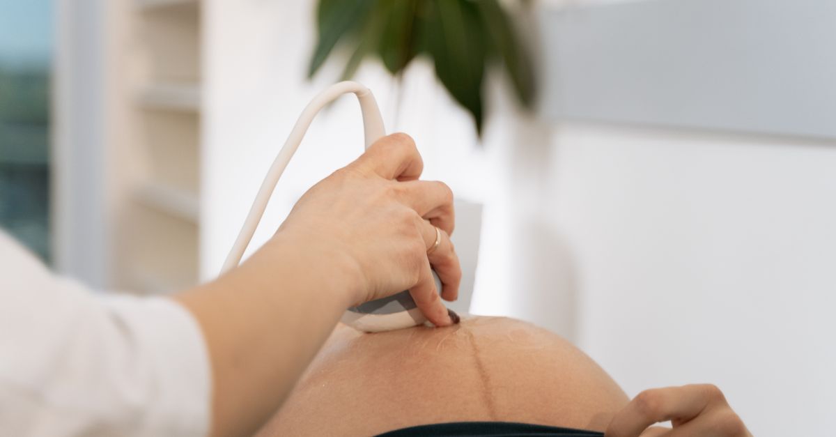
While physical examination is usually sufficient for diagnosis, diagnostic imaging is employed to evaluate the contents of the hernia sac and plan for surgical intervention, especially in obese patients where palpation is challenging.
| Imaging Modality | Clinical Utility |
|---|---|
| Abdominal Ultrasound | Non-invasive tool to confirm the fascial defect and identify trapped fat or fluid. |
| Computed Tomography (CT) Scan | Provides detailed cross-sectional imaging, crucial for identifying trapped bowel loops. |
| Magnetic Resonance Imaging | Used rarely, but helpful for complex abdominal wall reconstructions. |
11. Conservative Management Strategies
For small, completely reducible umbilical hernias that cause only rare, mild pain, watchful waiting is a medically acceptable strategy. The focus is on mitigating intra-abdominal pressure to prevent the defect from enlarging.
Patients are instructed to practice proper lifting mechanics, utilizing the legs rather than the core and back. Aggressive management of chronic coughs or constipation is essential, as these conditions subject the hernia to relentless daily strain.
While specialized hernia belts or binders are available commercially, they are generally discouraged for long-term use. They do not heal the hernia and can sometimes apply improper pressure, inadvertently increasing the risk of incarceration by trapping the tissue outside the fascial ring.
12. Surgical Repair Options
The only definitive treatment for an umbilical hernia is surgical closure of the fascial defect. Elective surgery is highly recommended for any hernia that causes persistent sharp pain, is enlarging, or has ever been incarcerated.
Open herniorrhaphy involves making a small semi-circular incision directly under the belly button. The surgeon pushes the protruding tissue back into the abdomen and sutures the strong fascial edges together.
For defects larger than two centimeters, a synthetic surgical mesh is typically utilized. The mesh is anchored beneath the muscle layer to bridge the gap, functioning as a permanent reinforcement that absorbs intra-abdominal pressure and drastically reduces the rate of recurrence.
13. Recovery and Post-Operative Care
Post-operative recovery from a routine umbilical hernia repair is generally rapid. Patients typically return home the same day. Pain is managed with oral analgesics and ice applied to the surgical site to minimize local swelling.
Strict adherence to activity restrictions is critical during the initial healing phase. Patients must avoid lifting anything heavier than ten pounds for a minimum of four to six weeks. This allows the fascial tissues to fuse solidly with the surgical mesh.
Engaging in premature strenuous activity before the biological integration of the mesh is complete is the leading cause of surgical failure and hernia recurrence.
14. Preventing Hernia Recurrence
Preventing the recurrence of a hernia requires permanent lifestyle modifications to control intra-abdominal pressure. Maintaining a healthy body weight is the most significant preventative measure. Excess visceral fat places an unyielding, continuous outward pressure on the anterior abdominal wall.
Patients must also avoid smoking. Nicotine impairs collagen synthesis, weakening the fascial tissues and delaying surgical healing. Furthermore, smoking induces a chronic cough, subjecting the repaired umbilicus to violent mechanical forces.
Engaging in targeted, safe core-strengthening exercises under the guidance of a physical therapist can improve the overall tone of the abdominal wall, providing better muscular support for the vulnerable umbilical region.
15. Identifying Red Flag Symptoms
Patients with an unrepaired umbilical hernia must remain vigilant for signs of clinical deterioration. If the sharp, intermittent pain suddenly changes into a continuous, severe, burning ache that radiates across the abdomen, the tissue is likely strangulated.
An inability to push the bulge back in when lying flat, a sudden change in the color of the skin over the belly button to deep red or purple, or the onset of severe nausea and vomiting are absolute medical emergencies.
These red flag symptoms demand immediate transport to the nearest emergency department for urgent surgical intervention to prevent bowel necrosis.
16. Frequently Asked Questions FAQ
1. Will an umbilical hernia heal on its own without surgery?
No, an umbilical hernia is a physical hole in the tough fascial layer of the abdominal wall. Tissues cannot spontaneously grow back together to close this gap. It requires surgical suturing or mesh placement for permanent closure.
2. Is it safe to exercise with a small umbilical hernia?
Light aerobic exercise like walking or cycling is generally safe. However, heavy weightlifting, intense core workouts, or exercises requiring the Valsalva maneuver must be avoided, as they will aggressively force tissue into the defect and cause sharp pain.
3. Why does my belly button hurt more when I am bloated?
Bloating distends the intestines with gas, rapidly increasing the internal pressure of the abdominal cavity. This increased volume pushes harder against the abdominal wall, forcing more tissue into the unyielding umbilical ring and exacerbating the pinching nerve pain.
4. How long does the pain last after umbilical hernia surgery?
Most patients experience moderate localized surgical pain for the first three to seven days, which is manageable with oral medications. Mild pulling sensations or twinges near the incision can persist for several weeks as the internal scar tissue matures and tightens.
5. Can pregnancy cause an umbilical hernia?
Yes, the massive expansion of the uterus during pregnancy exerts immense outward pressure on the abdominal wall, frequently stretching the umbilical ring and causing a new hernia or exacerbating a pre-existing, silent defect.
17. Bibliography
Disclaimer: The content is for informational purposes only and does not replace medical advice. Always consult your doctor for personalized treatment.