1. Introduction and Primary Treatment Focus
To relieve chronic pain in the ball of the foot, management must focus on offloading pressure from the metatarsal heads through the use of custom orthotics or metatarsal pads, transitioning to footwear with wide toe boxes and low heels, and implementing daily stretching protocols for the Achilles tendon. Persistent inflammation can be addressed with targeted icing and nonsteroidal anti-inflammatory medications, but long-term resolution requires identifying and correcting the underlying structural or biomechanical fault driving the pressure imbalance.
Chronic discomfort localized to the plantar aspect of the forefoot is clinically termed metatarsalgia. This symptom is not a specific disease entity but rather a manifestation of abnormal weight distribution, inflammatory processes, or neuropathic compression within the intricate structures of the forefoot.
Effective clinical resolution demands a systematic assessment of the patient gait, footwear habits, and foot architecture. Ignoring the root biomechanical cause while solely treating the pain inevitably leads to symptom recurrence and potential progressive deformity.
2. Anatomical Overview of the Forefoot
The forefoot comprises the five metatarsal bones and the corresponding phalanges of the toes. The metatarsal heads articulate with the proximal phalanges, forming the metatarsophalangeal joints, which serve as primary weight-bearing focal points during the push-off phase of the gait cycle.
Beneath the first metatarsal head lie two small, spherical bones known as the sesamoids. These structures are embedded within the flexor hallucis brevis tendon, functioning mechanically like pulleys to enhance the leverage of the great toe during propulsion.
A dense network of plantar ligaments, the plantar fascia, and intrinsic foot muscles support these osseous structures. Digital nerves run parallel to the metatarsals, bifurcating near the metatarsal heads to supply sensation to the toes, making them vulnerable to mechanical compression.
3. Understanding Metatarsalgia
Metatarsalgia typically presents as a diffuse, burning, or aching pain beneath the second, third, or fourth metatarsal heads. This discomfort is universally exacerbated by prolonged standing, walking barefoot on hard surfaces, or participating in high-impact activities.
The fundamental pathophysiology involves the excessive concentration of mechanical loading on the metatarsal heads. Over time, this repetitive microtrauma leads to inflammation of the periarticular tissues, capsulitis, and degradation of the protective plantar fat pad that normally cushions the bones.
Patients often describe the sensation as walking on a pebble or having a folded sock bunched up under the front of their foot, indicating the localized nature of the mechanical pressure.
4. Morton Neuroma and Nerve Entrapment
A common, specific etiology of forefoot pain is Morton neuroma. Despite the name, it is not a true tumor but rather a perineural fibrosis and thickening of the common digital nerve, most frequently occurring in the third web space between the third and fourth metatarsals.
This localized nerve entrapment results from repetitive shearing forces and compression between the adjacent metatarsal heads. The pain is distinctly neuropathic, characterized by sharp, shooting, or burning sensations radiating into the adjacent toes, often accompanied by numbness or tingling.
Clinical examination often reveals a positive Mulder sign, where lateral compression of the forefoot while applying upward pressure on the plantar web space elicits a palpable click and reproduces the neuralgic pain.
5. Sesamoiditis and Localized Inflammation
Pain specifically isolated under the base of the great toe suggests pathology involving the sesamoid apparatus. Sesamoiditis is an inflammatory condition affecting the tendons surrounding these tiny bones, typically provoked by activities requiring repetitive upward bending of the big toe, such as dancing or sprinting.
The constant mechanical friction leads to localized tendinopathy, chondromalacia of the sesamoid articular surface, or, in severe cases of acute loading, a stress fracture of the sesamoid bone itself.
Clinical differentiation involves precise palpation. Exquisite point tenderness directly plantar to the first metatarsal head, coupled with pain during resisted flexion of the great toe, strongly points toward a sesamoid etiology rather than general metatarsalgia.
6. Biomechanical Factors and Foot Arch Architecture
The structural architecture of the foot heavily dictates the distribution of plantar pressures. Pes cavus, or a high-arched foot, rigidly locks the midfoot, forcing the heel and the metatarsal heads to absorb a disproportionate amount of impact forces.
Conversely, pes planus, or flatfoot deformity, alters the propulsive mechanics of the foot. The destabilization of the first ray shifts weight laterally, overloading the lesser metatarsals, which are not anatomically designed for primary weight-bearing duties.
A shortened Achilles tendon is another critical biomechanical contributor. Limited ankle dorsiflexion forces the foot to lift off the ground prematurely during walking, drastically increasing the duration and magnitude of pressure exerted on the forefoot.
7. The Impact of Inadequate Footwear
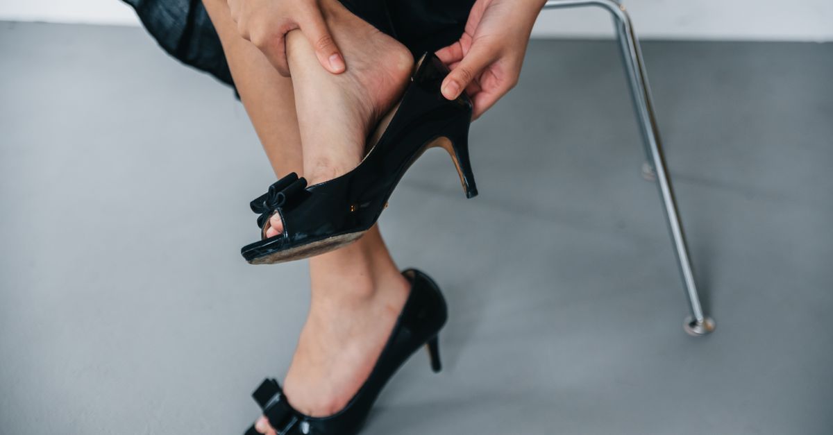
Footwear selection is perhaps the most significant modifiable risk factor for chronic forefoot pain. High-heeled shoes place the foot in profound plantarflexion, shifting nearly all the body weight directly onto the small surface area of the metatarsal heads.
Furthermore, shoes with narrow, tapering toe boxes compress the metatarsals laterally. This unnatural squeezing bows the transverse arch, promotes the development of neuromas by compressing the digital nerves, and exacerbates capsular inflammation.
Worn-out athletic shoes that have lost their midsole cushioning properties fail to attenuate impact forces. Proper shoe selection must prioritize a wide toe box, zero or minimal heel drop, and adequate forefoot cushioning. Modifying footwear is just as crucial here as it is for other structural foot issues, similar to strategies utilized in managing plantar fasciitis.
8. Orthotic Devices and Metatarsal Pads
The mechanical offloading of the inflamed structures is vital for healing. Metatarsal pads are simple, cost-effective devices placed proximal to the metatarsal heads, not directly underneath them. By elevating the metatarsal shafts, these pads reestablish the transverse arch and shift pressure away from the painful joints.
Custom orthotic inserts provide a more comprehensive biomechanical correction. A podiatrist can design an orthotic that supports the medial longitudinal arch, incorporates specific forefoot accommodations, and corrects abnormal pronation or supination mechanics.
Rocker-bottom shoes are another excellent biomechanical tool. The curved sole facilitates the transition from heel strike to toe-off, effectively bypassing the need for the metatarsophalangeal joints to bend forcefully, thereby reducing joint stress.
9. Stretching the Achilles Tendon and Plantar Fascia
Addressing tissue inflexibility is mandatory for resolving altered gait mechanics. A tight calf muscle complex exerts a continuous pull on the heel bone, which translates to increased pressure on the front of the foot during locomotion.
Consistent, daily stretching of the gastrocnemius and soleus muscles using wall stretches or slant boards improves ankle mobility. This increased range of motion allows the foot to remain flat on the ground longer during the stance phase of walking.
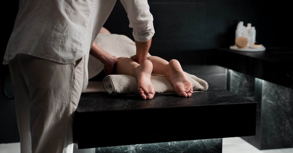
Additionally, gently massaging and stretching the plantar fascia helps maintain the elasticity of the soft tissues crossing the sole of the foot, preventing secondary stiffness and fascial contractures resulting from altered walking patterns.
10. Ice Massage and Topical Interventions
To manage daily inflammatory flare-ups, localized cryotherapy is highly effective. Freezing a small water bottle and rolling the sole of the foot over it provides a dual benefit: the cold temperature induces vasoconstriction to reduce tissue edema, while the rolling motion offers a deep tissue massage.
Topical nonsteroidal anti-inflammatory gels, such as diclofenac, can be applied directly to the dorsal and plantar aspects of the affected metatarsophalangeal joints. Topical agents deliver high local tissue concentrations of the medication with minimal systemic absorption.
These modalities are best utilized at the end of the day or immediately following periods of prolonged standing to arrest the inflammatory cascade before it causes overnight stiffness and pain.
11. Corticosteroid Injections for Refractory Cases
If conservative measures yield insufficient relief after several months, a targeted corticosteroid injection may be considered. Using ultrasound guidance, a physician can inject a localized dose of steroids directly into the inflamed joint capsule or the perineural space surrounding a neuroma.
This intervention rapidly suppresses acute inflammation, providing significant symptomatic relief. However, steroid injections in the foot must be used judiciously, as repeated administrations can cause atrophy of the protective plantar fat pad or weaken the joint capsules.
It is critical that the mechanical offloading protocols remain in place following an injection; otherwise, the underlying structural stress will quickly cause the inflammation to return once the medication wears off.
12. Surgical Considerations for Anatomical Correction
Surgical intervention is strictly reserved for patients with severe, chronic pain that is entirely refractory to all conservative biomechanical and pharmacological therapies. The surgical approach depends entirely on the specific anatomical pathology.
For a true Morton neuroma, surgical excision of the thickened nerve segment, known as a neurectomy, yields high success rates, although it results in permanent numbness between the affected toes.
In cases of severe metatarsophalangeal joint subluxation or disproportionate metatarsal length, an osteotomy may be performed. This involves precisely cutting and realigning the metatarsal bone to elevate the head, thereby permanently redistributing plantar pressures.
13. Diagnostic Imaging Modalities
Accurate diagnosis hinges on appropriate imaging. Standard weight-bearing radiographs of the foot are the initial imaging modality of choice. X-rays reveal the osseous architecture, identifying metatarsal length disparities, stress fractures, joint space narrowing indicative of arthritis, or sesamoid abnormalities.
If soft tissue pathology is suspected, such as a neuroma, plantar plate tear, or severe bursitis, diagnostic ultrasound or magnetic resonance imaging is utilized.
| Condition | Primary Diagnostic Finding | Imaging Modality |
|---|---|---|
| Metatarsal Stress Fracture | Cortical thickening or visible fracture line | X-ray or MRI |
| Morton Neuroma | Hypoechoic mass in the intermetatarsal space | Ultrasound or MRI |
| Plantar Plate Tear | Fluid in joint space, ligament discontinuity | MRI |
| Sesamoiditis | Bone marrow edema in sesamoid bones | MRI |
14. Frequently Asked Questions (FAQ)
1. Can walking barefoot make ball of foot pain worse?
Yes. Walking barefoot eliminates all shock absorption and arch support, forcing the metatarsal heads to bear the full impact of your body weight directly against hard surfaces, which rapidly worsens inflammation.
2. How long does a metatarsal stress fracture take to heal?
A metatarsal stress fracture typically requires six to eight weeks of strict immobilization in a stiff-soled surgical shoe or walking boot, followed by a gradual return to weight-bearing activities.
3. Will losing weight help with metatarsalgia?
Yes. Every step you take places forces equal to multiple times your body weight on your forefoot. Reducing total body mass directly decreases the mechanical load transmitted to the metatarsal heads.
4. Can calluses on the bottom of the foot cause pain?
Calluses are a symptom of excess friction and pressure. While the thickened skin itself can become painful and act like a hard stone under the foot, it is a clinical indicator that the bone beneath it is absorbing too much pressure.
5. Are steroid injections painful in the foot?
Injections in the plantar aspect of the foot can be quite uncomfortable due to the dense nerve supply. Physicians often use a local anesthetic block or a cooling spray prior to the injection to minimize discomfort.
15. Bibliography
- Read relevant clinical studies on PubMed
- Search for academic articles on Google Scholar
- Read relevant clinical studies on PubMed
- Search for academic articles on Google Scholar
Disclaimer: The content is for informational purposes only and does not replace medical advice. Always consult your doctor for personalized treatment.