1. Introduction
A severe tension headache at the base of the skull is typically caused by the sustained contraction and fatigue of the suboccipital muscles, often resulting from poor cervical posture or chronic stress. This muscle tension irritates the local fascial tissues and local nerves, creating a localized band of pressure. Correcting ergonomic habits and utilizing targeted physical therapy generally provide the most effective relief.
2. Anatomical Overview Of The Occipital Region
The base of the skull, known clinically as the occipital region, serves as a crucial junction between the cervical spine and the cranium. This area is densely populated with intricate layers of muscle, fascia, and connective tissue. The primary function of these structures is to support the heavy weight of the human head while allowing for a wide range of multidirectional movements.
3. The Role Of The Suboccipital Muscles
Deep within the back of the neck lie the suboccipital muscles. This specific group consists of four small muscle pairs that attach the upper cervical vertebrae directly to the occipital bone. These muscles are packed with sensory receptors that constantly monitor head position and balance. When subjected to continuous mechanical stress, they are highly prone to spasm and hypertonicity.
4. Mechanisms Of Muscular Tension And Spasm
A tension headache in this region begins when the suboccipital muscles are overworked. Prolonged contraction reduces local blood flow, leading to localized ischemia. This lack of adequate oxygen and accumulation of metabolic waste products, such as lactic acid, triggers pain receptors in the muscle tissue. The brain interprets this nociceptive signaling as a deep, aching pressure at the skull base.
5. The Impact Of Forward Head Posture
Forward head posture is one of the most common mechanical triggers for this specific type of headache. When staring at computer monitors or mobile devices, individuals often crane their necks forward. For every inch the head moves anterior to the shoulders, the weight sustained by the posterior neck muscles effectively doubles. This unrelenting biomechanical load forces the suboccipital muscles to remain contracted just to keep the eyes level.
6. Neurological Connections And Referred Pain
The base of the skull houses the greater and lesser occipital nerves, which emerge from the upper cervical spine and travel upward over the back of the head. When the suboccipital muscles become tense and inflamed, they can physically compress or irritate these nerve pathways. This nerve irritation can cause pain to radiate from the base of the skull up towards the crown, a phenomenon often associated with cervicogenic headache.
7. Psychological Stress And Muscle Guarding
Mental and emotional stress have a profound physiological impact on the musculoskeletal system. During periods of high anxiety, the body enters a subtle fight or flight state. A natural, subconscious response to stress is the elevation of the shoulders and the tightening of the neck muscles, known as muscle guarding. Over time, this chronic tightening solidifies into persistent occipital tension headaches.
8. Sleep Ergonomics And Cervical Support
The alignment of the cervical spine during sleep dictates the resting state of the neck muscles. Using a pillow that is either too high or too flat forces the neck into unnatural flexion or extension for several hours. This sustained awkward positioning prevents the suboccipital muscles from fully relaxing, ensuring the individual wakes up with a throbbing ache at the base of the skull.
9. Dental Factors And Jaw Clenching
Bruxism, which involves the unconscious clenching or grinding of teeth, frequently contributes to headaches at the back of the head. The temporomandibular joint is mechanically linked to the cervical spine. Intense contraction of the jaw muscles creates a chain reaction of tension that travels down into the neck and specifically targets the muscles anchoring the skull.
10. Differentiating Headache Types
It is important to distinguish a suboccipital tension headache from other cranial pain syndromes. The following table highlights key clinical differences.
| Headache Type | Typical Characteristics |
|---|---|
| Occipital Tension Headache | Dull, aching pressure; bilateral; localized to the skull base |
| Migraine | Throbbing pain; often unilateral; accompanied by nausea or photophobia |
| Occipital Neuralgia | Sharp, electric, or stabbing pain along the nerve pathways |
| Cluster Headache | Severe piercing pain around one eye; accompanied by tearing |
11. Myofascial Trigger Points
Trigger points are hyperirritable nodules located within a taut band of skeletal muscle. In the context of the upper neck, these microscopic areas of muscle spasm are exceptionally common. Pressing directly on these nodules at the base of the skull will often reproduce the exact headache pain the patient experiences, confirming their role in the symptom profile.
12. Physical Therapy And Targeted Stretching
Conservative management focuses on restoring normal muscle length and joint mechanics. Physical therapy programs emphasize stretching the pectorals and anterior neck muscles while strengthening the deep cervical flexors and upper back. This dual approach gradually corrects forward head posture, thereby removing the mechanical burden from the suboccipital region.
13. Application Of Thermal Modalities
Applying heat to the base of the skull is a highly effective, non-pharmacological intervention. Warmth induces vasodilation, increasing blood flow to the ischemic muscle tissue. This fresh blood supply delivers oxygen and washes away the accumulated pain-causing metabolites. Conversely, ice therapy may be applied if acute inflammation or nerve irritation is suspected to be the dominant driver of pain.
14. Pharmacological Interventions
Over the counter nonsteroidal anti-inflammatory drugs are typically the first line of chemical defense. These medications reduce both the pain signaling and the localized tissue inflammation. Muscle relaxants may be prescribed for short term use if severe spasms are present, though they do not address the underlying biomechanical faults causing the headache.
15. Ergonomic Workspace Modifications
Preventing recurrence requires modifying the daily environment. Computer monitors must be elevated so the top third of the screen is at eye level. Office chairs should support the natural curve of the lumbar spine, which indirectly promotes a neutral cervical spine. Frequent microbreaks to perform simple range of motion exercises can prevent muscle fatigue from accumulating.
16. When To See A Doctor
While most headaches at the base of the skull are benign, certain features require immediate medical attention. A sudden, thunderclap headache reaching maximum intensity within minutes is a medical emergency. Additionally, if the headache is accompanied by fever, neck stiffness, visual disturbances, or numbness in the arms, a comprehensive neurological evaluation is mandatory.
17. Frequently Asked Questions (FAQ)
1. Can dehydration cause a headache specifically at the base of the skull?
Yes, systemic dehydration reduces blood volume and can lead to muscle cramping and tissue irritability, often manifesting as tension in the highly active neck muscles.
2. How long do suboccipital tension headaches typically last?
Depending on the severity of the muscle spasm and the intervention used, these headaches can last anywhere from thirty minutes to several days if the underlying postural trigger is not removed.
3. Are massages effective for this type of headache?
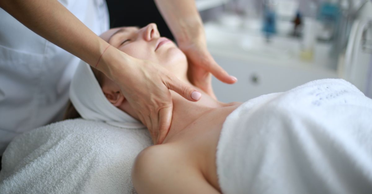
Targeted massage therapy, particularly myofascial release focused on the upper cervical spine, is highly effective for reducing muscle hypertonicity and relieving headache symptoms.
4. Can poor eyesight contribute to neck tension?
Yes, if you have uncorrected vision problems, you may unconsciously lean forward and squint to see clearly, which directly exacerbates forward head posture and neck strain.
5. Is it normal to feel a cracking sound in the neck with these headaches?
Mild crepitus or cracking sounds during neck movement are generally normal and relate to the release of gas bubbles in the facet joints or tendons sliding over bone, often noticed more when muscles are tight.
18. Bibliography
Disclaimer: The content is for informational purposes only and does not replace medical advice. Always consult your doctor for personalized treatment.