
1. Introduction
A traumatic hemothorax is a critical, often life-threatening condition defined by the rapid accumulation of blood within the pleural cavity following a physical injury to the chest. The thorax houses the most vital organs of the human body, supported by an intricate network of high-pressure blood vessels. When trauma breaches this anatomy, the resulting hemorrhage into the pleural space severely compromises both the respiratory and cardiovascular systems simultaneously.
This condition is a leading cause of mortality in both civilian and military trauma settings. The bleeding originates from lacerations to the lung tissue, disruption of the intercostal arteries beneath the ribs, or catastrophic tears to the major central vessels, such as the aorta. The rapid pooling of blood not only drains the circulatory system of its vital volume but also physically crushes the lung, rendering it incapable of gas exchange.
Management in the trauma bay relies on the rapid, protocol-driven execution of Advanced Trauma Life Support guidelines. The overarching clinical priorities are immediate securing of the airway, aggressive restoration of circulating blood volume, and rapid decompression of the chest cavity to restore cardiopulmonary mechanics and identify cases requiring emergency operative intervention.
2. Mechanisms of Injury: Blunt vs. Penetrating
Traumatic chest injuries are broadly classified by their physical mechanism, which directly influences the expected pattern of internal bleeding and organ damage.
Blunt thoracic trauma is overwhelmingly caused by high-velocity motor vehicle collisions, severe falls from a height, or crushing injuries. The sheer kinetic energy transferred to the chest wall during a deceleration impact forcefully compresses the internal structures. This compressive force can burst the fragile lung tissue (pulmonary contusion and laceration) or cause a sudden, lethal shearing of the major hilar vessels as they are violently thrown forward against the rigid spine.
Penetrating thoracic trauma, resulting from stab wounds, gunshot wounds, or impalement, involves the direct mechanical transection of tissue and blood vessels. Gunshot wounds are particularly devastating due to the unpredictable trajectory of the bullet and the massive kinetic energy wave, known as cavitation, which destroys a wide cylinder of tissue far beyond the actual path of the projectile.
3. The Role of Rib Fractures
Rib fractures are the most frequent structural injury associated with a traumatic hemothorax. The ribs form a protective, bony cage, but when they break, the sharp, jagged ends of the fractured bones become dangerous internal projectiles.
Beneath the inferior margin of every rib runs the neurovascular bundle, which contains a high-pressure intercostal artery. When a rib fractures and is displaced inward, it frequently lacerates this artery. Because the intercostal arteries branch directly from the aorta, a laceration can produce a rapid, persistent arterial bleed directly into the pleural cavity.
Furthermore, displaced rib fractures can plunge directly into the underlying lung parenchyma. This tears the highly vascular lung tissue, creating a combined injury where blood and air leak into the chest simultaneously, producing a hemopneumothorax.
4. Massive Hemothorax and Hypovolemic Shock
The most immediate threat to life following thoracic trauma is exsanguination. The pleural cavity is an expansive potential space; an adult can lose up to three liters of blood into a single side of the chest. This volume represents more than half of the total circulating blood supply of an average human.
When a massive hemothorax occurs, the patient enters profound hypovolemic shock. The dramatic loss of intravascular volume drastically reduces the amount of blood returning to the heart. Consequently, the heart cannot pump sufficient oxygenated blood to the brain, kidneys, and other vital organs.
Clinical signs of severe hemorrhagic shock include a markedly rapid heart rate (tachycardia), a severe drop in blood pressure (hypotension), a weak and thready pulse, extreme pallor, and an altered level of consciousness as the brain is starved of oxygen.
5. Tension Hemothorax Physiology
While less common than a tension pneumothorax, a tension hemothorax can develop if the internal bleeding is extraordinarily rapid and arterial in nature. As blood fills the rigid chest cavity, the intrapleural pressure rises relentlessly.
This escalating pressure completely collapses the lung on the injured side. As the cavity fills further, the sheer weight and hydrostatic pressure of the blood begin to push the mediastinum—the central compartment containing the heart, trachea, and great vessels—toward the uninjured side of the chest.
This mediastinal shift severely kinks the superior and inferior vena cava, preventing venous blood from returning to the right atrium of the heart. Cardiac output drops precipitously to near zero, leading to rapid cardiac arrest if the pressure is not immediately relieved.
6. Primary Survey and ATLS Protocols
Upon arrival at a trauma center, patients undergo a highly standardized primary survey based on Advanced Trauma Life Support (ATLS) protocols. The goal is to identify and treat immediate life-threatening injuries in a strict sequence: Airway, Breathing, and Circulation.
During the “Breathing” assessment, the trauma team looks for asymmetric chest wall movement, bruising, and penetrating wounds. The physician listens to both sides of the chest; absent breath sounds on one side strongly suggest a large hemothorax or pneumothorax. Percussion of the chest revealing a dull sound confirms the presence of fluid.
During the “Circulation” assessment, the team evaluates the blood pressure and heart rate. Identifying profound shock in the presence of absent breath sounds on one side prompts immediate therapeutic action before the patient is even transported for advanced imaging.
7. The eFAST Examination
In the acute trauma setting, the Extended Focused Assessment with Sonography for Trauma (eFAST) is an invaluable diagnostic tool. This rapid bedside ultrasound is performed within minutes of patient arrival.
The physician places the ultrasound probe over specific windows on the chest and abdomen. When assessing the thorax, the probe is placed at the lower lateral lung bases. Blood in the pleural space is easily identified as a prominent anechoic (black) fluid collection above the diaphragm.
The eFAST exam is highly sensitive, non-invasive, and does not require the patient to be moved from the resuscitation bay. It allows the trauma team to rapidly differentiate between shock caused by a massive hemothorax and shock caused by abdominal bleeding or cardiac tamponade.
8. Radiographic Evaluation in Trauma
For trauma patients who are hemodynamically stable, advanced imaging provides a detailed anatomical map of the injuries. A portable, supine chest X-ray is often the first film obtained. However, interpreting a supine X-ray is notoriously difficult. Blood layers out evenly across the back of the chest cavity, appearing only as a diffuse, hazy whitening over the entire lung field, rather than the classic meniscus seen on upright films.
A computed tomography scan with intravenous contrast is the gold standard diagnostic test for the stable trauma patient. A chest CT provides exquisite detail of pulmonary lacerations, precise rib fracture localization, and the exact volume of blood in the pleural space.
Crucially, the contrast dye can identify active arterial bleeding, known as a contrast blush. This highlights torn intercostal or internal mammary arteries, guiding the surgical team or interventional radiologist in planning definitive treatment.
9. Acute Fluid and Blood Resuscitation
Simultaneous with the diagnostic evaluation, aggressive resuscitation is initiated to combat hypovolemic shock. Large-bore intravenous lines are placed. While isotonic crystalloids may be given initially, current trauma resuscitation protocols strongly prioritize the early use of blood products for hemorrhagic shock.
Massive transfusion protocols are activated for patients with a suspected massive hemothorax. These protocols deliver packed red blood cells, fresh frozen plasma, and platelets in balanced ratios. This strategy prevents the lethal trauma triad of hypothermia, acidosis, and coagulopathy.
Administering whole blood or balanced blood products ensures that the patient receives not only the red cells necessary to carry oxygen but also the critical clotting factors required to help seal the torn blood vessels.
10. Chest Tube Insertion (Tube Thoracostomy)
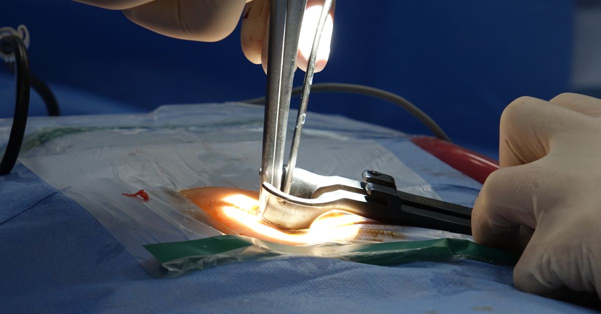
The primary therapeutic intervention for a traumatic hemothorax is the rapid insertion of a large-bore chest tube (tube thoracostomy). The tube is typically placed in the fourth or fifth intercostal space at the mid-axillary line.
The insertion of the tube serves three vital functions. First, it immediately evacuates the accumulated blood, relieving the mechanical compression on the lung and the mediastinum. Second, by emptying the pleural space, it allows the lung to re-expand; the expanded lung can often press against the chest wall, acting as a direct internal tamponade to stop minor bleeding.
Third, the chest tube provides a direct, continuous measurement of the rate of blood loss. The trauma surgeon closely monitors the initial output and the ongoing hourly drainage, as these numbers strictly dictate whether the patient requires emergency open surgery.
11. Indications for Urgent Thoracotomy
The vast majority of traumatic hemothoraces—approximately eighty-five percent—are successfully managed with a chest tube alone. The bleeding typically originates from low-pressure pulmonary vessels or minor chest wall tears that clot spontaneously once the lung is re-expanded.
However, specific output parameters mandate an urgent trip to the operating room for a thoracotomy. If the initial placement of the chest tube drains more than 1,500 milliliters of blood immediately, this represents a massive hemothorax and an active, high-volume bleed.
Furthermore, if the chest tube drains continuously at a rate of more than 200 milliliters per hour for two to four consecutive hours, or if the patient remains in severe shock despite aggressive blood transfusions, emergency surgical exploration is absolutely required to physically identify and ligate the ruptured blood vessels.
12. Operative Management and VATS
When surgical intervention is required, the approach depends on the stability of the patient. An open thoracotomy involves a large incision between the ribs, providing the trauma surgeon wide, immediate access to the entire chest cavity. This is necessary to repair catastrophic injuries to the heart, aorta, or major pulmonary vessels.
For patients who are stable but have ongoing moderate bleeding or a large retained clot that will not drain through the chest tube, Video-Assisted Thoracoscopic Surgery (VATS) is highly effective.
VATS is a minimally invasive approach using small incisions and a camera. The surgeon can thoroughly wash out the pleural cavity, evacuate thick, coagulated blood, and utilize specialized clips or electrocautery to secure bleeding intercostal arteries, significantly reducing the trauma of a major open operation.
13. Managing Coagulated Blood and Retained Hemothorax
One of the most frequent complications of a traumatic hemothorax is the incomplete drainage of blood. Blood in the pleural space rapidly coagulates, forming thick, gelatinous clots that cannot pass through the narrow lumen of a standard chest tube.
This condition is termed a retained hemothorax. If left untreated, the retained blood acts as a potent inflammatory irritant and an ideal breeding ground for bacteria. The patient is at high risk of developing a severe pleural infection, known as a post-traumatic empyema.
If imaging confirms a significant retained blood volume, early surgical intervention, typically using the VATS procedure within the first three to seven days, is recommended to evacuate the clot before it adheres firmly to the lung surface.
14. Long-Term Complications: Fibrothorax
If a retained hemothorax is not surgically evacuated, it undergoes a process of organization over several weeks. Fibroblasts migrate into the clotted blood and begin to lay down dense, inflexible scar tissue over the surface of the lung and the inside of the chest wall.
This thick, fibrous peel, often several centimeters thick, creates a condition known as a fibrothorax. The scar tissue acts like a rigid shell, physically trapping the underlying lung (trapped lung syndrome) and completely preventing it from expanding during inspiration.
Patients with a fibrothorax suffer from chronic shortness of breath and a restrictive lung defect. Treating this late complication requires a major, highly complex surgical procedure called a decortication, wherein the surgeon meticulously peels the thick scar tissue off the delicate surface of the lung to liberate it.
15. Frequently Asked Questions (FAQ)
1. How is a traumatic hemothorax different from a regular one?
A traumatic hemothorax is caused by a specific physical injury, such as a car crash, a fall, or a stab wound that violently tears the blood vessels inside the chest. A non-traumatic (spontaneous) hemothorax happens without an injury, usually due to bleeding disorders or bursting tumors.
2. Why do broken ribs cause so much bleeding in the chest?
Every rib has a relatively large, high-pressure artery running right along its bottom edge. When a rib breaks, the sharp bone edges frequently slice into this artery, causing it to bleed heavily and continuously directly into the chest cavity.
3. Does a chest tube hurt when it is put in?
Yes, placing a chest tube is a very painful procedure because the tissue between the ribs is highly sensitive. Trauma doctors use local numbing medicine and pain medications to minimize discomfort, but in extreme emergencies, it must be placed rapidly to save the patient’s life.
4. If I lost a lot of blood in my chest, will I need surgery?
Most of the time, no. About eighty-five percent of these injuries heal simply by draining the blood with a chest tube and allowing the body to form a natural clot. Surgery is only required if a major artery is cut and the bleeding refuses to stop.
5. What happens if the blood is not completely removed from my chest?
If a large amount of blood is left inside, it turns into a solid clot. This clot can easily become infected, or over time, it turns into a hard layer of scar tissue that acts like a cage, permanently trapping your lung and making it hard to breathe.
16. Bibliography
Disclaimer: The content is for informational purposes only and does not replace medical advice. Always consult your doctor for personalized treatment.

