1. Introduction
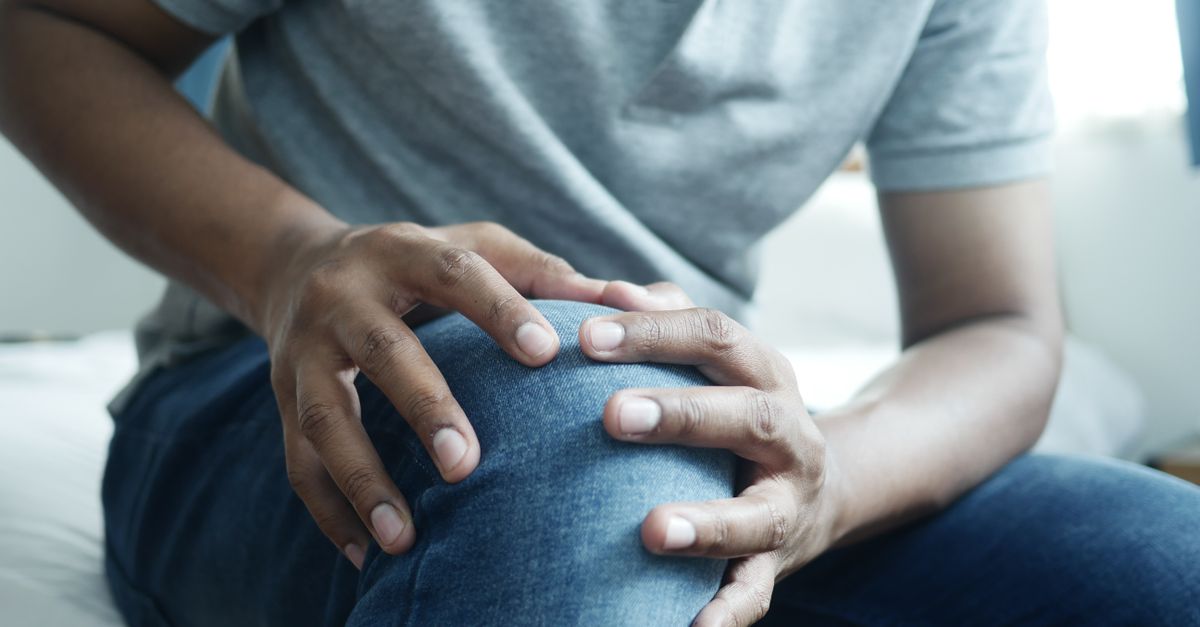
Chronic pain behind the kneecap is typically caused by abnormal tracking of the patella within its femoral groove, leading to conditions such as patellofemoral pain syndrome or chondromalacia patellae. Relief requires a comprehensive approach that includes reducing joint load, strengthening the supporting kinetic chain, and correcting biomechanical faults through targeted physical therapy. In refractory cases, medical interventions ranging from specialized bracing to pharmacological management become necessary to restore joint function.
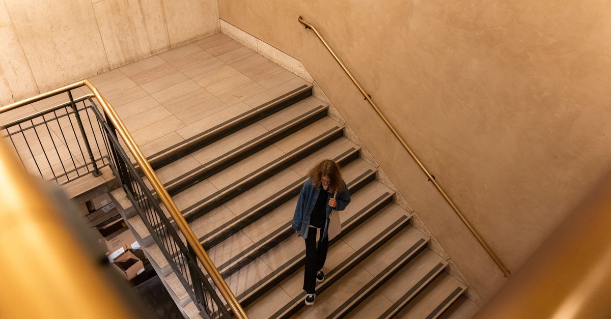
This specific type of anterior knee pain affects individuals across various activity levels, from sedentary office workers to competitive athletes. The patellofemoral joint experiences tremendous mechanical stress during daily activities like climbing stairs or standing up from a chair. When the articular cartilage beneath the kneecap is subjected to repetitive, uneven pressure, the resulting inflammation creates a persistent, dull ache.
Managing this condition is rarely about a single quick fix. Clinical evidence points to muscular imbalances, particularly weakness in the hip and quadriceps, as primary contributors to poor patellar tracking. Addressing these systemic mechanical issues is the most reliable pathway to achieving lasting pain relief and preventing early-onset osteoarthritis.
2. Anatomy of the Patellofemoral Joint
The knee joint is a complex hinge that relies on the interaction of three bones consisting of the femur, the tibia, and the patella. The patella, or kneecap, is a sesamoid bone embedded within the quadriceps tendon. Its primary biomechanical function is to act as a fulcrum, increasing the leverage and mechanical advantage of the quadriceps muscle group during knee extension.
As the knee bends and straightens, the patella glides up and down within a specific groove on the front of the femur, known as the trochlear groove. Smooth articular cartilage covers the undersurface of the patella and the trochlear groove, allowing for frictionless movement. This gliding mechanism relies on a delicate balance of muscular forces and ligamentous restraints.
If any component of this structural system is unbalanced, the patella may shift slightly off-center. Even a minor deviation alters the contact pressures between the bone surfaces. Over time, this uneven distribution of force leads to microscopic tissue damage and the onset of chronic pain.
3. Patellofemoral Pain Syndrome Mechanism
Patellofemoral pain syndrome is the broad clinical diagnosis for anterior knee pain that originates from the contact area between the kneecap and the femur. It is commonly described by patients as a diffuse, aching pain behind or around the patella. The exact source of the pain is often the highly innervated subchondral bone or the synovial lining of the joint, as the cartilage itself contains no nerve endings.
The mechanism of this syndrome is intrinsically linked to joint loading. Activities that involve deep knee flexion, such as squatting or running downhill, significantly multiply the compressive forces acting on the patellofemoral joint. When these forces exceed the physiological tolerance of the joint tissues, an inflammatory response is triggered.
Prolonged sitting with knees bent can also provoke symptoms, a phenomenon classically referred to as the movie theater sign. The static pressure of the tight quadriceps pulling the patella against the femur over hours creates localized ischemia in the joint tissues, leading to severe stiffness and pain upon standing.
4. Chondromalacia Patellae and Cartilage Wear
While patellofemoral pain syndrome describes the clinical symptom of pain, chondromalacia patellae refers to a specific structural change beneath the kneecap. It involves the softening, fraying, and eventual breakdown of the articular cartilage on the undersurface of the patella. This degenerative process is a direct consequence of chronic maltracking and excessive focal pressure.
Clinicians grade chondromalacia on a scale from one to four. Grade one involves mild softening of the cartilage, whereas grade four indicates severe cartilage loss exposing the underlying subchondral bone. As the protective cartilage layer diminishes, the mechanical stress is transferred directly to the sensitive bone beneath, causing sharp, localized pain during movement.
Patients with chondromalacia frequently experience a grinding or grating sensation, clinically termed crepitus, when extending the knee under load. While crepitus alone without pain is generally benign, painful crepitus strongly suggests active cartilage degradation and warrants clinical intervention to halt disease progression.
5. The Role of Malalignment and Tracking Issues
Proper patellar tracking depends on the structural alignment of the entire lower extremity. Variations in bony anatomy can predispose an individual to chronic knee pain. A common anatomical factor is a high Q-angle, which is the angle formed by a line drawn from the pelvis to the patella and another from the patella to the tibia. A larger Q-angle increases the lateral, or outward, pull on the kneecap.
Flat feet or excessive foot pronation can also disrupt knee mechanics. When the arch of the foot collapses inward during weight-bearing, it forces the tibia to rotate internally. This internal rotation throws the patellofemoral joint out of its natural alignment, increasing friction and sheer stress beneath the kneecap.
Structural anomalies such as a shallow trochlear groove or an abnormally high-riding patella, known as patella alta, further compromise joint stability. These inherent anatomical variations make conservative management more challenging but emphasize the need for meticulous strengthening of the active muscular stabilizers.
6. Quadriceps Weakness and Muscle Imbalance
The quadriceps muscle group plays the most direct role in controlling the movement of the patella. This group consists of four distinct muscles, with the vastus medialis obliquus acting as the primary medial stabilizer. The vastus medialis obliquus is responsible for pulling the patella inward, counteracting the strong outward pull of the vastus lateralis.
In many patients with chronic anterior knee pain, a distinct imbalance exists between these muscles. If the vastus lateralis is disproportionately strong or tight compared to a weakened vastus medialis obliquus, the patella is inevitably pulled laterally out of its groove. This muscular imbalance is a primary target during physical rehabilitation.
Furthermore, general quadriceps weakness diminishes the shock-absorbing capacity of the knee. Without adequate muscular support, the impact forces from walking or running are transferred directly to the articular cartilage and the underlying bone, accelerating wear and tear.
7. The Importance of Hip and Gluteal Strength
Modern biomechanical research highlights the crucial role of the hip musculature in patellofemoral health. The gluteus medius and gluteus maximus are responsible for stabilizing the pelvis and controlling the alignment of the femur during dynamic tasks. Weakness in these hip abductors and external rotators allows the femur to collapse inward during activities like single-leg squats or running.
When the femur rotates internally beneath the patella, it is equivalent to the patella tracking laterally. This relative maltracking significantly increases the lateral patellofemoral contact pressure. Therefore, treating the knee requires looking up the kinetic chain to ensure the hip joints are providing adequate structural support.
Clinical physical therapy programs now routinely prioritize hip strengthening alongside direct knee interventions. Enhancing gluteal activation not only improves lower extremity alignment but also redistributes mechanical loads, taking immense pressure off the vulnerable patellofemoral joint.
8. Initial Conservative Management Strategies
The initial phase of managing chronic pain behind the kneecap focuses on reducing acute inflammation and tissue irritability. The concept of relative rest is applied, meaning the patient avoids specific pain-provoking activities while maintaining general mobility. Completely immobilizing the knee is contraindicated, as it leads to rapid muscle atrophy and cartilage stiffening.
Activity modification involves substituting high-impact activities like running or jumping with low-impact alternatives such as swimming or stationary cycling. Bicycling must be performed with the seat elevated appropriately to prevent deep knee flexion angles that increase joint compression.
Ice therapy remains a valuable tool for symptomatic relief, especially following physical activity. Applying cold packs to the anterior knee for fifteen minutes helps constrict local blood vessels, reducing inflammatory exudate and temporarily numbing the nociceptive nerve fibers in the joint capsule.
9. Physical Therapy and Kinetic Chain Strengthening
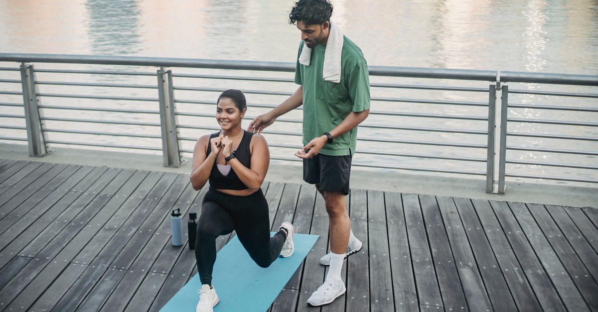
A formalized physical therapy program is the most evidence-based treatment for chronic patellofemoral pain. The physical therapist conducts a comprehensive evaluation to identify specific flexibility deficits and strength imbalances. The resulting rehabilitation protocol targets the entire lower kinetic chain, from the lumbar spine down to the foot.
Stretching protocols typically focus on releasing tension in the hamstrings, calves, and the iliotibial band. A tight iliotibial band can exert a strong lateral force on the patella, exacerbating maltracking. Myofascial release techniques and consistent stretching help restore normal tissue elasticity.
Strengthening exercises are introduced progressively. Closed kinetic chain exercises, such as controlled wall squats and leg presses in shallow ranges of motion, are favored because they closely mimic functional movements while minimizing shear forces across the joint. These exercises safely build quadriceps and gluteal endurance.
10. Orthotics and Taping Techniques
External mechanical supports can provide immediate symptomatic relief by altering the forces acting on the knee. Custom or over-the-counter foot orthotics are highly beneficial for patients demonstrating excessive foot pronation. By supporting the medial arch, orthotics prevent the internal rotation of the tibia, effectively realigning the patellofemoral joint from the ground up.
Patellar taping, commonly using the McConnell taping technique, involves applying rigid adhesive tape to physically pull the patella medially. This passive realignment reduces lateral contact pressures and often provides immediate pain reduction. Taping is particularly useful during physical therapy, allowing patients to perform strengthening exercises without pain.
Knee sleeves or braces with a patellar cutout offer proprioceptive feedback and mild structural support. While they do not physically hold the kneecap in place against strong muscular forces, the compression they provide enhances joint awareness and helps reduce localized swelling.
11. Pharmacological Pain Management
When mechanical pain is accompanied by significant inflammatory flare-ups, pharmacological intervention provides necessary relief. Nonsteroidal anti-inflammatory drugs, such as ibuprofen or naproxen, are commonly recommended to suppress synovial inflammation and reduce chemical pain mediators within the joint.
For patients who cannot tolerate oral anti-inflammatories due to gastrointestinal or cardiovascular risks, topical formulations offer a safer alternative. Topical diclofenac gel can be applied directly over the anterior knee, penetrating the skin to deliver targeted anti-inflammatory action to the superficial joint structures with minimal systemic absorption.
Acetaminophen may be utilized for pure analgesia in chronic management, though it lacks anti-inflammatory properties. All pharmacological strategies should be viewed as adjunctive treatments, designed to control symptoms sufficiently to allow for active participation in a physical rehabilitation program.
12. Medical Injections and Advanced Therapies
If oral medications and physical therapy fail to provide adequate relief, physicians may consider intra-articular injections. Corticosteroid injections deliver a potent anti-inflammatory medication directly into the knee joint capsule. While highly effective at resolving acute inflammatory flares, repetitive steroid injections are discouraged as they can weaken the articular cartilage over time.
Hyaluronic acid injections, also known as viscosupplementation, aim to improve the lubricating properties of the synovial fluid. This therapy can reduce friction beneath the kneecap, offering medium to long-term pain relief for patients with established chondromalacia. The clinical efficacy of viscosupplementation varies among individuals.
Emerging regenerative medicine therapies, including platelet-rich plasma injections, concentrate the body’s natural healing factors and deliver them to the damaged cartilage. Clinical studies suggest that platelet-rich plasma can modulate the inflammatory environment of the joint and potentially stimulate tissue repair, offering a promising avenue for chronic patellofemoral pain management.
13. Understanding Diagnostic Data
Distinguishing the specific factors contributing to anterior knee pain is vital for effective management. The table below outlines common clinical findings and their biomechanical impact.
| Biomechanical Factor | Clinical Finding | Impact on the Knee |
|---|---|---|
| Overpronation | Flattened foot arches during weight-bearing | Causes tibial internal rotation, increasing patellofemoral stress. |
| Gluteal Weakness | Pelvic drop or inward collapse of the knee during a squat | Fails to control femoral alignment, leading to lateral patellar maltracking. |
| Quadriceps Imbalance | Weak vastus medialis relative to lateral thigh muscles | Pulls the kneecap laterally against the trochlear groove during extension. |
14. Activity Modification and Load Management
Load management is a critical concept in resolving chronic tendon and joint pain. It involves finding the exact threshold of physical activity that the patellofemoral joint can tolerate without triggering a pain response. Consistently exceeding this threshold sustains the inflammatory cycle and prevents tissue adaptation.
Athletes returning to sport must employ a meticulously graded return-to-play protocol. Running mileage, jumping frequency, and lifting volumes should be increased incrementally, usually by no more than ten percent per week. Monitoring the joint’s response over a twenty-four-hour period dictates the safety of the current load.
For sedentary individuals, incorporating frequent movement breaks is essential. Interrupting prolonged sitting periods every thirty minutes to perform gentle knee extensions helps circulate synovial fluid and prevents the patella from becoming statically compressed against the femur.
15. Diagnostic Approaches and Imaging
A thorough clinical history and physical examination remain the gold standard for diagnosing patellofemoral pain. The clinician assesses lower limb alignment, muscular flexibility, and joint stability. Specific provocative tests, such as the patellar grind test, help isolate the pain source to the undersurface of the kneecap.
Standard weight-bearing X-rays, including a specialized view called the merchant or sunrise view, allow physicians to assess the alignment of the patella within the trochlear groove. Radiographs can also detect joint space narrowing, the presence of bone spurs, or underlying osteoarthritis.
When conservative treatment fails, magnetic resonance imaging is utilized to evaluate the soft tissues and the structural integrity of the articular cartilage. An MRI provides detailed cross-sectional images, confirming the extent of chondromalacia and ruling out other internal derangements, such as meniscal tears, which might mimic anterior knee pain.
16. Surgical Options for Refractory Cases
Surgery is strictly reserved for a small percentage of patients whose chronic pain does not resolve after several months of rigorous conservative management, and who present with identifiable structural abnormalities. The goal of surgical intervention is to mechanically realign the patella and reduce pathological contact pressures.
A lateral release is an arthroscopic procedure where the tight lateral retinaculum on the outside of the knee is carefully divided. This procedure releases the outward tension on the patella, allowing it to rest more centrally in the groove. However, it is only effective in highly specific clinical scenarios involving isolated lateral tightness.
For severe malalignment or a deficient trochlear groove, more complex realignment surgeries are necessary. The tibial tubercle osteotomy involves repositioning the bony attachment of the patellar tendon on the shinbone. By moving the attachment site medially or anteriorly, surgeons can drastically alter the tracking mechanics and relieve focal pressure on damaged cartilage areas.
17. Frequently Asked Questions (FAQ)
1. Is cycling good or bad for pain behind the kneecap?
Cycling can be highly beneficial as a low-impact strengthening exercise, provided the bicycle is fitted correctly. If the saddle is too low, the knee flexes too deeply, which drastically increases pressure behind the kneecap and exacerbates pain.
2. Will losing weight help my chronic knee pain?
Yes, weight reduction significantly decreases the mechanical load on the patellofemoral joint. Because the forces acting on the kneecap multiply during activities like stair climbing, even a moderate reduction in body weight translates to a massive reduction in joint stress.
3. Can chondromalacia patellae heal completely?
Cartilage has a poor blood supply and limited capacity to heal completely once structurally damaged. However, by correcting the mechanical forces causing the wear through physical therapy, patients can effectively manage the condition and eliminate the associated pain.
4. Should I stop exercising completely if my kneecap hurts?
Complete cessation of exercise leads to muscle atrophy, which ultimately worsens joint stability and pain. It is recommended to modify activities to avoid pain while continuing non-aggravating exercises to maintain strength in the quadriceps and hips.
5. Does cracking or popping in the knee mean I need surgery?
Joint noises like crepitus are extremely common and, if painless, are usually benign, caused by the movement of synovial fluid or normal tissue gliding. Surgery is considered only if the popping is associated with severe structural damage and chronic pain unresponsive to therapy.
18. Bibliography
Disclaimer: The content is for informational purposes only and does not replace medical advice. Always consult your doctor for personalized treatment.