1. Introduction to Nail Infections
A nail infection is a localized clinical condition affecting the structure and surrounding tissues of the fingernails or toenails. The immediate outcome is visible structural deformity, discoloration, and localized inflammation. Prompt clinical evaluation is necessary, as these infections rarely resolve without targeted therapeutic intervention and can cause significant functional impairment.
These infections are broadly classified into two primary categories based on the causative agent and the precise anatomical location. Onychomycosis involves a fungal invasion of the nail plate and bed, leading to slow, progressive damage. Conversely, paronychia is primarily a bacterial or yeast infection causing acute inflammation of the soft tissues immediately surrounding the nail margin.
Successful management requires identifying the precise pathogen and understanding the slow growth cycle of the nail unit. Treatment regimens are often prolonged and require high patient compliance to eradicate the underlying organism and restore normal anatomical function.
2. Anatomy of the Nail Unit
To understand how infections take hold, one must understand the unique anatomy of the nail unit. The hard, visible portion is the nail plate, composed of densely packed, dead keratinized cells. It rests upon the highly vascularized nail bed, which provides the necessary nutrients for structural maintenance.
The nail matrix, located beneath the skin at the base of the nail, is the primary center for cell division and growth. Any damage or infection reaching the matrix can result in permanent, irreversible deformity of the growing nail plate. The cuticle acts as a vital waterproof seal, protecting the matrix from environmental pathogens.
When the integrity of the cuticle is compromised by trauma or excessive moisture, a portal of entry is created. Microorganisms exploit this breach, migrating beneath the nail fold or directly invading the keratin structure of the nail plate, initiating the infectious process.
3. Fungal Pathogens and Dermatophytes
Onychomycosis is predominantly caused by dermatophytes, a specialized group of fungi that require keratin for growth. Trichophyton rubrum is the most frequently isolated pathogen in clinical practice. These organisms possess specialized enzymes capable of digesting the tough keratin structure, allowing them to tunnel through the nail plate.
The infection typically begins at the distal edge or lateral margins of the nail and slowly progresses proximally toward the cuticle. Because the nail plate itself lacks a blood supply and a direct cellular immune presence, the fungi can multiply unchecked within the keratin layers, shielded from systemic immune defenses.
Non-dermatophyte molds and certain yeast species, such as Candida albicans, can also cause onychomycosis. Yeast infections are more frequently observed in fingernails and are strongly associated with underlying immunosuppression or continuous exposure to wet environments.
4. Bacterial Infections and Acute Paronychia
Acute paronychia is a rapid-onset bacterial infection of the epidermal folds framing the nail. Staphylococcus aureus and Streptococcus pyogenes are the usual culprits. These bacteria enter through microscopic tears in the cuticle or lateral nail folds, often secondary to nail-biting, aggressive manicuring, or minor trauma.
The clinical presentation is acute and highly localized. The affected tissue becomes erythematous, tensely swollen, and exquisitely tender to palpation. The rapid multiplication of bacteria and the influx of immune cells frequently lead to the formation of a localized abscess filled with purulent material along the nail margin.
Unlike the slow, insidious progression of fungal infections, acute bacterial paronychia develops over a matter of hours or days. It requires immediate clinical attention to relieve the painful pressure and prevent the infection from tracking deeper into the digital pulp or tendon sheaths.
5. Environmental and Lifestyle Risk Factors
The development of nail infections is heavily influenced by environmental exposures and individual habits. Fungi thrive in warm, dark, and hyperhidrotic environments. Consequently, toenails are far more frequently affected than fingernails due to prolonged enclosure in non-breathable footwear.
Frequent microtrauma to the nails, common in athletes or individuals wearing improperly fitted shoes, causes minor lifting of the nail plate from the bed. This creates a microscopic void where moisture and pathogens accumulate. Similarly, habits like aggressive cuticle trimming destroy the natural barrier, predisposing the area to bacterial invasion.
Occupations requiring prolonged submersion of the hands in water, such as dishwashing or professional cleaning, lead to maceration of the skin and cuticle degradation. This constant moisture exposure is the primary risk factor for developing chronic paronychia, characterized by recurrent inflammation and yeast colonization.
6. Structural Damage and Nail Discoloration
The clinical hallmarks of onychomycosis involve profound changes to the visual appearance and structural integrity of the nail. Initially, patients notice localized discoloration, presenting as white, yellow, or brown streaks within the nail plate. This discoloration indicates the physical presence of the fungal colonies and the accumulation of their metabolic byproducts.
As the dermatophytes aggressively digest the keratin, the nail plate becomes hyperkeratotic, resulting in significant thickening. The structural breakdown leads to a brittle, friable texture, causing the nail to crumble easily at the edges. Subungual hyperkeratosis, the accumulation of debris beneath the nail, further lifts the plate from the underlying bed.
In advanced stages, onycholysis occurs, which is the complete separation of the nail plate from the nail bed. This not only causes functional discomfort but also provides an expansive reservoir for secondary bacterial colonization. Similar presentations are discussed in our overview of fungal skin conditions.
7. Localized Pain and Inflammation
While fungal infections of the nail plate are generally painless in the early stages, significant thickening can cause pressure-related pain when wearing shoes. The physical deformation of the nail alters the normal mechanics of walking, leading to localized discomfort and secondary callusing of the surrounding skin.
In contrast, acute bacterial paronychia is characterized by severe, throbbing pain. The intense inflammatory response within the confined space of the tight digital tissues generates substantial pressure. The pain is often exacerbated by a dependent position, allowing blood to pool in the inflamed digit.
Chronic paronychia presents with a less intense, dull ache. The nail folds remain persistently swollen, indurated, and red, with intermittent episodes of acute exacerbation. The continuous inflammation eventually affects the underlying matrix, leading to horizontal ridging and irregular growth of the nail plate.
8. Clinical Evaluation and Diagnosis
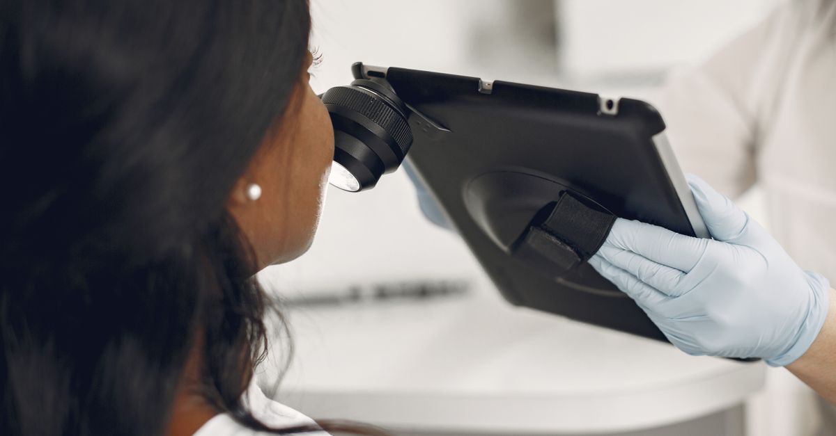
Accurate diagnosis begins with a meticulous clinical examination of all twenty nails and the surrounding skin. The physician notes the specific pattern of discoloration, the presence of thickening or friability, and the exact anatomical location of the inflammation. The concurrent presence of tinea pedis, or athlete’s foot, strongly supports a fungal etiology.
For paronychia, the clinician assesses the degree of localized fluctuance to determine if a discrete abscess has formed. The neurovascular status of the digit must be evaluated to ensure the swelling has not compromised arterial perfusion or compressed the digital nerves.
Visual inspection alone is insufficient for definitively diagnosing onychomycosis, as numerous inflammatory skin conditions can mimic its appearance. Precise identification of the pathogen is legally and clinically required before initiating prolonged courses of systemic antifungal medications, due to their potential for adverse hepatic effects.
9. Laboratory Testing and Fungal Cultures
To confirm a fungal infection, the clinician must obtain high-quality samples of the affected tissue. This involves aggressively clipping the abnormal nail plate and using a small curette to scrape the subungual debris closest to the active proximal border of the infection.
The collected material is subjected to a potassium hydroxide preparation. This chemical dissolves the hard keratin, allowing the microscopic visualization of fungal hyphae under direct microscopy. While this provides rapid confirmation of a fungal presence, it cannot identify the specific species.
For definitive identification, the specimen is sent for fungal culture or polymerase chain reaction testing. Culturing dermatophytes is notoriously slow, often requiring several weeks for visible growth. Polymerase chain reaction assays offer a highly sensitive and rapid alternative, precisely detecting the DNA of specific fungal pathogens within days.
10. Structured Diagnostic Differentiation
The table below outlines the primary clinical differences between the major types of nail infections, aiding in accurate diagnosis and treatment planning.
| Condition | Primary Pathogen | Clinical Presentation | Onset and Duration |
|---|---|---|---|
| Onychomycosis (Fungal) | Dermatophytes | Thickened, yellow/brown brittle nails, subungual debris. | |
| Acute Paronychia | Bacteria | Red, swollen, painful nail fold, pus accumulation. | |
| Chronic Paronychia | Mixed Flora/Yeast | Persistent boggy swelling of cuticle, loss of cuticle barrier. |
11. Topical Antifungal Therapies
Topical antifungal agents are utilized for early, superficial fungal infections that do not involve the lunula or the nail matrix. Specialized medicated nail lacquers, containing active ingredients like ciclopirox or amorolfine, are applied daily to the surface of the nail plate.
These formulations are designed to penetrate the dense keratin structure, though their efficacy is inherently limited by the physical barrier of the thick nail. To improve penetration, patients are instructed to regularly file down the surface of the affected nail to thin the plate and remove heavily infected material.
Topical therapy requires immense patient compliance, often necessitating daily application for 48 weeks or longer. Due to their limited penetration, topical treatments are frequently used as an adjunct to systemic therapy rather than a standalone cure for moderate to severe cases.
12. Oral Medications and Systemic Treatment
Systemic oral antifungal medications represent the gold standard for treating deep, widespread onychomycosis. Agents such as oral terbinafine or itraconazole are absorbed into the bloodstream and are actively deposited directly into the nail matrix and the growing nail plate.
By impregnating the newly forming nail with antifungal compounds, the medication physically prevents the fungus from advancing. The therapy must be continued for 6 to 12 weeks for toenails. However, clinical cure is only visible months later, once the entire infected portion of the nail has grown out and been clipped away.
Before prescribing oral antifungals, clinicians mandate baseline hepatic function panels, as these medications are processed by the liver and carry a small risk of hepatotoxicity. Patients must avoid alcohol consumption and be closely monitored for potential drug interactions throughout the therapeutic course.
13. Surgical Nail Removal Options
In scenarios where the fungal infection is completely refractory to systemic medications, or if a severely thickened nail is causing debilitating pain, surgical intervention is indicated. Complete or partial avulsion of the nail plate removes the infected tissue entirely, exposing the nail bed for direct topical treatment.
For acute bacterial paronychia complicated by an abscess, the primary treatment is prompt surgical incision and drainage. A scalpel is used to carefully elevate the nail fold or incise the abscess to release the trapped purulent fluid. This immediately relieves the painful pressure and expedites resolution, often without the need for systemic antibiotics.
If a patient suffers from recurrent, intractable paronychia, surgical excision of the lateral nail fold or permanent destruction of a lateral strip of the nail matrix via chemical matricectomy may be performed. This permanently narrows the nail plate, preventing future ingrowths and localized infections.
14. Home Care and Hygiene Maintenance
Daily hygiene practices are critical for supporting clinical treatments. Patients are advised to wash their feet daily with antimicrobial soap, ensuring the interdigital spaces are thoroughly dried. Moisture control is paramount, and the use of absorbent powders in footwear can significantly reduce fungal proliferation.
For acute paronychia in its earliest stages, warm water soaks performed multiple times a day can increase local blood flow and promote spontaneous drainage of minor superficial infections. However, prolonged soaking should be avoided in chronic paronychia, as it exacerbates the destruction of the cuticle barrier.
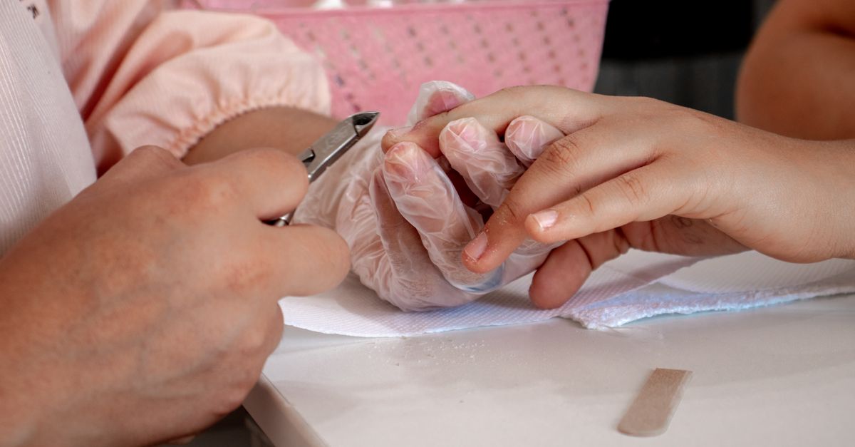
Proper nail grooming techniques are essential. Nails should be trimmed straight across, avoiding aggressive rounding of the corners, which promotes ingrown nails. Tools used for clipping infected nails must be thoroughly disinfected with alcohol to prevent auto-inoculation to healthy adjacent digits.
15. Preventing Recurrence
The recurrence rate for fungal nail infections is notoriously high, demanding sustained preventive strategies. Patients must systematically discard or aggressively disinfect old, highly contaminated footwear. Antifungal shoe sprays or ultraviolet light sanitizers can reduce the environmental reservoir of dormant spores.
In communal bathing environments, such as gym locker rooms or public pools, protective footwear is absolutely mandatory to prevent acquiring new dermatophyte infections. Cotton or moisture-wicking socks should be worn daily and changed immediately after intense physical activity.
Individuals prone to chronic paronychia must rigorously protect their hands from wet environments. The diligent use of cotton-lined rubber gloves during household chores and the frequent application of robust emollient hand creams will help restore the natural lipid barrier of the skin and maintain cuticle integrity.
16. Frequently Asked Questions (FAQ)
1. Why do fungal nail infections take so long to cure?
The treatment takes a long time because the medication can only protect the new nail as it grows from the base. Toenails grow very slowly, and it can take up to a year for the completely new, healthy nail to replace the infected portion.
2. Should I cut away the thickened, yellow part of my nail?
Yes, gently filing down or clipping the heavily infected, thick parts of the nail removes the bulk of the fungus and allows topical treatments to penetrate the underlying tissue more effectively.
3. What is the difference between a fungal infection and paronychia?
A fungal infection attacks the hard nail plate itself, causing it to thicken and turn yellow slowly over months. Paronychia is usually a bacterial infection of the skin surrounding the nail, causing sudden, painful swelling and redness.
4. Can I wear nail polish while treating a fungal infection?
Standard cosmetic nail polish creates a non-breathable seal that traps moisture and encourages fungal growth. It should be avoided unless you are using a specific, medically prescribed antifungal nail lacquer.
5. Are the oral pills for nail fungus dangerous for the liver?
While generally safe for healthy individuals, the oral medications are processed by the liver. Your doctor will require a blood test before starting and potentially during treatment to monitor your liver function and ensure safety.
17. Bibliography
Disclaimer: The content is for informational purposes only and does not replace medical advice. Always consult your doctor for personalized treatment.