1. Introduction
Thick, yellowed toenails that become exceedingly hard to cut are the hallmark clinical presentation of onychomycosis, a chronic fungal infection of the nail apparatus. This condition occurs when microscopic dermatophyte fungi invade the hard keratin structure of the nail plate or the soft tissue of the nail bed beneath it. As the fungi multiply, they actively digest the keratin proteins, physically destroying the architectural integrity of the nail. The body responds to this chronic localized infection by ramping up cellular production, leading to a massive buildup of abnormal, discolored tissue that transforms a normally thin, transparent nail into a dense, opaque, and structurally deformed plate.
Onychomycosis is a progressive dermatological disease rather than a simple cosmetic flaw. Left untreated, the fungal organisms embed themselves deeply into the matrix where the nail is formed, ensuring that all new growth is immediately infected. The resulting mechanical thickening can cause significant pain while wearing footwear and increases the risk of secondary bacterial infections, particularly in vulnerable populations. Understanding the biological behavior of these specific fungal pathogens and the environmental factors that promote their survival is critical for executing an effective, long-term medical eradication strategy.
2. Anatomy of the Nail Unit
To understand how a fungal infection causes such profound structural changes, one must examine the anatomy of the toenail. The visible hard surface is the nail plate, composed of tightly packed, dead keratin cells. This plate rests upon the highly vascular nail bed, which provides nutrition and the pink coloration of a healthy nail.
At the base of the nail, hidden beneath the cuticle, lies the nail matrix. The matrix is the biological factory where living cells divide, keratinize, and are pushed forward to create the nail plate. Fungal infections typically begin at the distal edge, the tip of the nail, and slowly work their way backward toward this vital matrix structure.
3. The Pathophysiology of Onychomycosis
The primary infectious agents driving thick, yellow toenails are dermatophytes, with Trichophyton rubrum being the most prevalent species globally. These highly specialized fungi possess enzymes called keratinases, which allow them to actively dissolve and consume the tough keratin protein that makes up human hair, skin, and nails.
The infection typically initiates when the fungal spores gain entry through a microscopic separation between the nail plate and the nail bed, a space known as the hyponychium. Once inside this warm, dark, and protected environment, the fungus extends thread-like structures called hyphae deep into the keratin network, establishing a chronic, entrenched infection.
4. Dermatophyte Fungi Mechanics
As the dermatophytes consume the nail, they leave behind metabolic waste products and digested cellular debris. This biological debris accumulates in large volumes directly beneath the nail plate, a condition clinically termed subungual hyperkeratosis.
This massive accumulation of crumbly, chalky material physically lifts the nail plate away from the vascular bed, a process known as onycholysis. It is this combination of the fungal debris pushing upward and the nail plate reacting to the chronic inflammation that creates the profound, unyielding thickness that renders the nail nearly impossible to trim with standard clippers.
5. Structural Damage and Yellow Discoloration
The striking yellow or brownish discoloration associated with onychomycosis stems from two distinct factors. First, the fungal organisms themselves, along with their metabolic byproducts, carry distinct pigments that stain the surrounding tissues.
Second, the structural destruction of the nail plate alters how light passes through the keratin. A healthy nail is translucent, appearing pink because of the blood flowing beneath it. As the fungus bores microscopic tunnels through the plate and the subungual debris separates the nail from the bed, the plate becomes completely opaque, locking in the dull, yellowed appearance characteristic of dead, infected keratin.
6. Distinguishing from Psoriatic Nails
Accurate clinical diagnosis requires distinguishing fungal infections from other systemic diseases that cause nail dystrophy. Psoriasis is a chronic autoimmune condition that frequently attacks the nail matrix, causing the nails to become thick, yellow, and prone to lifting from the nail bed.
However, psoriatic nails often present with deep, distinct pitting across the surface of the nail plate, resembling the surface of a thimble. Furthermore, patients with nail psoriasis typically exhibit classic red, scaly skin plaques on their knees, elbows, or scalp. Fungal infections, conversely, lack deep pitting and are often accompanied by athlete’s foot, a fungal infection of the surrounding skin.
7. Aging and Peripheral Circulation
The incidence of onychomycosis increases exponentially with advancing age. As the human body ages, peripheral blood circulation to the toes naturally diminishes. This reduced blood flow impairs the delivery of vital immune cells to the distal extremities, crippling the body’s ability to fight off superficial infections.
Additionally, the rate of nail growth slows significantly in older adults. A slow-growing nail provides the invading dermatophytes ample time to establish themselves and spread toward the matrix before the infected sections can be pushed outward and clipped away, leading to higher rates of chronic, intractable infections in the elderly.
8. Diabetes Mellitus and Immune Susceptibility
Patients with diabetes mellitus face a dramatically elevated risk of developing severe onychomycosis. Chronic hyperglycemia damages the microvasculature and the peripheral nerves, leading to diminished circulation and a loss of protective sensation in the feet.
A thick, fungal nail in a diabetic patient is a serious medical concern. The rigid, deformed nail plate can exert immense mechanical pressure on the underlying compromised tissue, creating deep, painless ulcers. These subungual ulcers serve as entry portals for dangerous bacterial pathogens, potentially leading to severe cellulitis or even osteomyelitis, a bone infection requiring surgical intervention.
9. Trauma and Micro-Injury
Fungal spores are ubiquitous in the environment, but they cannot penetrate a healthy, intact nail plate. The initial infection always requires a portal of entry. Acute physical trauma, such as dropping a heavy object on the toe, breaks the protective seal around the cuticle or the tip of the nail, allowing spores immediate access to the vulnerable nail bed.
Chronic, repetitive micro-trauma is an equally potent catalyst. Athletes, particularly runners, constantly subject their toes to repeated impact against the inside of their footwear. This continuous physical stress creates microscopic fissures in the keratin and lifts the nail plate slightly, establishing the perfect micro-environment for a fungal colony to flourish.
10. Environmental Risk Factors
Dermatophytes thrive in warm, dark, and highly humid environments. The modern practice of wearing closed-toe, non-breathable synthetic footwear for prolonged hours traps heat and sweat directly against the skin and nails, creating a tropical microclimate ideal for fungal proliferation.
Communal, damp environments present significant exposure risks. Public swimming pools, locker rooms, and shared showers harbor massive quantities of infectious fungal spores shed by other individuals. Walking barefoot in these high-traffic areas allows the spores to adhere to the skin of the feet, frequently establishing an initial skin infection that eventually migrates beneath the toenails.
11. Clinical Diagnosis and Mycological Testing
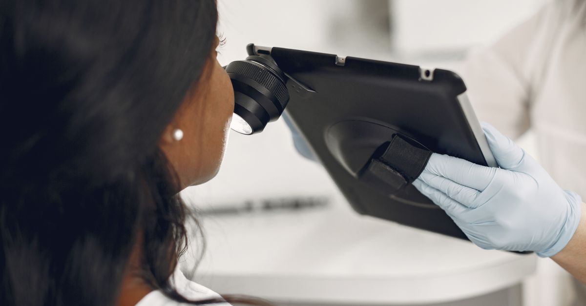
Because up to fifty percent of thickened, discolored nails are caused by non-fungal etiologies, empirical treatment without definitive testing is medically discouraged. A physician will utilize a specialized tool to scrape the crumbling debris from deep beneath the affected nail plate.
This tissue sample undergoes microscopic examination utilizing a potassium hydroxide preparation, which dissolves the human keratin and visually exposes the distinct fungal hyphae. Furthermore, a fungal culture may be performed in a laboratory to identify the exact species of the invading organism, allowing the physician to tailor the pharmacological therapy specifically to that pathogen.
| Diagnostic Feature | Fungal Infection (Onychomycosis) | Autoimmune Disorder (Psoriasis) |
|---|---|---|
| Visual Appearance | Yellow/brown, thick, opaque, crumbling debris. | Yellow/orange oil spots, deep pitting, lifting. |
| Associated Conditions | Athlete’s foot, ringworm. | Red scaly skin plaques, joint pain. |
| Microscopic Testing | Positive for fungal hyphae. | Negative for infectious organisms. |
12. Topical Antifungal Therapies
For superficial infections that have not yet reached the proximal nail matrix, topical antifungal lacquers may be prescribed. These medicated varnishes, containing agents like ciclopirox or efinaconazole, are painted directly onto the surface of the nail plate daily.
However, topical therapies have historically demonstrated low cure rates for thick toenails. The active medication struggles to physically penetrate through the dense layers of infected keratin to reach the deep fungal colony hiding in the nail bed. Consistent application over a period of ten to twelve months is strictly required to achieve any meaningful clinical resolution.
13. Systemic Oral Medications
The clinical gold standard for eradicating moderate to severe onychomycosis is the administration of oral antifungal medications. Drugs such as terbinafine or itraconazole are absorbed into the systemic bloodstream and delivered directly to the actively growing nail matrix.
The medication incorporates itself into the newly forming keratin cells, creating a toxic barrier that the fungus cannot penetrate. As the new, healthy nail grows forward over several months, it physically pushes the old, infected, yellowed portion out. Because these medications are metabolized by the liver, physicians require baseline blood tests to ensure healthy hepatic function prior to initiating the multi-month prescription protocol.
14. Laser Therapy and Surgical Options
Recent advancements in dermatological technology have introduced specific laser therapies for the treatment of onychomycosis. Specialized lasers emit wavelengths of light that safely penetrate the hard nail plate, generating localized thermal energy that destroys the fungal cells beneath without burning the surrounding human tissue.
In the most severe, recalcitrant cases where the deformed nail causes intractable physical pain or chronic bacterial infections, surgical intervention is considered. A podiatrist performs a complete nail avulsion, physically removing the entire infected nail plate under local anesthesia, allowing direct application of topical antifungals to the exposed, infected nail bed.
15. When to Seek Medical Evaluation
The development of thick, yellow, brittle toenails warrants a scheduled evaluation by a podiatrist or dermatologist. Early clinical intervention yields vastly superior outcomes; attempting to treat a deeply entrenched infection that has reached the cuticle is exceptionally difficult and time-consuming.
Immediate medical assessment is absolutely critical for patients with diabetes, compromised immune systems, or peripheral vascular disease. In these vulnerable populations, a deformed fungal nail can rapidly trigger secondary bacterial cellulitis, presenting as spreading red, hot, swollen skin surrounding the toe, which requires urgent systemic antibiotic therapy to prevent severe tissue necrosis.
16. Frequently Asked Questions (FAQ)
1. Can soaking my feet in vinegar cure thick yellow toenails?
While highly acidic solutions like vinegar create an inhospitable environment for fungi on the skin surface, they cannot physically penetrate the dense, hard keratin of the nail plate to reach and kill the deep fungal infection causing the thickness.
2. Will cutting the yellow part off get rid of the fungus?
Trimming away the loose, infected portions of the nail reduces the total fungal burden and provides symptomatic relief, but it does not cure the infection, as the microscopic fungus remains deeply embedded in the underlying nail bed tissue.
3. Is toenail fungus contagious to my family members?
Yes, dermatophyte fungi are highly contagious; shedding infected skin scales or nail clippings in shared environments, particularly damp areas like bathroom floors and shower mats, can easily transmit the infection to others.
4. Why does treatment for toenail fungus take so long?
Toenails grow exceptionally slowly, averaging only one millimeter per month; even after oral medication kills the fungus, you must wait up to a full year for the damaged, yellowed nail to completely grow out and be replaced by healthy tissue.
5. Does wearing nail polish cause the nails to turn yellow and thick?
Leaving dark nail polish on for prolonged periods can temporarily stain the superficial surface of the nail yellow, but cosmetics alone cannot cause the structural deformity, crumbling, and massive thickness characteristic of a true biological fungal infection.
17. Bibliography
Disclaimer: The content is for informational purposes only and does not replace medical advice. Always consult your doctor for personalized treatment.