1. Introduction
A fungal infection is indeed the most common clinical cause of toenails becoming thick and yellow. This condition, medically diagnosed as onychomycosis, occurs when microscopic fungi invade the nail bed and the structural tissue of the nail plate. As the fungal organisms proliferate, they consume keratin, the primary protein composing the nail, resulting in profound structural and cosmetic changes.
The infection typically progresses slowly, beginning as a subtle discoloration at the distal edge of the nail before migrating proximally toward the cuticle. The immune system’s localized reaction, combined with the accumulation of fungal debris, physically lifts and thickens the nail plate.
Recognizing these symptoms early is critical for effective management. Onychomycosis is a chronic and stubborn infection that rarely resolves without targeted pharmacological intervention. Unmanaged nail fungus can lead to pain, difficulty in walking, and an increased risk of secondary bacterial infections in the surrounding tissues.
2. Understanding Onychomycosis
Onychomycosis is a chronic fungal infection of the nail apparatus. The nail apparatus comprises the nail matrix where the nail is formed, the nail bed upon which the plate rests, and the nail plate itself. Fungal pathogens exploit any micro-trauma or weakness in the seal surrounding the nail to gain entry into this protected environment.
Once established beneath the nail plate, the fungi are shielded from topical cleansing and basic immune surveillance. The warm, dark, and humid environment inside footwear provides ideal conditions for their sustained growth.
The structural damage caused by the infection is progressive. The fungi excrete enzymes called keratinases, which actively break down the hard keratin network. This enzymatic destruction compromises the integrity of the nail, leading to the classic crumbling and thickening observed in advanced clinical cases.
3. Common Dermatophyte Pathogens
The vast majority of onychomycosis cases are caused by a specific group of fungi known as dermatophytes. Trichophyton rubrum and Trichophyton mentagrophytes are the primary pathogens responsible for most toenail infections. These organisms are highly adapted to surviving on human host tissues and are the same fungi responsible for tinea pedis, or athlete’s foot.
In addition to dermatophytes, non-dermatophyte molds and yeasts, such as Candida species, can also infect the nails. Yeast infections are more frequently seen in fingernails or in patients with compromised immune systems, whereas dermatophytes predominantly attack the toenails of healthy individuals.
Dermatophytes are highly contagious and persist in the environment. They are commonly contracted by walking barefoot in public communal areas such as locker rooms, swimming pool decks, and public showers, where infected skin cells and nail debris have been shed by others.
4. The Mechanism of Nail Thickening
The pronounced thickening of the toenail, clinically referred to as subungual hyperkeratosis, is a direct result of both the fungal invasion and the host’s defensive response. As the dermatophytes consume the keratin in the nail bed, the localized epidermal cells react to the damage by rapidly proliferating.
This hyperproliferation results in a massive buildup of abnormal keratinocytes and inflammatory cells directly beneath the rigid nail plate. Because the nail plate restricts upward expansion, this accumulation densely packs together, forcefully pushing the nail upward and creating the appearance of a severely thickened nail.
Over time, this thickened mass becomes incredibly rigid and difficult to trim with standard nail clippers. The abnormal pressure exerted by this hyperkeratotic tissue against the sensitive underlying nail bed is often the primary source of pain associated with the infection.
5. Causes of Discoloration and Keratin Debris
The yellow, brown, or opaque discoloration of the infected toenail is a hallmark of onychomycosis. This color change is not merely superficial; it reflects the deep structural decay within the nail plate and the accumulation of foreign biological material.
As the fungi metabolize the keratin, they leave behind an array of metabolic byproducts and necrotic debris. This subungual debris traps dirt, dead skin cells, and bacteria. The mixture of destroyed keratin and fungal colonies creates the characteristic opaque, yellowish-brown hue that obscures the healthy pink color of the underlying vascular nail bed.
In certain types of infections, such as white superficial onychomycosis, the fungi directly colonize the surface of the nail plate. This results in crumbly, chalky white islands that gradually spread across the superficial layer of the nail, further altering its natural appearance.
6. Differentiating Fungal Infections from Other Conditions
While onychomycosis is the primary suspect for thick, yellow nails, it is critical to differentiate it from other dermatological and systemic conditions that mimic this presentation. Psoriasis frequently affects the nails, causing pitting, discoloration, and thickening that closely resembles a fungal infection.
Repeated micro-trauma from restrictive footwear or aggressive sports activities can cause a condition called onycholysis, where the nail lifts from the bed and thickens in response to the injury. As the lifted nail dries out, it often turns yellow or opaque.
Normal physiological aging also causes a gradual thickening and yellowing of the toenails due to reduced peripheral circulation and slower keratinocyte turnover. Accurate clinical differentiation requires laboratory confirmation, as treating psoriasis or trauma with antifungal medication is entirely ineffective.
7. Risk Factors for Developing Nail Fungus
Several physiological and lifestyle factors significantly increase the susceptibility to developing onychomycosis. Age is a major determinant; the prevalence of nail fungus increases dramatically in older adults. Reduced peripheral blood circulation, a slower rate of nail growth, and cumulative years of exposure to fungi all contribute to this higher risk.
Underlying systemic diseases, particularly diabetes mellitus, compromise peripheral immunity and microvascular circulation. Diabetic patients are exceptionally vulnerable to severe fungal infections, which can easily escalate into dangerous bacterial cellulitis or foot ulcers.
Individuals with a history of athlete’s foot are at a very high risk of the infection spreading to the nails. Furthermore, trauma to the nail, whether acute from dropping a heavy object or chronic from tight shoes, breaks the protective seal of the cuticle, allowing fungal spores an easy entry point.
8. The Impact of Environmental Moisture
The role of environmental moisture cannot be overstated in the pathogenesis of onychomycosis. Fungi require sustained moisture to germinate and proliferate. The microclimate inside a closed shoe, especially during prolonged physical activity or in warm climates, provides optimal incubation conditions.
Individuals who sweat heavily or who wear non-breathable synthetic socks and shoes trap moisture directly against the skin and nails. This macerates the surrounding epidermis, weakening its structural defenses and facilitating fungal invasion.
Occupational hazards also play a role. Workers required to wear waterproof, rubber, or heavy steel-toed boots for extended shifts frequently develop fungal infections due to the unavoidable accumulation of trapped sweat throughout the workday.
9. Clinical Signs and Variations
Onychomycosis presents in several distinct clinical patterns, depending on how the fungus invades the nail apparatus. Understanding these variations assists clinicians in selecting the most appropriate therapeutic approach.
| Clinical Subtype | Visual Presentation |
|---|---|
| Distal Lateral Subungual | Yellowing and thickening beginning at the tip and sides, moving backward. The most common form. |
| White Superficial | Chalky white patches directly on the top surface of the nail plate. |
| Proximal Subungual | Infection starts near the cuticle and moves outward; often associated with immunocompromise. |
| Total Dystrophic | The entire nail is thickened, opaque, crumbly, and severely deformed. |
10. Laboratory Diagnostics and Cultures
Because multiple conditions mimic the appearance of onychomycosis, clinical observation alone is insufficient for a definitive diagnosis. Pharmacological treatment is often lengthy and carries potential systemic side effects; therefore, confirming the presence of fungi is clinically mandatory.
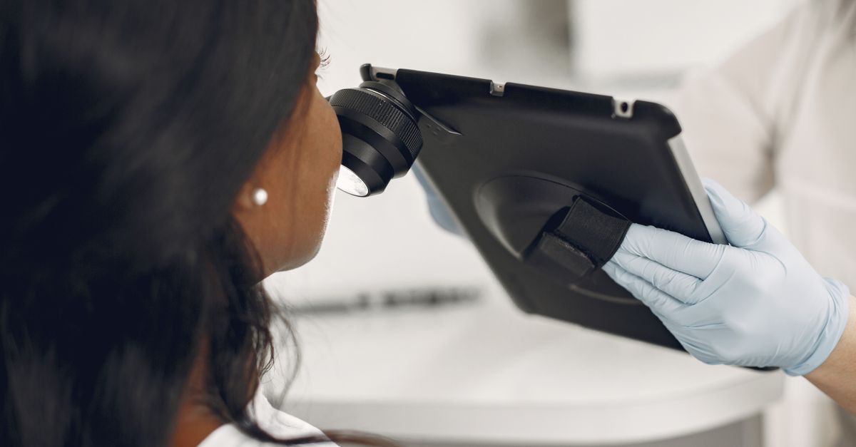
The standard diagnostic approach involves taking clippings of the affected nail and scraping the subungual debris. These samples are examined under a microscope using a potassium hydroxide preparation, which dissolves keratin and highlights the fungal hyphae.
For a more precise identification of the exact pathogen, the sample may be sent for a fungal culture or advanced Polymerase Chain Reaction testing. Identifying the specific organism is crucial for selecting the most effective targeted antifungal medication.
11. Topical Antifungal Therapies
Topical treatments are generally reserved for mild to moderate cases of onychomycosis, particularly when the infection is limited to the distal portion of the nail and does not involve the nail matrix. Specialized antifungal lacquers, such as ciclopirox or amorolfine, are painted onto the nail surface daily or weekly.
The primary limitation of topical therapy is the poor penetration of the medication through the thick, hyperkeratotic nail plate to reach the fungal colonies residing deep in the nail bed. To improve efficacy, clinicians often recommend concurrent physical debridement—filing the nail down to reduce its thickness.
While topical treatments possess an excellent safety profile with virtually no systemic side effects, the cure rates are relatively low compared to oral medications, and treatment must often be continued for nearly a year to see full resolution.
12. Oral Antifungal Medications
Oral antifungal therapy is considered the gold standard for treating moderate to severe onychomycosis. Medications such as terbinafine and itraconazole circulate through the bloodstream and are deposited deep within the nail matrix. From there, the drug is incorporated into the new nail cells as they grow, effectively pushing the infection out.
A standard course of oral therapy for toenails typically lasts for twelve weeks. Because these medications are metabolized by the liver, clinicians usually order baseline liver function tests before initiating therapy, and monitor hepatic enzymes during treatment to ensure safety.
Oral medications offer significantly higher cure rates than topical lacquers. However, complete visual clearing of the nail is not immediate; it takes several months for the healthy, uninfected nail to grow out entirely and replace the damaged, yellowed plate.
13. Laser Therapy and Advanced Modalities
In recent years, light-based therapies have emerged as an alternative or adjunct treatment for onychomycosis. Specific wavelengths of laser energy are directed through the nail plate to superheat and destroy the fungal pathogens residing in the nail bed.
This localized thermal damage attempts to eradicate the fungi without causing significant injury to the surrounding human tissue. Laser therapy is particularly appealing for patients who cannot tolerate the potential hepatic side effects of oral medications or those who have contraindications to systemic drugs.
While laser treatments offer a very high safety profile, their long-term clinical efficacy and mycological cure rates are still under investigation, and they are frequently used in combination with topical agents for optimal results.
14. Surgical Debridement or Removal
In severe, chronic cases where the nail has become grotesquely thickened, painful, and completely unresponsive to pharmacological therapy, mechanical or surgical intervention may be required. Routine podiatric debridement involves safely grinding away the diseased, hyperkeratotic nail tissue. This immediately relieves pressure pain and significantly improves the penetration of topical medications.
If the infection causes intractable pain or recurrent secondary bacterial cellulitis, surgical avulsion, or complete removal of the nail plate, may be necessary. The procedure is performed under local anesthesia.
Once the infected plate is removed, antifungal medications can be applied directly to the exposed nail bed to eradicate any remaining spores before the new nail grows back. In extreme cases, the nail matrix may be permanently destroyed to prevent the nail from ever regrowing.
15. Long-Term Prevention Strategies
Onychomycosis has a high rate of recurrence even after a successful clinical cure. Implementing stringent preventative strategies is essential to maintain healthy nails. The fundamental rule of prevention is managing moisture. Patients must dry their feet meticulously after bathing and change socks promptly if they become damp.
Using prophylactic antifungal powders in shoes helps control the fungal population in the footwear environment. Regularly disinfecting shoes, particularly athletic footwear, prevents reinoculation.
In public areas such as gym locker rooms and hotel showers, wearing protective footwear like flip-flops provides a critical physical barrier against infectious spores. Finally, treating any signs of athlete’s foot aggressively prevents the fungi from migrating back to the vulnerable nail tissues.
16. Frequently Asked Questions (FAQ)
1. Can I get a fungal nail infection from a nail salon?
Yes, if a salon fails to properly sterilize their instruments, pedicure tubs, or files between clients, highly contagious fungal spores can easily be transferred into your nail apparatus.
2. Will painting over the yellow nail with polish make the fungus worse?
Standard nail polish creates an impermeable barrier that traps moisture and darkens the environment, which actually accelerates fungal growth. It is advisable to avoid polish during treatment or use specialized medicated, breathable polishes.
3. Is it possible for a thick yellow nail to heal on its own?
No, clinical onychomycosis is a chronic, progressive infection that will not resolve spontaneously. The immune system cannot clear the fungi hidden deep within the avascular nail keratin.
4. Why does treatment for toenail fungus take so long?
Medications only protect the new nail growth. Because toenails grow very slowly (approximately 1 to 1.5 millimeters per month), it takes up to a year or more for the entirely new, healthy nail plate to completely replace the diseased one.
5. Can apple cider vinegar or bleach cure a thick fungal nail?
While acidic or harsh chemical soaks might alter the surface environment, they cannot penetrate the thick keratin of the nail plate to reach the actual fungal colonies in the nail bed, making them clinically ineffective for advanced infections.
17. Bibliography
Disclaimer: The content is for informational purposes only and does not replace medical advice. Always consult your doctor for personalized treatment.