1. Introduction
The peeling of skin, clinically designated as desquamation, is a visible manifestation of epidermal distress or accelerated cellular turnover. In a healthy physiological state, the outermost layer of the epidermis, the stratum corneum, continuously sheds microscopic dead skin cells (corneocytes) invisibly. This highly regulated, constant renewal process maintains the structural integrity and barrier function of the skin. However, when the skin is subjected to intense environmental trauma, severe localized inflammation, or systemic pharmacological reactions, this orderly process is violently disrupted.
Macroscopic peeling—where large, visible flakes or sheets of skin detach—indicates that the intercellular bonds holding the corneocytes together have been prematurely degraded. Evaluating clinical desquamation requires distinguishing between benign environmental damage, such as a severe sunburn, and life-threatening systemic pathologies, such as drug-induced epidermal necrolysis. Accurate diagnosis dictates the appropriate clinical intervention, which ranges from simple barrier repair with emollient therapy to aggressive immunomodulatory treatments.
2. The Pathophysiology of the Stratum Corneum
The stratum corneum operates as the body’s primary interface with the external environment. It is composed of terminally differentiated, anucleated cells (corneocytes) tightly bound together by specialized protein structures known as corneodesmosomes. These cells are embedded in a highly organized lipid matrix consisting of ceramides, cholesterol, and free fatty acids.
Desquamation is normally controlled by a delicate balance of proteolytic enzymes that carefully dissolve the corneodesmosomes, allowing single cells to shed imperceptibly. When inflammation occurs, cytokines flood the epidermal tissue, accelerating the basal cell replication rate. The rapidly produced cells reach the surface before they are fully mature, and the enzymatic degradation fails to keep pace. Consequently, the corneocytes remain clumped together and shed in large, visible scales or peeling sheets.
3. Environmental Trauma: Sunburn and Thermal Injury
Ultraviolet radiation is the most common cause of acute environmental desquamation. A severe sunburn induces massive, localized DNA damage within the epidermal keratinocytes. When the damage is beyond cellular repair, the cells undergo apoptosis, or programmed cell death.
This massive cellular die-off triggers a profound inflammatory response, causing the characteristic redness, heat, and pain. Within a few days, the body forces the detachment of the entire damaged epidermal layer to eliminate the mutated cells and prevent malignant transformation. The dead tissue visibly peels away, revealing the newly formed, hyper-sensitive epidermis beneath. Mechanical trauma, chemical burns, and extreme cold exposure (frostbite) trigger an identical restorative peeling process.
4. Inflammatory Dermatoses: Eczema and Psoriasis
Chronic skin peeling is a hallmark of prevalent inflammatory dermatoses. In atopic dermatitis (eczema), a genetic defect in the filaggrin protein severely compromises the skin barrier. The skin fails to retain moisture, leading to intense dryness, micro-fissures, and subsequent inflammation. The chronic scratching associated with eczema physically tears the compromised stratum corneum, resulting in continuous scaling and peeling.
Psoriasis represents an entirely different mechanism of peeling. It is an autoimmune condition where T-cells mistakenly attack healthy skin, triggering profound hyperproliferation. In psoriasis, the cellular journey from the basal layer to the surface, which normally takes roughly twenty-eight days, is compressed into three to five days. The immature cells rapidly stack upon the surface, creating thick, silvery plaques that continuously peel and flake.
5. Infectious Etiologies: Fungal and Bacterial
Cutaneous infections frequently present with localized, well-demarcated skin peeling. Dermatophyte infections, such as tinea pedis (athlete’s foot) or tinea corporis (ringworm), rely on the keratin in the stratum corneum for nutrition. As the fungus consumes the structural proteins, the skin barrier collapses, resulting in severe peeling, maceration, and intense pruritus.
Bacterial infections can induce peeling through the secretion of powerful exotoxins. Staphylococcal Scalded Skin Syndrome, primarily affecting infants and young children, is caused by specific strains of Staphylococcus aureus. The bacteria release epidermolytic toxins that systematically destroy the desmoglein-1 proteins holding the upper epidermis together. This results in the rapid, agonizing peeling of large sheets of skin, mimicking a widespread thermal burn and requiring immediate intravenous antibiotic intervention.
6. Drug Eruptions and Severe Systemic Reactions
Desquamation can be the primary presentation of a severe, life-threatening adverse pharmacological reaction. Stevens-Johnson Syndrome (SJS) and Toxic Epidermal Necrolysis (TEN) lie on a clinical spectrum of acute, immune-mediated destruction of the epidermis, most commonly triggered by antibiotics, anticonvulsants, or non-steroidal anti-inflammatory drugs.
In these catastrophic conditions, cytotoxic T-cells initiate massive apoptosis of the basal keratinocytes. The entire epidermis detaches from the underlying dermis, leading to widespread blistering and the sloughing (peeling) of massive sheets of skin. These patients require treatment in specialized burn units, as the loss of the epidermal barrier results in massive fluid loss, profound hypothermia, and a highly elevated risk of lethal systemic sepsis.
7. Kawasaki Disease and Pediatric Peeling
In pediatric medicine, specific patterns of peeling skin serve as vital diagnostic clues. Kawasaki disease is an acute, self-limiting systemic vasculitis (inflammation of the blood vessels) that predominantly affects children under five. It is the leading cause of acquired heart disease in children.
A classic, delayed clinical sign of Kawasaki disease is periungual desquamation—the deep peeling of skin starting at the tips of the fingers and toes, usually occurring in the second or third week of the illness. Identifying this specific peeling pattern, often following a prolonged high fever, red eyes, and a “strawberry tongue,” is essential for diagnosing the disease and initiating intravenous immunoglobulin therapy to prevent the formation of fatal coronary artery aneurysms.
8. Exfoliative Keratolysis and Dyshidrosis
Exfoliative keratolysis is a common, localized, and benign condition characterized by recurrent, asymptomatic peeling on the palms of the hands and soles of the feet. It frequently begins as small, air-filled blisters that burst, leaving behind circular areas of peeling skin with collarettes of scale. While the exact etiology is unknown, it is frequently exacerbated by excessive sweating, friction, or exposure to harsh detergents.
This condition must be clinically differentiated from dyshidrotic eczema, which also affects the palms and soles but is characterized by deeply seated, intensely pruritic (itchy) vesicles. While exfoliative keratolysis primarily requires simple barrier protection and avoidance of irritants, dyshidrotic eczema often demands the application of potent topical corticosteroids to suppress the intense local inflammation.
9. Structural Data: Diagnostic Differentiation of Peeling Skin
Categorizing the underlying cause of desquamation ensures the appropriate therapeutic modality is applied.
| Clinical Presentation | Common Etiology | Primary Mechanism of Peeling |
|---|---|---|
| Post-erythema peeling (widespread) | Severe Sunburn. | Massive apoptosis of UV-damaged keratinocytes. |
| Silvery plaques, thick scaling | Psoriasis. | Autoimmune-driven cellular hyperproliferation. |
| Peeling between toes with itching | Tinea Pedis (Fungal). | Enzymatic destruction of keratin by dermatophytes. |
| Widespread sloughing, mucosal involvement | Toxic Epidermal Necrolysis (TEN). | Severe drug-induced cytotoxic immune reaction. |
10. The Role of Barrier Repair Therapy
When skin peeling is driven by environmental damage or chronic conditions like eczema, the primary therapeutic objective is the restoration of the epidermal barrier. The raw, exposed skin is highly susceptible to secondary bacterial infections and massive transepidermal water loss.
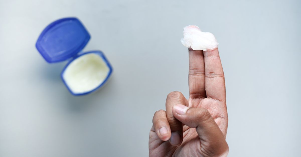
Clinical barrier repair involves the aggressive application of thick, occlusive emollients. Unlike standard lotions, which contain high water content and can further dry the skin through evaporation, clinical emollients are rich in ceramides, humectants (like glycerin or hyaluronic acid), and occlusives (like petrolatum). These agents artificially replace the damaged lipid matrix, sealing in moisture and providing the physiological environment required for the basal cells to regenerate a healthy stratum corneum.
11. Keratolytic Agents in Clinical Practice
In conditions characterized by hyperkeratosis—where the skin peeling is thick, rigid, and tightly adhered, such as in severe psoriasis or ichthyosis—physicians deploy keratolytic agents. These topical medications actively dissolve the intercellular bonds holding the dead skin cells together, facilitating their removal.
Common keratolytic agents include salicylic acid, urea, and alpha-hydroxy acids (such as lactic acid). By chemically breaking down the thick layers of scale, these agents not only smooth the skin but also dramatically increase the penetration and efficacy of concurrent topical treatments, such as corticosteroids or topical immunomodulators, ensuring the medication reaches the active site of inflammation.
12. Managing Pruritus and Secondary Infection
Peeling skin is frequently accompanied by intense pruritus. The mechanical act of scratching exacerbates the epidermal damage and introduces bacteria, commonly Staphylococcus aureus, into the compromised tissue. Recognizing and treating these secondary infections is paramount.
If a peeling area begins to ooze yellow fluid, form golden crusts, or become highly erythematous and painful, topical or oral antibiotics are required. To manage the pruritus and halt the itch-scratch cycle, clinicians frequently prescribe oral antihistamines to be taken at night, aiding in sleep, alongside mid-to-high potency topical corticosteroids to rapidly suppress the localized inflammatory cascade driving the urge to itch.
13. Systemic Therapies for Severe Desquamation
When localized peeling transitions into widespread, uncontrollable desquamation, as seen in severe erythrodermic psoriasis or severe generalized eczema, topical therapies are insufficient. The massive loss of skin scales causes profound metabolic disruptions, including dangerous protein loss and the inability to regulate core body temperature.
These acute cases require aggressive systemic intervention. Dermatologists deploy systemic immunosuppressants, such as methotrexate or cyclosporine, to halt the hyperactive immune response. Increasingly, targeted biologic therapies are utilized. These monoclonal antibodies specifically intercept the inflammatory cytokines responsible for the cellular chaos, providing rapid and sustained clearance of the severe peeling without the broad toxicity of older systemic drugs.
14. Preventative Strategies and Skin Care Regimens
Preventing recurrent pathological skin peeling relies on establishing a protective, long-term dermatological regimen. Patients prone to eczema or contact dermatitis must switch to gentle, fragrance-free syndet (synthetic detergent) cleansers that maintain the skin’s naturally acidic pH. Traditional alkaline soaps violently strip the lipid barrier and exacerbate peeling.
Furthermore, patients must adopt the “soak and seal” method. This involves bathing in lukewarm water for no more than ten minutes and immediately applying a thick ceramide-based emollient to damp skin within three minutes of exiting the water. This traps the environmental moisture within the epidermis, maintaining barrier elasticity and significantly reducing the frequency of inflammatory flares and subsequent desquamation.
15. When to Seek Urgent Medical Evaluation
While most instances of skin peeling are benign and easily managed, certain clinical presentations demand immediate emergency evaluation. If skin peeling is accompanied by a sudden, high fever, widespread severe pain, or if the peeling involves the mucosal membranes (the eyes, inside of the mouth, or the genitals), it strongly suggests a severe drug reaction or a systemic infection.
Similarly, if large sheets of skin rub off with minor friction (a positive Nikolsky sign), the patient must be evaluated immediately in a hospital setting. Rapid diagnosis and the immediate discontinuation of the offending medication, combined with intensive supportive care, are absolutely critical for survival in cases of toxic epidermal necrolysis or severe staphylococcal toxemia.
16. Frequently Asked Questions (FAQ)
1. Why does my skin peel after a severe sunburn?
A sunburn causes massive DNA damage to your skin cells. To protect you from developing skin cancer, your body triggers those mutated, damaged cells to die off. The peeling is the mechanical shedding of this dead layer to reveal the new, healthy skin beneath.
2. Is it safe to pull or pick at peeling skin?
No. Pulling at peeling skin can prematurely tear away cells that are still attached to living tissue. This causes bleeding, delays the healing process, and significantly increases your risk of developing a severe bacterial infection or permanent scarring.
3. What is the best way to treat dry, peeling skin on the hands?
Avoid washing your hands with hot water and harsh antibacterial soaps. Use a gentle, fragrance-free cleanser, and immediately apply a thick, petrolatum or ceramide-based ointment while your hands are still slightly damp to seal in the moisture.
4. Can a skin infection cause my skin to peel?
Yes. Fungal infections, like athlete’s foot, eat away at the keratin in your skin, causing intense peeling and itching. Certain severe bacterial infections can also release toxins that cause the skin to blister and peel away in large sheets.
5. When is peeling skin a medical emergency?
Skin peeling becomes a medical emergency if it is accompanied by a high fever, severe pain, occurs suddenly after starting a new medication (like an antibiotic), or if it spreads to your eyes, mouth, or genital areas, which may indicate a life-threatening allergic reaction.
17. Bibliography
Disclaimer: The content is for informational purposes only and does not replace medical advice. Always consult your doctor for personalized treatment.