1. Introduction
Aching in the soles of the feet after brief periods of standing is primarily caused by mechanical overload on the plantar fascia and the intrinsic foot muscles. Without adequate structural support or neuromuscular conditioning, continuous weight-bearing stretches these connective tissues beyond their endurance limit. Gravity pulls the body downward, and the arches of the feet are forced to absorb and distribute this static load. When the supporting structures begin to fatigue, the tension shifts entirely to the inelastic fascial bands, producing a deep, relentless ache along the bottom of the foot.
This rapid onset of fatigue is a clear indication that the biomechanical architecture of the foot is failing to manage the gravitational stress. Standing still is actually more demanding on the localized tissues of the feet than dynamic walking. During a walk, the mechanical load is distributed and shared among different muscle groups as the foot moves through the gait cycle.
Addressing this structural ache requires a thorough evaluation of the foot’s anatomy, specifically focusing on the medial longitudinal arch, the integrity of the plantar fat pad, and the capacity of the intrinsic muscles. By identifying the exact point of biomechanical failure, clinical interventions can be implemented to properly support the foot and prevent chronic tissue degradation.
2. The Architecture of the Foot
The human foot is an extraordinary feat of biological engineering, composed of twenty-six bones, thirty-three joints, and a complex web of over a hundred muscles, tendons, and ligaments. This architecture is arranged into three primary arches: the medial longitudinal arch, the lateral longitudinal arch, and the transverse arch.
These arches form a resilient, dome-like structure designed to absorb the shock of body weight and adapt to uneven terrain. The bony alignment is passively supported by thick, fibrous ligaments that bind the bones tightly together. This passive skeletal support is the foundation of weight-bearing.
Active support is provided by the intrinsic foot muscles, which originate and insert entirely within the foot, and the extrinsic muscles, which originate in the lower leg and send tendons down into the foot. These muscles dynamically contract to maintain the integrity of the arches when the foot is loaded.
3. The Role of the Plantar Fascia
The plantar fascia is a thick, pearlescent band of connective tissue that originates at the calcaneus, or heel bone, and fans out to attach to the base of each toe. It is the primary passive stabilizer of the medial longitudinal arch. The fascia acts much like the string of a bow; as weight presses down on the arch of the foot, the fascia stretches to prevent the arch from collapsing entirely.
Because the plantar fascia is composed of dense collagen fibers, it is highly tensile but remarkably inelastic. It is designed to sustain tension, not to stretch significantly. When the foot is forced to bear weight continuously without relief, the fascia is held in a state of maximum tension.
This continuous stretching pulls forcefully against its attachment point on the heel bone and creates microscopic mechanical strain along its central band. The resulting sensation is a deep, broad ache across the sole of the foot, signaling that the connective tissue is actively resisting structural failure.
4. Static Standing versus Dynamic Walking
It is a common clinical paradox that many individuals can walk for miles without pain but experience agonizing aches after standing still for just one hour. The physiology of movement explains this discrepancy. Walking is a dynamic process involving muscle pumping, weight shifting, and alternating periods of load and rest for each foot.
When standing statically, the body weight remains fixed over the same anatomical structures continuously. The blood flow to the bottom of the foot is significantly reduced due to the constant capillary compression, leading to mild tissue ischemia. The intrinsic muscles are forced into a prolonged isometric contraction, where they generate force without changing length.
Isometric contractions are metabolically expensive and highly fatiguing. As the muscles tire and weaken, they stop supporting the arch. The entirety of the mechanical load is subsequently dumped onto the non-contractile plantar fascia, rapidly accelerating the onset of pain and structural irritation.
5. Plantar Fasciitis and Tissue Overload
When the mechanical strain on the plantar fascia becomes chronic, it develops into plantar fasciitis. This is the most common cause of pain in the sole of the foot. Despite the suffix “itis,” modern histological studies reveal that the condition is primarily degenerative rather than purely inflammatory.
Repeated overloading causes micro-tears within the collagen matrix of the fascia, specifically near the heel attachment. The body attempts to repair this damage, but if the standing stress continues, the repair process becomes disorganized. The tissue thickens, loses its organized structure, and becomes highly sensitive to tension.
Individuals developing early plantar fasciitis frequently notice that the aching ache after standing progresses to a sharp, stabbing pain, particularly during the first few steps after a period of rest. This sharp pain occurs because the compromised, thickened fascia is abruptly stretched.
6. Plantar Fat Pad Atrophy
Beneath the heel bone and the metatarsal heads (the ball of the foot) lies the plantar fat pad. This specialized tissue is composed of dense adipose lobules enclosed within tough, fibrous septa. Its sole biological purpose is to provide hydraulic shock absorption and to cushion the underlying bones and nerves from the hard ground.
With advancing age, or due to repetitive high-impact trauma, this fat pad naturally begins to thin and degrade, a condition known as fat pad atrophy. The fibrous compartments break down, and the shock-absorbing capacity of the foot is severely diminished.
When an individual with a thinned fat pad stands on a hard surface for an hour, the bony prominences of the foot press directly against the skin and the hard floor. This creates severe, localized compressive pain, often described as feeling like standing directly on a bruised bone.
7. Foot Arch Biomechanics and Pronation
The natural resting posture of the foot dictates how mechanical loads are distributed across the sole. Pronation is the natural inward rolling of the foot that helps absorb shock. However, overpronation, often associated with flexible flat feet, forces the medial arch to collapse completely against the ground.
This collapse drastically elongates the plantar fascia and twists the biomechanical axis of the foot. The constant, extreme tension required to prevent total structural collapse exhausts the intrinsic muscles rapidly. Individuals with severe overpronation will inevitably experience a deep, burning ache in the midfoot after brief standing.
Conversely, supination refers to a high, rigid arch. A highly arched foot lacks mobility and fails to absorb shock effectively. The weight is concentrated entirely on the heel and the ball of the foot, leading to rapid, intense focal pressure and aching in those specific areas.
8. Intrinsic Muscle Fatigue
The intrinsic muscles of the foot, including the abductor hallucis and the flexor digitorum brevis, form the active muscular core of the foot. In populations accustomed to wearing stiff, highly supportive shoes, these intrinsic muscles frequently become neurologically dormant and physically weak.
When called upon to support the arch during prolonged standing, these deconditioned muscles fatigue within minutes. As they fail, the foot physically sags. The aching sensation in the sole is the direct sensory feedback from these small, exhausted muscles cramping and struggling to maintain structural integrity.
Rehabilitating foot pain requires waking up these dormant muscles through targeted strengthening. A robust intrinsic muscular core acts as an active suspension system, absorbing the static load and protecting the plantar fascia from excessive mechanical strain.
9. The Impact of Inadequate Footwear
The interface between the sole of the foot and the ground determines the rate of tissue fatigue. Standing barefoot or in completely flat, unsupportive shoes on unyielding surfaces like tile or concrete provides zero external assistance to the foot’s structural mechanisms.
Shoes that lack a rigid shank flex too easily in the middle, failing to support the arch. Footwear without a contoured footbed concentrates all the body weight on the heel and forefoot, skipping the midfoot entirely. This uneven pressure distribution accelerates fat pad compression and fascial strain.
Transitioning to footwear designed for occupational standing—featuring a supportive heel counter, a defined arch, and a rocker-bottom sole—can immediately alter the biomechanics of standing. Proper footwear redistributes the gravitational load across the entire surface area of the sole, drastically increasing standing endurance.
10. Neurological Nerve Entrapment
If the aching in the sole is accompanied by burning, tingling, or localized numbness, a neurological entrapment must be considered. Tarsal tunnel syndrome is a condition where the posterior tibial nerve is compressed as it travels through a narrow anatomical space on the inside of the ankle.
When the foot overpronates during prolonged standing, the structures on the inside of the ankle are stretched tightly. This tension can compress the tibial nerve against the surrounding ligamentous bands. The resulting nerve irritation radiates a diffuse, burning ache along the bottom of the foot, often extending into the toes.
Unlike purely mechanical fascial pain, neuropathic pain from nerve entrapment often worsens at night and may cause the foot to feel subjectively hot or cold. Differentiating nerve pain from structural strain is critical for appropriate clinical management.
11. Differential Diagnosis of Sole Pain
Determining the exact pathology driving the aching soles requires separating overlapping symptoms. Aching concentrated perfectly in the center of the heel pad is indicative of fat pad atrophy or a heel spur, which is a bony growth resulting from chronic fascial tension.
Pain localized exclusively to the ball of the foot is termed metatarsalgia. This frequently results from standing in elevated footwear or having a shortened Achilles tendon, which shifts the body’s center of gravity forward, placing disproportionate crushing force on the delicate metatarsal heads.
If the pain is widespread and feels like a deep, cramping fatigue through the entire arch, it is a combination of intrinsic muscle failure and diffuse plantar fascia strain. Mapping the precise location of the pain points the clinician toward the failing structural component.
12. Clinical Assessment Methods
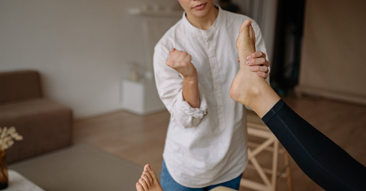
A thorough clinical examination evaluates the dynamic and static posture of the foot. The clinician will perform the windlass test, pulling the big toe upward to engage the plantar fascia. If this maneuver reproduces the sharp pain in the heel or arch, it strongly indicates plantar fasciitis.
The clinician will observe the patient’s standing posture to assess the degree of pronation and arch collapse. Palpation of the sole helps differentiate between a bruised fat pad, a tender fascial band, or a specific inflamed joint.
| Clinical Pathology | Primary Pain Location | Distinguishing Feature |
|---|---|---|
| Plantar Fasciitis | Medial heel and arch. | Pain is sharpest during the first steps after resting. |
| Fat Pad Atrophy | Directly under the heel bone. | Feels like standing on a bruise; lacks sharp morning pain. |
| Metatarsalgia | Ball of the foot. | Pain worsens when leaning forward or wearing heels. |
| Tarsal Tunnel Syndrome | Diffuse burning across the sole. | Accompanied by tingling or numbness radiating to toes. |
Diagnostic ultrasound is an excellent tool for visualizing the thickness of the plantar fascia and assessing the integrity of the fat pad. Identifying abnormal tissue thickening confirms the presence of chronic degenerative changes requiring targeted intervention.
13. Rehabilitative Strategies
The immediate clinical strategy to improve standing tolerance focuses on passive external support. Over-the-counter or custom orthotic inserts provide a physical shelf for the arch to rest upon. This instantly relieves the tension on the plantar fascia and allows the exhausted intrinsic muscles to recover.
Long-term resolution requires active tissue remodeling. Calf stretching is vital, as a tight Achilles tendon restricts ankle mobility and forces the foot to overpronate to compensate, dragging the arch down. Improving ankle dorsiflexion reduces the mechanical load on the sole significantly.
Strengthening the intrinsic foot muscles through exercises like towel scrunches and marble pickups rebuilds the active arch support. A stronger muscular core inherently absorbs more gravitational force, protecting the inelastic fascial tissues and expanding the individual’s capacity to stand pain-free.
14. Frequently Asked Questions FAQ
1. Are gel insoles the best way to stop my feet from aching when I stand?
Gel insoles provide temporary cushioning, which can help a bruised fat pad, but they provide zero structural support for the arch. If your pain is caused by the arch collapsing and pulling the plantar fascia, a firm, rigid orthotic is vastly superior to soft gel.
2. Should I stand on an anti-fatigue mat at work?
Yes, anti-fatigue mats are highly effective. They introduce subtle instability to the surface, which forces your leg and foot muscles to make micro-adjustments constantly. This micro-movement acts as a muscle pump, improving blood flow and preventing the isometric fatigue that causes severe aching.
3. Will rolling my foot on a frozen water bottle heal the pain?
Rolling the foot on ice provides excellent temporary relief by numbing the inflamed tissues and creating a gentle stretch along the fascia. However, it only treats the symptoms. It will not heal the condition unless you also address the footwear and muscle weakness causing the overload.
4. Why does the bottom of my foot throb for hours after I finally sit down?
When you sit down and remove your body weight, the blood vessels in the bottom of your foot that were compressed for an hour suddenly dilate. This rapid rush of blood back into the tired, inflamed tissues causes a throbbing, vascular ache as the area attempts to clear metabolic waste.
15. Bibliography
Disclaimer: The content is for informational purposes only and does not replace medical advice. Always consult your doctor for personalized treatment.