1. Introduction
Feeling a sharp pain in the heel when walking is not a normal physiological condition and typically signals structural inflammation or biomechanical strain within the foot. The heel bone is designed to act as the primary shock absorber for the entire human body, effortlessly bearing the repetitive impact of the gait cycle. When sharp, localized pain occurs, it indicates that the complex network of connective tissues, bones, or nerves supporting this area has been compromised or overloaded.
The most frequent etiology behind this specific presentation is plantar fasciitis, a degenerative irritation of the thick ligament running along the bottom of the foot. However, due to the intricate anatomy of the foot and ankle, accurate diagnosis requires differentiating fascial strain from tendinopathy, nerve compression, or structural bone issues.
Addressing this pain promptly prevents the development of compensatory gait abnormalities that can ultimately damage the knees, hips, and lumbar spine. A structured clinical evaluation focuses on pinpointing the exact anatomical origin of the pain, guiding the implementation of targeted physical therapies and orthotic interventions to restore pain-free mobility.
2. Anatomy of the Plantar Fascia
The plantar fascia is a dense, fibrous band of connective tissue spanning the arch of the foot. It originates at the medial tubercle of the calcaneus, the large heel bone, and fans out anteriorly to insert into the bases of the toes. This tissue is incredibly robust and acts as a dynamic bowstring, supporting the longitudinal arch of the foot.
During the gait cycle, the plantar fascia performs a critical mechanical function. As a person steps forward and weight transfers to the ball of the foot, the toes extend. This extension actively winds the plantar fascia around the metatarsal heads, pulling it taut and elevating the arch, a mechanism known as the windlass effect.
This windlass mechanism locks the bones of the midfoot into a rigid lever, allowing the calf muscles to effectively propel the body forward. Consequently, the insertion point of the fascia at the heel sustains massive tensile forces with every single step, making it highly susceptible to mechanical overload.
3. Biomechanics of the Gait Cycle
Normal walking is a complex sequence of precisely timed muscular contractions and joint articulations. The cycle begins with the heel strike, where the calcaneus absorbs the initial shock of body weight hitting the ground. The foot then rapidly pronates, rolling slightly inward to unlock the joints and adapt to uneven terrain.
If the biomechanics are flawed, such as in cases of severe overpronation or rigid high arches, the distribution of force is altered. Overpronation causes the arch to collapse excessively, forcefully elongating the plantar fascia beyond its physiological limit and pulling aggressively on its attachment at the heel bone.
Conversely, a rigid, highly arched foot lacks adequate shock absorption. The rigid structure forces the heel pad to bear a disproportionate amount of the impact during the heel strike. Both of these biomechanical extremes lead to chronic micro-trauma of the connective tissues surrounding the calcaneus.
4. Pathophysiology of Plantar Fasciitis
Despite its name implying an inflammatory condition, chronic plantar fasciitis is more accurately described as fasciosis—a non-inflammatory, degenerative process. The relentless mechanical overload causes microscopic tears in the collagen fibers of the fascia, specifically near its origin at the heel bone.
Under normal circumstances, the body repairs these micro-tears during periods of rest. However, if the mechanical strain continues daily, the rate of damage exceeds the rate of cellular repair. The collagen matrix degrades, becoming disorganized and weakened.
This localized tissue degeneration creates a focal point of intense sensitivity. The sharp pain experienced during walking is the neurological result of placing body weight directly onto this degraded, structurally compromised insertion point on the calcaneus.
5. The Morning Pain Phenomenon
A hallmark clinical sign of plantar fasciitis is the intense, sharp pain experienced during the very first steps out of bed in the morning. This phenomenon is a direct consequence of the tissue’s healing mechanics and nocturnal foot posture.
During sleep, the feet naturally rest in a state of plantarflexion, with the toes pointing slightly downward. In this relaxed position, the plantar fascia shortens. The body attempts to repair the micro-tears by laying down fragile, new collagen fibers across the shortened tissue.
Upon standing in the morning, the sudden dorsiflexion of the foot forces the fascia to stretch back to its full length. This rapid mechanical stretch abruptly rips the fragile, newly formed healing tissue, resulting in a sudden, sharp, tearing sensation in the heel. As the individual continues to walk, the tissue warms up and slightly stretches, generally causing the acute pain to dull into a persistent ache.
6. Heel Spurs and Bony Exostoses
A heel spur is a calcified, bony outgrowth that forms on the underside of the calcaneus, exactly where the plantar fascia attaches. Historically, these spurs were blamed as the primary source of sharp heel pain, with physicians believing the bony point was stabbing into the surrounding flesh.
Current clinical understanding recognizes that the heel spur is actually a secondary structural adaptation. In response to the chronic, relentless pulling of the tight plantar fascia, the bone reacts by laying down extra calcium deposits to reinforce the attachment site, creating the spur.
Therefore, the spur itself is rarely the direct source of the pain; rather, the pain originates from the degenerative fascial tissue pulling on the bone. Many individuals possess large heel spurs without experiencing any symptoms, while others have excruciating plantar fasciitis with completely smooth heel bones.
7. Achilles Tendinopathy Considerations
If the sharp pain is localized to the posterior aspect of the heel, precisely where the shoe rubs, the underlying pathology likely involves the Achilles tendon rather than the plantar fascia. The Achilles is the thickest tendon in the body, connecting the powerful calf muscles to the back of the calcaneus.
Achilles tendinopathy is a degenerative condition resulting from overuse, often seen in runners or individuals suddenly increasing their walking distance. The collagen fibers degenerate, and the tendon thickens and becomes exquisitely tender to palpation.
Pain from Achilles involvement is typically sharpest during the push-off phase of walking, when the calf muscles violently contract to lift the heel off the ground. Managing posterior heel pain requires entirely different rehabilitative protocols focused on eccentric calf strengthening rather than fascial stretching.
8. Tarsal Tunnel Syndrome and Neuropathy
When sharp heel pain is accompanied by a tingling, burning, or electrical sensation that radiates into the arch of the foot, clinicians must consider a neurological etiology. Tarsal tunnel syndrome is a compression neuropathy akin to carpal tunnel syndrome in the wrist.
The posterior tibial nerve travels down the inside of the ankle and passes through a narrow anatomical corridor called the tarsal tunnel before branching into the heel and sole of the foot. If the surrounding tissues swell due to injury, arthritis, or severe flat feet, the space narrows, compressing the nerve.
This compression disrupts the nerve’s ability to transmit signals accurately, resulting in sharp, shooting pain during weight-bearing activities. Tarsal tunnel syndrome mandates a careful neurological examination to prevent permanent nerve damage.
9. Fat Pad Atrophy
The calcaneus is protected by a specialized, dense layer of adipose tissue known as the heel fat pad. This pad is constructed of tightly packed fat cells held within a tough, fibrous matrix, designed to absorb and dissipate the immense compressive forces of the heel strike.
With advancing age, or following multiple corticosteroid injections into the heel, the structural integrity of this fibrous matrix can break down. The fat pad loses its elasticity and thickness, a condition termed fat pad atrophy.
Without this vital shock absorber, the calcaneus strikes the ground with minimal cushioning. Patients with fat pad atrophy report a deep, bruising, sharp pain located directly in the center of the heel, which is noticeably exacerbated by walking barefoot on hard surfaces like tile or hardwood floors.
10. Risk Factors and Occupational Hazards
The development of sharp heel pain is strongly correlated with specific occupational and lifestyle factors. Individuals whose occupations require prolonged periods of standing on hard, unforgiving surfaces, such as factory workers or retail employees, are at an exceptionally high risk for fascial overload.
A rapid increase in body weight places an immediate, disproportionate mechanical burden on the arches of the feet, severely stressing the plantar fascia. Similarly, athletes who drastically increase their training volume or switch to running on asphalt without adequate preparation frequently develop acute symptoms.
Inadequate footwear is a ubiquitous contributing factor. Wearing unsupportive, flat shoes, or athletic shoes whose internal cushioning has completely deteriorated, forces the foot’s internal structures to bear the entire impact of the gait cycle without assistance.
11. Differential Diagnoses of Heel Pain
Accurate diagnosis relies on pinpointing the precise location and nature of the sharp pain.
| Pain Location and Nature | Primary Trigger | Probable Clinical Diagnosis |
|---|---|---|
| Bottom of heel, sharp tearing | First steps in the morning | Plantar Fasciitis |
| Back of heel, sharp pulling | Pushing off toes to walk | Achilles Tendinopathy |
| Center of heel, deep bruising | Walking barefoot on hard floors | Fat Pad Atrophy |
| Inside of ankle/heel, burning/tingling | Prolonged standing, nerve compression | Tarsal Tunnel Syndrome |
Charting these specific characteristics guides the clinician away from generic treatments and toward precise, etiology-specific interventions.
12. Diagnostic Imaging and Clinical Assessment
The clinical evaluation of heel pain relies heavily on the physical examination. The physician palpates the specific attachment points of the fascia and tendons to isolate the origin of the tenderness. They evaluate the range of motion of the ankle joint, as a tight calf muscle is a primary driver of foot pathology.
Radiographic imaging, specifically plain X-rays, is utilized primarily to rule out stress fractures of the calcaneus or to identify the presence of bony spurs. However, X-rays cannot visualize soft tissue damage.
If the diagnosis remains ambiguous, or if conservative treatment fails to yield improvement, magnetic resonance imaging or high-frequency ultrasound is deployed. These modalities allow the physician to visualize the actual thickening and structural tearing of the plantar fascia or the Achilles tendon in high detail.
13. Conservative Management and Stretching
The initial approach to resolving sharp heel pain involves a structured, conservative rehabilitative protocol. For plantar fasciitis, the cornerstone of therapy is diligent, specific stretching.
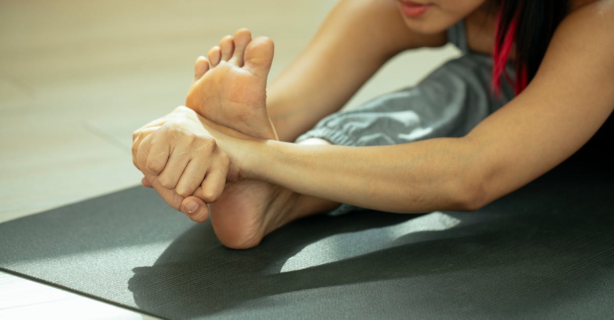
Patients must actively stretch both the plantar fascia and the entire posterior chain, specifically the gastrocnemius and soleus calf muscles. A tight calf muscle anchors the heel bone, preventing normal ankle motion and forcing the foot to compensate by overstretching the arch.
Night splints are highly effective conservative devices. Worn during sleep, the splint maintains the foot in a strictly 90-degree angle. This prevents the fascia from shortening and healing in a contracted position overnight, effectively eliminating the severe, sharp tearing sensation experienced during the first morning steps.
14. Orthotic Interventions and Footwear
Correcting flawed foot biomechanics is essential for long-term resolution. Supportive footwear with a robust arch and a slightly elevated, cushioned heel reduces the tensile strain on the fascia and provides necessary shock absorption for the calcaneus.
Custom or high-quality over-the-counter orthotic inserts act to physically support the longitudinal arch and control excessive pronation during the gait cycle. By redistributing the mechanical load evenly across the entire foot, orthotics relieve the isolated stress point at the heel insertion.
Patients are instructed to strictly avoid walking barefoot, even indoors, during the recovery phase, as hard floors offer zero shock absorption and perpetuate the micro-trauma to the damaged tissues.
15. Advanced Pharmacological and Interventional Therapies
If rigorous conservative management fails to relieve the sharp pain after several months, advanced interventions are considered. Extracorporeal shock wave therapy is a non-invasive procedure that uses high-energy sound waves to stimulate localized blood flow and reignite the cellular healing process in chronic, stagnant fasciosis.
Corticosteroid injections can provide rapid, potent suppression of localized inflammation. However, they are used judiciously, as repeated steroid exposure weakens the collagen matrix of the fascia, increasing the risk of complete rupture, and promotes the permanent degradation of the protective heel fat pad.
In severe, recalcitrant cases where the damaged tissue simply will not heal, a surgical plantar fasciotomy is performed to partially cut the fascial band, releasing the mechanical tension and permanently altering the biomechanics of the arch.
16. Frequently Asked Questions FAQ
1. Why does my heel hurt so much more during the first few steps in the morning?
While you sleep, your foot relaxes and points downward, causing the inflamed plantar fascia on the bottom of your foot to shorten and tighten. When you stand up, your weight violently forces the tight tissue to stretch back out, causing micro-tears and a sharp, severe pain.
2. Will a heel spur require surgery to remove?
Usually no. The bone spur itself is rarely the actual cause of the pain. The pain comes from the tight, damaged fascia pulling on the bone. Treating the tight tissue through stretching and orthotics almost always resolves the pain, leaving the spur in place harmlessly.
3. Is walking barefoot bad if my heel hurts?
Yes, walking barefoot on hard surfaces like tile or hardwood floors removes all shock absorption. This forces your damaged heel tissues and fat pad to bear the entire impact of your body weight, significantly worsening the inflammation and delaying healing.
4. Can being overweight cause sudden heel pain?
Yes, a rapid increase in body weight places massive mechanical stress on the arches of your feet. The connective tissues must suddenly support a heavier load during every step, which frequently leads to the mechanical overload and tearing seen in plantar fasciitis.
5. How long does it take for plantar fasciitis to completely heal?
Plantar fasciitis is a chronic degenerative condition, not a simple acute injury. Because you must continue to walk on the damaged tissue daily, complete healing often takes several months to a full year of diligent stretching, proper footwear, and biomechanical support.
17. Bibliography
Disclaimer: The content is for informational purposes only and does not replace medical advice. Always consult your doctor for personalized treatment.