1. Introduction
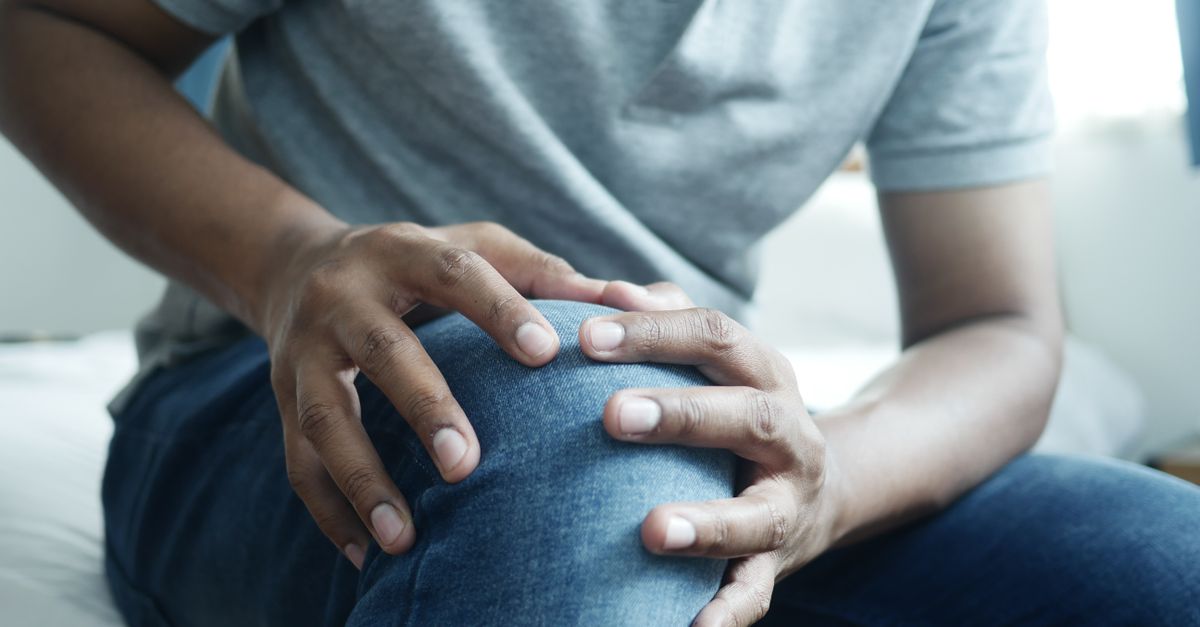
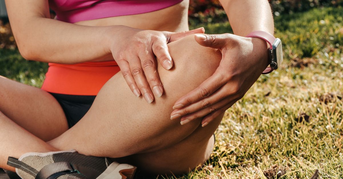
Physical pain in the kneecaps during deep walking lunges is primarily caused by elevated patellofemoral joint pressure and improper biomechanical tracking of the patella. As the knee flexes deeply to lower the body toward the floor, the quadriceps muscle generates massive tension. This tension acts as a biological pulley, driving the kneecap forcefully against the underlying femur. If the joint is slightly misaligned or the surrounding musculature is imbalanced, this crushing force severely irritates the articular cartilage and the highly innervated soft tissues encasing the joint.
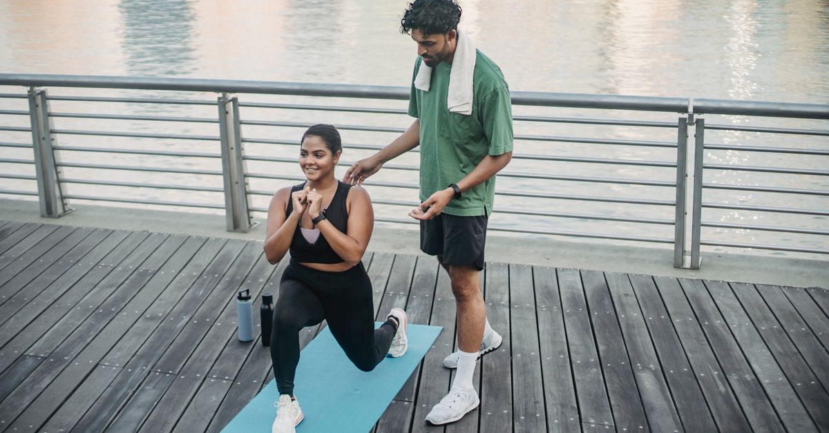
The walking lunge is an exceptional closed-kinetic-chain exercise for developing lower body strength and dynamic stability. However, it subjects the knee joint to immense shear and compressive forces. Unlike a stationary squat where the torso remains relatively centered, a walking lunge requires decelerating the entire forward momentum of the body while simultaneously dropping into deep flexion on a single leg.
When structural flaws exist—such as weak gluteal muscles allowing the knee to collapse inward, or tight quadriceps increasing the baseline pressure—the delicate cartilaginous surfaces of the knee are subjected to brutal mechanical friction. The resulting pain is typically a sharp, localized ache behind or directly under the kneecap, acting as a profound warning sign of mechanical overload.
Addressing patellofemoral pain during lunges requires a rigorous analysis of movement mechanics. By modifying the depth of the lunge, strengthening the lateral pelvic stabilizers, and ensuring proper foot placement, individuals can execute this powerful exercise safely, preserving the longevity and health of the knee joint.
2. Anatomy of the Patellofemoral Joint
The knee is an intricate hinge joint, but the specific source of anterior pain during a lunge is the patellofemoral joint. This is the articulation where the patella (kneecap) glides within the trochlear groove, a V-shaped depression on the front of the femur (thigh bone).
The patella is embedded within the thick tendon of the quadriceps muscle. Its primary biomechanical purpose is to act as a fulcrum, increasing the leverage and mechanical advantage of the thigh muscles as they extend the lower leg. The underside of the patella is coated in a thick, smooth layer of articular cartilage, allowing it to glide friction-free under massive pressure.
The stability of the patella within this groove is dictated by passive restraints (ligaments) and active dynamic restraints (muscles). The vastus medialis oblique on the inside of the thigh and the vastus lateralis on the outside must pull with perfectly balanced tension.
If this delicate muscular balance is disrupted, the patella tracks improperly. Instead of gliding smoothly down the center of the groove, it rubs harshly against the raised bony ridges of the femur, setting the stage for acute pain and eventual cartilage degradation.
3. Biomechanics of the Walking Lunge
A walking lunge is a dynamic, high-load movement divided into distinct phases. As the lead foot strikes the ground, the body must absorb significant forward kinetic energy. The quadriceps of the front leg engage aggressively in an eccentric contraction—lengthening under tension—to act as the primary brake and control the descent of the hips.
As the knee bends past sixty degrees, the contact area between the patella and the femur reaches its maximum. Simultaneously, the tension required by the quadriceps to halt the downward movement spikes.
This combination of maximum contact area and maximum muscular tension generates profound compressive forces across the patellofemoral joint. In a deep lunge where the back knee nearly touches the floor, the front knee is subjected to compressive forces that can exceed three to four times the individual’s body weight.
If the knee travels far forward past the toes during the descent, the lever arm changes, drastically amplifying this compressive joint force. This mechanical reality explains why the lunge feels uniquely punishing to the kneecaps compared to standard walking or light jogging.
4. Compressive Forces in Deep Flexion
The degree of knee flexion directly correlates with joint pressure. When the leg is straight, the patella rests completely above the trochlear groove with zero compressive load.
As flexion initiates, the patella is drawn down into the groove. Between thirty and ninety degrees of flexion, the compressive forces multiply rapidly. The deeper the lunge, the tighter the quadriceps tendon is stretched over the joint hinge, pressing the kneecap brutally into the bone beneath it.
If the articular cartilage is completely healthy, it absorbs this pressure like a sponge, weeping synovial fluid to lubricate the joint. However, if the cartilage is already slightly softened or damaged, it cannot buffer this intense pressure.
The highly sensitive subchondral bone beneath the cartilage registers this immense, unprotected load. The central nervous system interprets this mechanical stress as a deep, severe ache localized squarely behind the kneecap.
5. Patellar Maltracking and Cartilage Irritation
Patellar maltracking is the most common functional cause of lunge-induced knee pain. Perfect tracking requires the patella to remain precisely centered in the femoral groove.
In many individuals, the lateral structures of the thigh, notably the iliotibial band and the vastus lateralis, become chronically tight. Simultaneously, the medial stabilizing muscle, the vastus medialis oblique, becomes weak and delayed in its firing pattern.
This lateral dominance acts like a misaligned steering wheel. As the knee bends during the lunge, the tight lateral tissues physically drag the kneecap outward. The underside of the patella grinds fiercely against the lateral ridge of the femur.
This abnormal friction rapidly inflames the cartilage and the synovial lining of the joint capsule. The runner experiences a sharp, grinding pain, often accompanied by a distinct crunching or popping sound as the kneecap struggles to snap back into its proper groove.
6. Quadriceps Weakness and Imbalance
Counterintuitively, weak quadriceps are a massive risk factor for knee pain during lunges. The quadriceps are the body’s primary shock absorbers. During the descent of a lunge, they must decelerate the body smoothly.
If the quadriceps lack the absolute strength or endurance to manage the load, the muscle fails to control the descent. The body effectively “drops” into the bottom of the lunge.
This rapid, uncontrolled drop removes the muscular buffering system. The kinetic shock of arresting the fall is transferred instantly and entirely into the passive joint structures—the ligaments, the menisci, and the patellofemoral cartilage.
Building eccentric quadriceps strength ensures that the muscle fibers can absorb the kinetic energy smoothly, shielding the vulnerable joint surfaces from catastrophic mechanical spikes.
7. The Role of the Gluteal Muscles
The alignment of the knee joint is dictated almost entirely by the hip. The gluteus medius and gluteus maximus are the primary stabilizers of the pelvis and the femur.
During a walking lunge, the individual is momentarily balancing on a single leg. If the gluteus medius is weak, the pelvis drops, and the femur rotates internally and collapses inward toward the midline of the body. This inward collapse is known as knee valgus.
When the knee collapses into valgus during a deep lunge, the biomechanical tracking of the patella is instantly destroyed. The entire joint is twisted while under heavy compressive load.
The vast majority of anterior knee pain in athletes originates from this silent gluteal weakness. Strengthening the lateral hip muscles prevents the knee from caving inward, preserving the perfect vertical alignment required for pain-free lunging. Readers managing overlapping structural instability may consult our guide on hips out of alignment.
8. Patellar Tendinopathy Considerations
If the pain is sharp and localized specifically to the thick tendon directly below the kneecap, rather than a deep ache behind it, the issue is likely patellar tendinopathy.
Also known as “jumper’s knee,” this condition involves microscopic tearing and structural degradation of the collagen fibers within the patellar tendon due to repetitive explosive loading or heavy resistance training without adequate recovery.
During the lowest point of a deep lunge, the patellar tendon is placed under maximum stretch while simultaneously attempting to contract forcefully to lift the body back up.
A degraded tendon cannot handle this massive tensile force. The pain spikes sharply during the transition from lowering to lifting. Resolving tendinopathy requires pausing deep lunges and implementing heavy, slow, isometric holds to rebuild the tendon’s structural integrity.
9. Chondromalacia Patellae
Chondromalacia patellae is a clinical condition characterized by the softening, fraying, and breakdown of the articular cartilage on the underside of the kneecap. It frequently affects young adults and is a direct consequence of chronic patellar maltracking.
When healthy, the cartilage is firm and smooth. As it breaks down, it becomes fibrillated, resembling the rough texture of crabmeat.
When a person with chondromalacia performs a deep lunge, this roughened, frayed cartilage is forced to bear immense body weight while scraping against the femur. This generates intense, burning pain and loud, gravelly crepitus (grinding sounds).
If the cartilage is severely degraded, deep lunges may need to be permanently removed from the training program to prevent the complete erosion of the protective layer, which inevitably leads to early-onset osteoarthritis.
10. Identifying Anterior Knee Pain
Categorizing the specific nature and location of the pain helps differentiate the mechanical failure points within the knee.
| Pain Profile | Primary Mechanism | Identifying Features |
|---|---|---|
| Deep, diffuse ache behind the kneecap | Patellofemoral Pressure / Maltracking | Worse the deeper the knee bends, aches after workout |
| Sharp pain precisely below the kneecap | Patellar Tendinopathy | Tender to direct touch on the tendon, hurts changing direction |
| Grinding, gravel-like sensation with pain | Chondromalacia Patellae | Loud crunching sounds, chronic dull ache when sitting |
| Pain on the outer edge of the knee | Iliotibial Band Friction | Snapping sensation over the lateral knee bone |
11. Correcting Lunge Mechanics
The immediate solution to lunge-induced knee pain involves a strict correction of exercise mechanics. The stride length dictates the distribution of force.
Taking too short of a step forces the front knee to drive far out over the toes, maximizing the angle of flexion and exponentially increasing patellofemoral compression. Taking too long of a step strains the hip flexor of the trailing leg and alters pelvic alignment.
The ideal walking lunge stride allows the front shin to remain relatively vertical, with the knee tracking directly over the laces of the shoe, never collapsing inward.
Focus heavily on the trailing leg. A significant portion of the body’s weight should be distributed to the back foot, dropping the back knee straight down toward the floor, rather than throwing the entire torso weight forward onto the front kneecap.
12. Modifying the Depth of the Lunge
If perfect form still produces a deep ache, the depth of the lunge must be restricted. Pain is a clear indicator that the joint cannot handle the compressive force at maximum flexion angles.
Limit the descent of the lunge to the point just before the pain triggers. For many, stopping when the front knee reaches a seventy-degree angle, rather than dropping a full ninety degrees to the floor, keeps the patellofemoral pressure within a safe, tolerable limit.
Utilizing a suspension trainer or holding lightly onto a structural support can assist in unloading a percentage of body weight, allowing the individual to train the movement pattern and build muscular strength without subjecting the cartilage to destructive full-load compression.
13. Alternative Exercises for Leg Strength
If walking lunges cause persistent pain despite form corrections, they should be substituted with exercises that develop equivalent leg strength with vastly different joint mechanics.
The Bulgarian split squat, performed with the rear foot elevated on a bench, shifts more mechanical focus to the glutes and hamstrings while keeping the torso upright, reducing the sheer anterior force on the knee.
Step-ups onto a plyometric box are highly effective. Because the movement initiates from a static, supported position, it builds tremendous concentric strength without the violent eccentric braking forces required to arrest forward momentum during a walking lunge.
Reverse lunges are another excellent alternative. Stepping backward naturally keeps the front shin vertical and shifts the center of gravity rearward, significantly unloading the patellofemoral joint compared to the aggressive forward step of a walking lunge.
14. Strengthening the Medial Quadriceps
Fixing the patellar tracking issue requires targeted strengthening of the vastus medialis oblique (VMO), the teardrop-shaped muscle on the inner thigh just above the knee.
The VMO is notoriously difficult to isolate, but it responds well to closed-kinetic-chain exercises performed in the final thirty degrees of knee extension. Terminal knee extensions, utilizing a resistance band attached behind the knee to force the leg perfectly straight, highly recruit the VMO.
By building the endurance and responsiveness of this specific muscle, the inner thigh can effectively counteract the powerful outward pull of the lateral quad, ensuring the kneecap remains safely centered in the trochlear groove during heavy, deep bending movements.
15. When to Seek Orthopedic Evaluation
While anterior knee pain during lunges is overwhelmingly a functional, biomechanical issue, specific warning signs require professional medical evaluation.
If the knee swells visibly, becoming puffy and warm to the touch after a workout, an acute joint effusion (fluid buildup) has occurred, indicating structural trauma to the cartilage or a meniscus tear.
If the sharp pain is accompanied by a sudden “giving way” or a feeling that the knee has momentarily collapsed under your weight, this indicates severe mechanical instability or ligamentous failure.
An orthopedic specialist can perform a physical examination to test the integrity of the ligaments and order an MRI to assess the health of the articular cartilage, ensuring that continuing to exercise does not result in irreversible joint destruction.
16. Frequently Asked Questions (FAQ)
1. Is it bad if my knee goes past my toes during a lunge?
Allowing the knee to travel past the toes is not inherently dangerous for healthy knees with excellent mobility. However, it dramatically increases the compressive pressure on the kneecap. If you already experience patellofemoral pain, you must keep the shin vertical to protect the joint.
2. Why do my knees crack loudly when I lunge, even if it doesn’t hurt?
Painless cracking is usually joint cavitation—harmless gas bubbles releasing in the synovial fluid—or a tight tendon snapping smoothly over a bone. As long as there is zero pain associated with the sound, it is a normal mechanical phenomenon.
3. Will wearing knee sleeves stop the pain during lunges?
Neoprene knee sleeves provide warmth, which increases local blood flow, and mild compression, which improves joint proprioception (awareness). They can make the joint feel subjectively more stable, but they cannot fix poor form or prevent a tracking issue from causing pain.
4. Can doing too many lunges permanently damage my cartilage?
Yes. If you consistently push through sharp pain and severe grinding, you are physically destroying the articular cartilage under the kneecap. Once this cartilage is severely eroded, it does not grow back, leading to early-onset patellofemoral osteoarthritis.
5. Should I stretch my quads before lunging to stop the pulling pain?
Static stretching before a heavy lift can actually weaken the muscle slightly. Instead, perform a dynamic warm-up and foam roll the front and sides of your thighs. Releasing tight fascia allows the kneecap to move freely without being pulled aggressively off-center during the lunge.
17. Bibliography
Disclaimer: The content is for informational purposes only and does not replace medical advice. Always consult your doctor for personalized treatment.