
1. Introduction
A fetus classified as large for gestational age (LGA) or exhibiting fetal macrosomia represents a condition where the intrauterine growth of the fetus significantly exceeds established normative percentiles. Clinically, an LGA fetus is defined as having an estimated fetal weight at or above the 90th percentile for a specific gestational age. Macrosomia is a related term, generally utilized to describe a fetus with an absolute birth weight exceeding 4,000 grams or 4,500 grams, regardless of the gestational age.
While many large infants are constitutionally healthy and born without complication, accelerated fetal growth introduces substantial mechanical and metabolic variables into the birthing process. An oversized fetus dramatically alters the physical dynamics of labor, significantly increasing the risk of prolonged delivery, severe maternal tissue trauma, and catastrophic mechanical entrapment during vaginal birth.
Modern obstetric management focuses heavily on the early identification of maternal risk factors and the utilization of serial ultrasonography to monitor the fetal growth trajectory. By anticipating macrosomia, the clinical team can implement rigorous metabolic controls during pregnancy and meticulously plan the safest route of delivery, ensuring the optimal health of both the mother and the neonate.
2. Defining Large for Gestational Age
The clinical terminology surrounding accelerated fetal growth requires precise definition. “Large for gestational age” is a relative metric. It compares the estimated weight of the fetus to a standardized growth chart based on the specific week of pregnancy. If a fetus is at the 92nd percentile at 32 weeks, it is LGA, indicating that it is heavier than 92 percent of typical fetuses at that exact stage of development.
“Fetal macrosomia” is an absolute metric applied universally, regardless of the gestational week. The American College of Obstetricians and Gynecologists generally defines macrosomia as an estimated fetal weight greater than 4,500 grams (approximately 9 pounds, 15 ounces).
Distinguishing between these terms is important. A fetus born at 42 weeks might weigh 4,100 grams, which classifies as macrosomic by some definitions, but it may actually fall into the 80th percentile for that late gestational age, meaning it is not technically LGA. Both classifications alert the obstetrician to heightened procedural risks.
3. Maternal Diabetes and Fetal Growth
The most profound and clinically significant catalyst for accelerated fetal growth is poorly controlled maternal diabetes. This encompasses both pre-existing type 1 or type 2 diabetes mellitus and gestational diabetes, which develops exclusively during pregnancy.
The pathophysiology is driven by the transplacental transfer of glucose. Maternal glucose crosses the placenta freely, but maternal insulin does not. When the mother experiences chronic hyperglycemia, the fetus is flooded with excess sugar. In response, the fetal pancreas ramps up its own production of insulin to metabolize this heavy glucose load.
Fetal insulin acts as a powerful, primary growth hormone. The persistent hyperinsulinemia forcefully drives the excess glucose into the fetal cells, converting it rapidly into heavy stores of adipose (fat) tissue and glycogen. This insulin-driven growth is asymmetrical, primarily causing excessive fat deposition around the fetal abdomen and shoulders, a highly specific pattern that severely complicates vaginal delivery.
4. Maternal Obesity and Weight Gain
Independent of glucose metabolism, maternal body composition significantly influences fetal size. Maternal obesity prior to conception is a prominent, independent risk factor for delivering a macrosomic infant.
The physiological mechanisms are complex and not completely understood, but they likely involve altered lipid metabolism, chronic low-grade systemic inflammation, and elevated circulating levels of maternal amino acids and free fatty acids, which provide abundant building blocks for accelerated fetal tissue synthesis.
Furthermore, excessive gestational weight gain—gaining weight rapidly and well above the guidelines recommended by the Institute of Medicine during the pregnancy—substantially elevates the risk of an LGA fetus. Maintaining appropriate, monitored weight gain throughout all three trimesters is a critical preventative strategy.
5. Genetic and Constitutional Factors
Not all LGA fetuses are the result of maternal metabolic derangement; genetics dictate a significant proportion of fetal size. Tall, heavy parents are genetically predisposed to produce constitutionally large infants. Male fetuses, on average, weigh slightly more than female fetuses at any given gestational age and are more frequently classified as macrosomic.
Maternal parity also plays a recognized role. The birth weight of subsequent infants tends to increase progressively with each successive pregnancy. Therefore, a woman who has previously delivered a large infant is at a high statistical risk of delivering an equally large or heavier infant in subsequent pregnancies.
In exceedingly rare instances, fetal macrosomia is a manifestation of an underlying genetic syndrome. Beckwith-Wiedemann syndrome and Sotos syndrome are congenital overgrowth disorders characterized by macrosomia, an enlarged tongue, and specific abdominal wall defects, requiring specialized pediatric genetic evaluation immediately following birth.
6. Post-Term Pregnancy and Placental Function
The duration of the gestation directly correlates with the final birth weight. A term pregnancy is considered 39 to 40 weeks. If a pregnancy extends beyond 41 weeks, it is classified as late-term, and beyond 42 weeks, it is post-term.
As long as the placenta remains healthy and continues to deliver adequate oxygen and nutrients, the fetus will continue to grow, adding approximately 200 grams of weight per week during the final stages of pregnancy.
Consequently, prolonging a pregnancy significantly beyond the estimated due date increases the likelihood that a normally growing fetus will cross the threshold into macrosomia, elevating the mechanical risks associated with labor. This is a primary clinical reason why elective induction of labor is frequently recommended before 41 weeks of gestation.
7. Maternal Clinical Signs and Fundal Height
Predicting fetal size prior to delivery presents a significant clinical challenge, as there are no direct maternal symptoms indicating an oversized fetus. The diagnostic process relies heavily on objective clinical measurements obtained during routine prenatal visits.
The primary screening tool is the measurement of the symphysis-fundal height. The obstetrician uses a flexible measuring tape to determine the distance in centimeters from the maternal pubic bone to the top of the uterus (the fundus).
After 24 weeks of gestation, the fundal height in centimeters should roughly equal the number of weeks of pregnancy. If the fundal height consistently measures three or more centimeters larger than the expected gestational age, the physician will suspect accelerated fetal growth or excess amniotic fluid (polyhydramnios), prompting a detailed ultrasound evaluation.
8. Diagnostic Ultrasonography
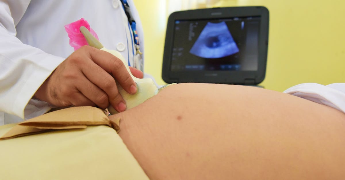
When clinical suspicion of an LGA fetus arises, obstetric ultrasonography is the standard diagnostic modality. The ultrasound technician takes multiple highly specific biometric measurements of the fetal anatomy.
These core measurements include the biparietal diameter (the width of the fetal head), the head circumference, the femur length (the length of the thigh bone), and crucially, the abdominal circumference.
These biometric values are inputted into a standardized mathematical algorithm that calculates the Estimated Fetal Weight (EFW). By plotting this calculated weight on a standardized growth curve, the obstetrician determines the exact growth percentile, confirming whether the fetus meets the criteria for being large for its specific gestational age.
9. Accuracy and Limitations of Fetal Weight Estimation
While ultrasonography is the most advanced tool available, parents must understand its significant inherent limitations. Ultrasound provides an estimate, not a definitive weight. The mathematical algorithms rely heavily on the measurement of the fetal abdominal circumference, which can be highly variable.
The accuracy of ultrasound estimation decreases significantly in the third trimester as the fetus becomes crowded within the uterus, making precise measurements difficult. Furthermore, the margin of error increases substantially at the extremes of fetal weight.
An estimated fetal weight near term can carry a margin of error of plus or minus 15 percent. This means an ultrasound predicting a 4,500-gram fetus could represent a baby weighing anywhere from 3,825 grams to 5,175 grams. This significant margin of error forces obstetricians to make critical delivery decisions based on imperfect data.
10. Data Summary: Risk Factors and Clinical Implications
Understanding the distinct maternal risk factors helps the obstetric team anticipate complications and prepare appropriate delivery resources.
| Primary Risk Factor | Underlying Mechanism | Clinical Implication for Fetus |
|---|---|---|
| Poorly Controlled Diabetes | Fetal hyperinsulinemia drives fat storage | Asymmetrical growth, high risk of shoulder dystocia |
| Maternal Obesity | Altered lipid/amino acid metabolism | Overall macrosomia, increased risk of prolonged labor |
| Previous Macrosomic Infant | Genetic/constitutional predisposition | High probability of recurrence in subsequent pregnancies |
| Post-Term Gestation | Continued placental nutrient transfer | Progressive weight gain beyond safe vaginal delivery limits |
11. Maternal Risks During Labor
Vaginal delivery of a macrosomic infant significantly elevates the risk of severe complications for the mother. The sheer physical size of the fetus often leads to a prolonged or arrested active phase of labor, as the fetal head and torso struggle to descend through the maternal pelvis.
This failure to progress frequently results in a high rate of emergency operative interventions, including the application of vacuum extractors or delivery forceps, or an urgent transition to a cesarean section after hours of exhausting labor.
If a vaginal delivery is completed, the mechanical stretching required to accommodate the oversized infant dramatically increases the risk of severe perineal lacerations. These lacerations can tear deeply through the vaginal wall and the anal sphincter, requiring complex surgical repair and leading to long-term issues with pelvic floor dysfunction and incontinence.
12. Fetal and Neonatal Risks During Delivery
The risks to the LGA fetus during a prolonged vaginal delivery are substantial. The intense mechanical compression against the maternal pelvic bones can result in a higher incidence of fetal skull fractures or intracranial hemorrhage.
Furthermore, the prolonged stress of an obstructed labor can compromise blood flow through the umbilical cord, leading to fetal distress, characterized by a dropping heart rate and potential hypoxic injury.
Because of these severe mechanical and physiological stressors, infants born macrosomic have a statistically higher rate of requiring immediate admission to a Neonatal Intensive Care Unit (NICU) for respiratory support and neurological monitoring following a traumatic birth.
13. The Risk of Shoulder Dystocia
The most feared and catastrophic complication associated with fetal macrosomia is shoulder dystocia. This constitutes a severe obstetric emergency. It occurs during a vaginal delivery when the fetal head successfully passes through the birth canal, but the broad, oversized fetal shoulders become wedged tightly behind the maternal pubic bone.
The infant becomes physically trapped, unable to breathe, and the umbilical cord is frequently compressed, rapidly cutting off oxygen. The delivering physician must execute a rapid sequence of highly specific physical maneuvers—manipulating the mother’s legs and applying precise suprapubic pressure—to dislodge the shoulders within a matter of minutes to prevent severe brain damage or death.
Even when these maneuvers are executed perfectly, the immense traction forces required can permanently damage the infant’s brachial plexus nerve network (Erb’s palsy), resulting in permanent paralysis of the arm, or cause fractures of the fetal clavicle or humerus.
14. Pre-Labor Management and Glucose Control
When a fetus is identified as tracking large for gestational age early in the third trimester, aggressive medical management is initiated to normalize the growth curve. If the mother has pre-existing or gestational diabetes, the absolute priority is achieving rigid, meticulous control of maternal blood glucose levels.
This requires rigorous dietary modifications, overseen by a registered dietician, to eliminate simple carbohydrates and manage portion sizes. The mother must test her blood glucose multiple times a day. If diet and exercise fail to maintain strict glycemic targets, the immediate initiation of insulin therapy or oral hypoglycemic agents is mandatory to halt the fetal hyperinsulinemia driving the accelerated growth.
Mothers are also counseled regarding healthy activity levels to mitigate excessive gestational weight gain, closely monitoring total caloric intake while ensuring adequate nutrition for fetal development.
15. Obstetric Delivery Planning
Delivery planning for a suspected macrosomic fetus requires a careful risk-benefit analysis, shared between the obstetrician and the parents. If the estimated fetal weight approaches dangerous thresholds—typically >4,500 grams in a diabetic mother, or >5,000 grams in a non-diabetic mother—a planned, scheduled cesarean delivery is strongly recommended to eliminate the risk of shoulder dystocia and severe perineal trauma.
Elective induction of labor prior to 39 weeks strictly to prevent macrosomia is generally not recommended, as the risk of neonatal respiratory complications from prematurity outweighs the benefits.
However, if the mother reaches 39 weeks of gestation and the cervix is favorable, induction of labor may be offered to prevent the fetus from continuing to grow into the high-risk, post-term macrosomic categories. If you are experiencing sudden, severe lower abdominal cramping during late pregnancy, understanding the mechanics of pelvic pain is vital for differentiating normal labor progress from potential mechanical obstruction.
16. Neonatal Care Post-Delivery
The medical management of an LGA infant does not end with delivery. These oversized neonates require vigilant pediatric surveillance during the first forty-eight hours of life.
Because they have been exposed to high maternal glucose levels, their own pancreas is accustomed to producing massive amounts of insulin. Once the umbilical cord is cut, the maternal glucose supply abruptly ends, but the neonate’s insulin levels remain dangerously high. This frequently causes severe neonatal hypoglycemia (low blood sugar), requiring immediate, frequent blood glucose testing and often intravenous glucose infusions to prevent neurological seizures.
Additionally, macrosomic infants have a higher incidence of polycythemia (an abnormally high red blood cell count), which makes their blood thick and sluggish, leading to a higher risk of severe newborn jaundice requiring prolonged phototherapy.
17. When to Consult a High-Risk Obstetrician
Pregnant individuals should seek consultation with a Maternal-Fetal Medicine specialist (a high-risk obstetrician) if their fundal height consistently measures significantly ahead of schedule, or if an ultrasound reveals an estimated fetal weight above the 90th percentile.
Specialist consultation is absolutely mandatory if a patient has poorly controlled pre-existing diabetes, requires high doses of insulin to manage gestational diabetes, or has a documented history of delivering a previous infant that suffered from shoulder dystocia or a brachial plexus injury.
Collaborating with a high-risk specialist ensures that the patient receives the most advanced serial ultrasound monitoring and that a highly coordinated, multidisciplinary delivery plan is established well before the onset of active labor.
18. Frequently Asked Questions (FAQ)
1. Does eating too much sugar cause a large baby?
If you have gestational or pre-existing diabetes, yes. Excess sugar in your blood crosses the placenta and forces the baby to produce insulin, which acts as a powerful growth hormone, causing the baby to store excess fat.
2. Can an ultrasound guarantee exactly how much my baby weighs?
No. Ultrasounds provide an estimate based on bone and abdominal measurements. In the third trimester, this estimate has a significant margin of error and can be off by more than a pound in either direction.
3. If my baby is measuring large, will I be forced to have a C-section?
You are never forced, but a C-section will be strongly recommended if the baby’s estimated weight exceeds specific safety thresholds (usually over 4,500g for diabetic mothers, or 5,000g for non-diabetic mothers) to prevent catastrophic trauma during delivery.
4. Can I safely diet to stop my baby from growing too big?
You should never aggressively restrict calories or diet to lose weight during pregnancy without strict medical supervision, as this can starve the baby of essential nutrients. Management focuses on controlling blood sugar and slowing the rate of weight gain, not weight loss.
5. Are big babies healthier?
Not necessarily. While a healthy weight is good, macrosomic babies frequently face severe complications after birth, including dangerous drops in their own blood sugar, severe jaundice, and breathing difficulties requiring NICU admission.
19. Bibliography
Disclaimer: The content is for informational purposes only and does not replace medical advice. Always consult your doctor for personalized treatment.

