1. Introduction to Nail Pathology
Nail problems encompass a wide spectrum of disorders affecting the structural integrity, color, and growth of the nails on the fingers and toes. Far from being merely cosmetic concerns, abnormalities of the nail unit frequently serve as critical clinical indicators of underlying dermatological diseases, systemic conditions, or nutritional deficiencies.
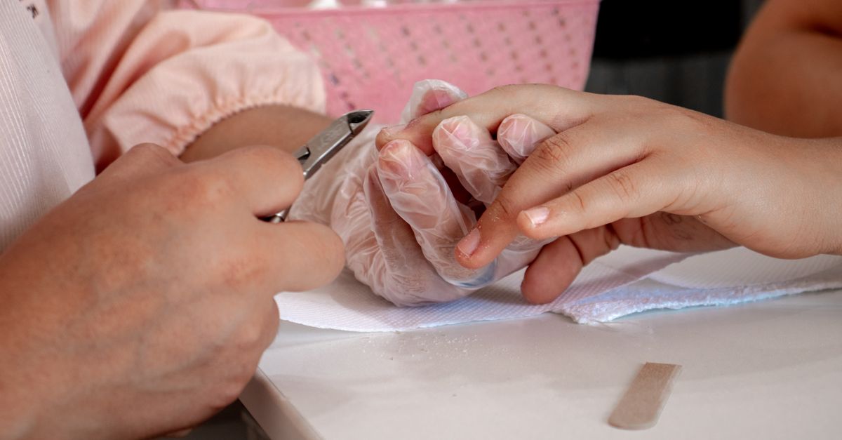
The nails are complex appendages of the skin composed primarily of hard keratin. They serve to protect the sensitive distal phalanges, enhance tactile sensation, and facilitate fine motor tasks. Because the nail unit grows continuously, any physiological disruption—whether from acute trauma, chronic infection, or systemic illness—leaves a visible, historical record on the nail plate.
A meticulous clinical evaluation of the nails is an essential component of a comprehensive physical examination. Proper diagnosis requires differentiating between benign mechanical injuries, localized fungal or bacterial infections, immune-mediated inflammatory conditions, and potentially life-threatening malignancies such as subungual melanoma.
2. Anatomy of the Nail Unit
To accurately diagnose nail pathology, one must understand the anatomy of the nail unit. The visible, hard portion of the nail is the nail plate. It rests upon the highly vascularized nail bed, which provides the plate with nutrients and gives healthy nails their pinkish hue.
The growth of the nail occurs primarily in the nail matrix, a specialized tissue located beneath the skin at the base of the nail. The white, crescent-shaped area visible at the base of some nails, known as the lunula, represents the distal portion of the nail matrix. Any injury or inflammation affecting the matrix will result in defective formation of the nail plate.
The surrounding skin structures are equally important. The proximal nail fold covers the matrix, while the cuticle (eponychium) forms a watertight seal between the nail plate and the proximal fold, protecting the matrix from environmental pathogens. The lateral nail folds support the sides of the nail plate. Disruption of these anatomical barriers directly leads to localized infections and chronic nail dystrophy.
3. Fungal Infections and Onychomycosis
Onychomycosis is the most prevalent disease of the nail unit, accounting for approximately half of all nail abnormalities. It is a fungal infection primarily caused by dermatophytes, particularly Trichophyton rubrum, though yeasts and non-dermatophyte molds can also be implicated. The condition predominantly affects the toenails due to the dark, warm, and moist environment provided by footwear.
Clinical presentation typically involves thickening, yellow-brown discoloration, and brittleness of the nail plate. The fungus frequently invades the nail bed, causing subungual hyperkeratosis (accumulation of debris under the nail) and onycholysis (separation of the nail plate from the bed). Without intervention, the infection can destroy the entire nail and spread to surrounding skin (tinea pedis).
Because structural nail changes from trauma or psoriasis can mimic onychomycosis, clinical diagnosis must be confirmed by laboratory testing before initiating systemic antifungal therapy, which carries the risk of hepatotoxicity.
4. Inflammatory Disorders of the Nail
Several systemic inflammatory and autoimmune skin conditions profoundly affect the nail unit. Nail psoriasis is highly common, affecting up to eighty percent of patients with psoriatic skin disease. Classic signs include irregular pitting of the nail plate, localized onycholysis surrounded by an erythematous border, and yellowish-brown discolorations known as “oil drop” or “salmon” patches on the nail bed.
Lichen planus is another inflammatory condition that can severely damage the nail matrix. It presents with longitudinal ridging, thinning of the nail plate, and the formation of a pterygium—a V-shaped extension of the proximal nail fold that fuses with the nail bed, eventually leading to permanent scarring and total loss of the nail.
Alopecia areata, an autoimmune disease that primarily causes hair loss, can also manifest with fine, geometric pitting of the nail plate or a roughened, sandpaper-like appearance termed trachyonychia. These inflammatory conditions require targeted immunomodulatory treatments directed at the underlying systemic disease process.
5. Traumatic and Mechanical Nail Injuries
The nail unit is highly susceptible to physical trauma. Acute injuries, such as crushing a finger in a door, frequently result in a subungual hematoma. This presents as a painful, dark red or black discoloration beneath the nail plate caused by localized bleeding. If the pressure is severe, the nail may eventually detach.
Chronic mechanical micro-trauma, often from ill-fitting shoes or repetitive athletic activities, leads to traumatic onycholysis, where the nail repeatedly lifts from the bed. This creates a subungual space that is highly prone to secondary bacterial colonization, particularly by Pseudomonas aeruginosa, which imparts a striking green-black discoloration to the nail.
Onychocryptosis, commonly known as an ingrown toenail, occurs when the sharp edge of the nail plate penetrates the lateral nail fold. This initiates a severe foreign-body inflammatory response, characterized by pain, erythema, swelling, and the formation of hypergranulation tissue. Ingrown nails are frequently exacerbated by improper nail cutting techniques and tight footwear.
6. Nail Manifestations of Systemic Disease
The nails provide invaluable diagnostic clues regarding systemic health. Clubbing of the nails is a classic sign characterized by an increased curvature of the nail plate and bulbous enlargement of the distal fingertip. While occasionally hereditary, acquired clubbing is strongly associated with chronic hypoxia resulting from pulmonary diseases (such as lung cancer or cystic fibrosis) or severe cardiovascular disorders.
Splinter hemorrhages are fine, longitudinal dark red lines beneath the nail plate, representing microscopic bleeding from the nail bed capillaries. While they can result from simple trauma, multiple splinter hemorrhages are a traditional hallmark of infectious endocarditis, requiring immediate cardiovascular evaluation.
Beau’s lines are deep, transverse grooves that run across the nail plate. They represent a temporary cessation of nail matrix activity caused by a severe systemic insult, such as a high fever, severe psychological stress, or the administration of cytotoxic chemotherapy. The distance of the groove from the cuticle allows clinicians to estimate when the systemic illness occurred.
7. Nutritional Deficiencies and Nail Changes
Proper nutrition is vital for the synthesis of hard keratin. Deficiencies in specific vitamins, minerals, and proteins manifest rapidly in the nail unit. Koilonychia, or “spoon nails,” is a condition where the nail plate becomes thin, flat, and eventually concave, capable of holding a drop of water. This is a classic, specific clinical sign of severe, chronic iron deficiency anemia.
Leukonychia refers to a white discoloration of the nails. While minor white spots (leukonychia punctata) are harmless and typically result from minor matrix trauma, widespread white banding can indicate severe hypoalbuminemia (low protein levels) associated with advanced liver cirrhosis or nephrotic syndrome.
Deficiencies in biotin (Vitamin B7), zinc, and essential fatty acids contribute to generalized onychoschizia, which is the splitting, peeling, and excessive brittleness of the distal nail plate. Restoring the specific nutritional deficit through diet or supplementation usually resolves these functional and structural abnormalities over several months as new nail tissue grows.
8. Pigmented Lesions and Subungual Melanoma
The appearance of a new, dark pigmented band running longitudinally along the nail (melanonychia) requires rigorous clinical evaluation. While longitudinal melanonychia is frequently benign, particularly in individuals with darker skin phototypes, it is also the primary clinical presentation of subungual melanoma, a rare but highly aggressive form of skin cancer.
Clinical red flags for subungual melanoma include a band that is rapidly widening, irregular in color, or developing blurred borders. The Hutchinson sign—where the dark pigment extends from the nail plate into the skin of the proximal or lateral nail folds—is highly suspicious for melanoma and necessitates urgent dermatological assessment.
In addition to melanoma, benign nevi (moles), lentigines, and friction-induced melanocytic activation can cause dark bands. Because distinguishing benign from malignant lesions is clinically challenging, an early matrix biopsy is strictly indicated for any suspicious pigmented lesion to ensure timely oncological intervention if necessary.
9. Clinical Evaluation and Dermoscopy
The diagnostic evaluation of a nail disorder begins with a comprehensive physical examination. The clinician observes the color, thickness, curvature, and surface texture of all twenty nails. The examination includes assessing the surrounding skin, hair, and mucous membranes for signs of concurrent inflammatory or systemic diseases.
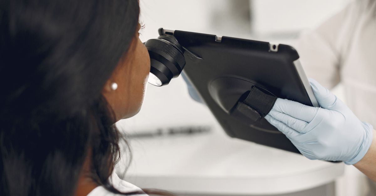
Onychoscopy (dermoscopy of the nail) has become an indispensable tool in modern dermatological practice. Using a handheld, illuminated magnification device, the clinician can visualize micro-structures within the nail plate and nail bed that are invisible to the naked eye.
Onychoscopy is particularly valuable in differentiating the source of nail pigmentation. It easily distinguishes the purple-red, clotted appearance of a subungual hematoma from the structured, parallel, grey-brown lines of a benign melanocytic nevus, or the irregular, disorganized patterns highly characteristic of a subungual melanoma, thereby preventing unnecessary surgical biopsies.
10. Diagnostic Laboratory Testing
To secure a definitive diagnosis, particularly for infectious etiologies, laboratory testing is mandatory. For suspected onychomycosis, clippings of the affected nail plate and subungual debris are collected. A Potassium Hydroxide (KOH) preparation allows the clinician to quickly visualize fungal hyphae under the microscope in the office.
For definitive identification of the pathogen, the sample is sent for fungal culture or Polymerase Chain Reaction (PCR) testing. PCR has largely revolutionized nail diagnostics, providing highly sensitive and rapid identification of specific dermatophyte DNA within a few days, compared to the several weeks required for traditional fungal cultures.
If an autoimmune or systemic disease is suspected, targeted blood tests are ordered. This may include a complete blood count and ferritin levels to investigate koilonychia, thyroid function tests, or specific autoantibody panels to correlate nail findings with systemic rheumatological conditions.
11. Data: Common Nail Abnormalities
The following table categorizes frequent nail abnormalities, their distinct clinical appearances, and their most common underlying medical etiologies.
| Clinical Sign | Appearance | Common Associated Conditions |
|---|---|---|
| Onycholysis | Painless separation of the nail plate from the nail bed. | Trauma, psoriasis, fungal infection, thyroid disease. |
| Koilonychia | Thin, concave, spoon-shaped nails. | Severe iron deficiency anemia, hemochromatosis. |
| Beau’s Lines | Deep, transverse horizontal grooves across the nail. | Severe systemic illness, chemotherapy, high fever. |
| Nail Pitting | Small, ice-pick-like depressions on the nail surface. | Nail psoriasis, alopecia areata, eczema. |
12. Topical and Systemic Pharmacotherapy
Treatment strategies for nail problems depend entirely on the precise etiology. For confirmed onychomycosis, topical antifungal lacquers (such as ciclopirox or efinaconazole) are generally effective only for mild, superficial infections. Moderate to severe infections, particularly those involving the matrix, mandate oral systemic therapy with agents like terbinafine or itraconazole. Treatment is lengthy, often requiring three to six months of continuous medication.
Inflammatory conditions like nail psoriasis are notoriously challenging to treat due to the poor penetration of topical agents through the hard nail plate. High-potency topical corticosteroids or calcipotriol are utilized, often applied under occlusion. In severe, refractory cases, intralesional steroid injections directly into the nail matrix or systemic biologic therapies are required to halt the inflammatory destruction.
Bacterial infections, such as acute paronychia (infection of the nail fold), are managed with warm compresses, topical antibiotics, and occasionally oral antibiotics if there is surrounding cellulitis. If an abscess has formed, mechanical drainage is curative.
13. Surgical Interventions for the Nail Unit
When pharmacological therapies fail or anatomical correction is required, surgical intervention is indicated. For recurrent, severe ingrown toenails (onychocryptosis), a partial nail avulsion combined with chemical matrixectomy is the definitive treatment. This involves surgically removing the offending border of the nail and applying phenol to the lateral matrix, permanently destroying the tissue to prevent regrowth of the problematic edge.
In cases of extensive, painful fungal infections that are refractory to oral medications, complete surgical avulsion (removal) of the nail plate may be performed. This allows direct application of antifungal agents to the infected nail bed while a healthy new nail slowly regenerates over several months.
Surgical biopsy of the nail matrix or nail bed is a delicate procedure requiring specialized training. It is absolutely indicated for evaluating suspicious pigmented longitudinal bands to rule out melanoma. The surgeon must carefully extract the tissue while minimizing permanent scarring or dystrophy to the future nail plate.
14. When to See a Doctor
While minor nail injuries or localized peeling frequently resolve independently as the nail grows out, persistent or unusual changes demand professional medical evaluation. Patients should consult a dermatologist if they notice thickening, yellowing, or crumbling of the nail plate that does not improve with standard foot hygiene, as early intervention prevents the spread of fungal infections.
Immediate clinical assessment is vital if a patient discovers a new, dark longitudinal stripe on the nail, especially if it affects a single digit, is widening, or involves the surrounding skin. This is the primary presentation of subungual melanoma, and early diagnosis is critical for a favorable outcome.
Furthermore, systemic signs such as severe clubbing of the fingertips, widespread concavity (spooning), or recurrent splinter hemorrhages without a history of trauma warrant an urgent visit to a primary care physician to rule out significant underlying cardiovascular, pulmonary, or hematological diseases.
15. Frequently Asked Questions (FAQ)
1. What causes thick, yellow toenails?
Thick, yellow, and brittle toenails are most commonly caused by a fungal infection called onychomycosis. However, psoriasis and chronic trauma from tight shoes can cause similar changes, so a laboratory test is necessary for an accurate diagnosis.
2. Are white spots on the nails a sign of a calcium deficiency?
No. The common, small white spots on the nails (leukonychia punctata) are typically the result of minor, everyday micro-trauma to the base of the nail. They are entirely harmless and are not related to calcium or vitamin deficiencies.
3. Why do my nails keep splitting and peeling?
Nail splitting (onychoschizia) is frequently caused by repeated cycles of wetting and drying the hands, exposure to harsh household chemicals, or aggressive use of nail polish removers. It can also indicate a deficiency in iron or biotin.
4. How is an ingrown toenail permanently fixed?
If an ingrown toenail recurs, a physician can perform a minor surgical procedure to remove the side of the nail and apply a chemical (phenol) to the root. This permanently stops that specific edge of the nail from growing back and digging into the skin.
5. Should I be worried about a dark line under my nail?
While many dark lines are benign moles or natural pigmentation, any new, expanding, or irregular dark stripe running down the nail should be evaluated by a dermatologist immediately to rule out subungual melanoma, a rare form of skin cancer.
Bibliography
Disclaimer: The content is for informational purposes only and does not replace medical advice. Always consult your doctor for personalized treatment.