1. Introduction
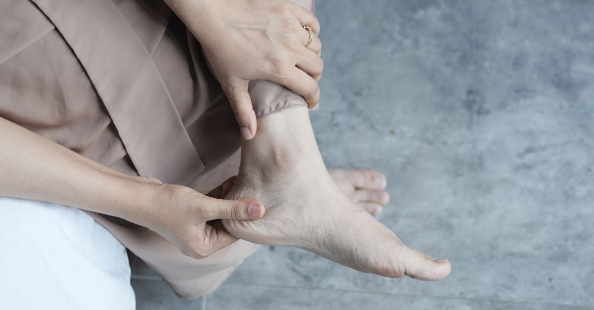
Sudden severe pain in the back of the heel usually indicates an acute injury to the Achilles tendon, such as a partial or complete rupture, or an acute flare-up of retrocalcaneal bursitis. This symptom occurs when the thick fibrous band connecting the calf muscle to the heel bone is subjected to abrupt mechanical stress, causing immediate tissue tearing and localized inflammation. The pain is typically localized precisely at the posterior aspect of the calcaneus and can radiate upward into the lower calf.
The human foot and ankle must absorb substantial mechanical forces during daily ambulation. The posterior heel serves as a critical junction where muscular power is translated into forward propulsion. When this biomechanical system fails due to fatigue, degenerative changes, or sudden overload, the resulting pain is sharp, immediate, and often debilitating.
Identifying the exact anatomical structure involved requires careful observation of the injury mechanism. Whether the pain began after a sudden sprint, a change in footwear, or developed following a minor stumble, tracing the onset provides crucial clinical clues to direct the appropriate rehabilitation strategy.
2. Anatomy of the Posterior Heel
The posterior aspect of the heel is dominated by the calcaneus, the largest bone in the foot. Attaching to the posterior surface of this bone is the Achilles tendon, the thickest and strongest tendon in the human body. This tendon serves as the confluence of the gastrocnemius and soleus muscles of the calf.
Situated between the Achilles tendon and the calcaneus is the retrocalcaneal bursa. A bursa is a small, fluid-filled sac designed to reduce friction between moving anatomical structures. Another bursa, the subcutaneous calcaneal bursa, lies just beneath the skin over the tendon attachment site.
These structures work in perfect synchronization to facilitate plantar flexion, the motion required to push off the ground during walking or running. Because these tissues operate under constant tension, any structural weakness or mechanical interference rapidly generates significant pain.
3. Achilles Tendon Rupture
The most urgent clinical cause of a sudden, sharp pain in the back of the heel is an Achilles tendon rupture. This injury frequently occurs during athletic activities that require sudden acceleration or abrupt changes in direction. Patients classically describe feeling as though they were struck or kicked in the back of the leg.
A rupture involves a complete or partial tearing of the tendon fibers. This mechanical failure is often accompanied by an audible popping or snapping sound. Following the tear, the patient typically experiences a profound loss of power in the affected leg and becomes unable to stand on their toes.
The pain is immediate and intense, followed rapidly by swelling and bruising around the heel and ankle. A visible or palpable gap may appear in the tendon just above the heel bone, signaling a complete structural disconnection that requires prompt orthopedic evaluation.
4. Acute Achilles Tendinopathy
Achilles tendinopathy encompasses both inflammation and microscopic degeneration of the tendon fibers. While often a chronic condition, it can present acutely if the tendon is suddenly overloaded. This frequently happens when an individual abruptly increases their exercise volume or intensity.
The sudden mechanical stress creates microtears in the collagen matrix of the tendon. The body initiates an inflammatory response to repair the damage, leading to localized swelling and severe pain. The pain is usually worst upon waking or when taking the first steps after a period of rest.
Unlike a complete rupture, the tendon remains intact, but its structural integrity is compromised. The area feels tender to the touch, and the patient may notice a grating sensation, known as crepitus, when moving the ankle.
5. Retrocalcaneal Bursitis
Retrocalcaneal bursitis involves the acute inflammation of the bursa located between the Achilles tendon and the heel bone. This condition is typically triggered by repetitive friction or direct pressure against the posterior heel.
When irritated, the bursa produces excess fluid, causing it to swell significantly. This swelling compresses the adjacent Achilles tendon and surrounding nerve fibers. The pain is characterized by a deep, throbbing ache directly at the back of the heel, which intensifies when the ankle is flexed upward.
This condition is frequently exacerbated by wearing rigid footwear that rubs against the heel. The skin over the back of the heel may appear red and feel warm to the touch, indicating active localized inflammation.
6. Haglunds Deformity and Mechanical Friction
Haglunds deformity is a bony enlargement on the back of the heel bone. This structural anomaly can be congenital or develop over time due to chronic irritation. The bony prominence creates a mechanical conflict with the Achilles tendon and the retrocalcaneal bursa.
When wearing shoes with a stiff back, the rigid material presses the soft tissues directly against the enlarged bone. This continuous friction triggers acute episodes of bursitis and tendinopathy, resulting in sudden, severe pain flares.
The condition is often colloquially referred to as a pump bump. Managing the pain requires identifying this specific anatomical variance, as standard treatments for tendon strain will fail if the underlying bony friction is not addressed.
7. Sever Disease in Adolescents
In children and young adolescents, sudden posterior heel pain is frequently caused by Sever disease, clinically termed calcaneal apophysitis. This condition involves the inflammation of the growth plate located at the back of the heel bone.
During periods of rapid skeletal growth, the heel bone may grow faster than the calf muscles and the Achilles tendon. This creates a state of continuous tension on the fragile growth plate. Physical activity, particularly running and jumping sports, places excessive traction on this area, leading to acute pain.
The pain is localized strictly to the back of the heel and worsens with weight-bearing activities. Squeezing the heel bone from both sides typically elicits a sharp pain response, confirming the diagnosis in the appropriate age group.
8. Differentiating Plantar Fasciitis
It is crucial to differentiate posterior heel pain from plantar fasciitis. While both conditions affect the heel, their anatomical locations and pain presentations differ significantly. Plantar fasciitis primarily causes pain on the bottom of the heel and along the arch of the foot.
The pain of plantar fasciitis is usually sharpest during the first steps in the morning but is located underneath the foot, where the thick fascial band attaches to the calcaneus. In contrast, Achilles-related pain is felt exclusively at the back of the heel and the lower calf.
Understanding this distinction is vital, as the stretching protocols and orthotic interventions for plantar fasciitis differ entirely from those required for posterior tendon injuries.
9. Inflammatory Arthropathies
In less common scenarios, sudden pain in the back of the heel serves as an early indicator of systemic inflammatory conditions. Ankylosing spondylitis and psoriatic arthritis belong to a group of diseases that frequently cause enthesitis, which is the inflammation of the site where a tendon attaches to a bone.
The Achilles tendon insertion is a classic site for enthesitis. The pain is severe, often bilateral, and typically accompanied by significant morning stiffness that improves gradually with movement.
If posterior heel pain occurs alongside chronic lower back pain, joint swelling, or skin rashes, a rheumatological evaluation is necessary to identify underlying systemic autoimmune activity.
10. Sural Nerve Entrapment
The sural nerve travels down the back of the calf and supplies sensation to the outer border of the heel and foot. If this nerve becomes compressed or irritated by surrounding scar tissue or tight footwear, it can produce a sudden, sharp pain in the posterior heel.
Nerve-related pain is distinctly different from musculoskeletal pain. It is frequently described as burning, electrical, or shooting in nature. The pain may be accompanied by a tingling sensation or numbness along the outer edge of the foot.
Tapping lightly along the course of the nerve behind the outer ankle bone may reproduce the shooting pain, a clinical sign that helps differentiate nerve entrapment from a primary tendon injury.
11. Risk Factors and Biomechanical Faults
Several intrinsic and extrinsic factors increase the risk of developing acute posterior heel pain. Extrinsic factors include abrupt changes in training surfaces, such as transitioning from grass to concrete, or wearing worn-out footwear that lacks adequate heel support.
Intrinsic factors involve individual biomechanics. Tight calf muscles constantly pull on the Achilles tendon, leaving it vulnerable to overload. Overpronation, where the foot rolls inward excessively during walking, places asymmetrical stress on the tendon fibers.
Addressing these underlying risk factors is essential to prevent recurrent episodes of pain. Modifying footwear and correcting biomechanical faults serve as fundamental components of long-term management.
12. Differential Diagnosis Table
A structured assessment aids in pinpointing the specific cause of posterior heel pain.
| Potential Diagnosis | Pain Characteristics | Key Clinical Signs |
|---|---|---|
| Achilles Tendon Rupture | Sudden, sharp tearing sensation. | Audible pop, inability to stand on toes. |
| Acute Tendinopathy | Aching pain, worse after rest. | Tendon thickening, morning stiffness. |
| Retrocalcaneal Bursitis | Deep throbbing at the heel bone. | Visible swelling, worsened by stiff shoes. |
| Sever Disease | Pain during sports in adolescents. | Tenderness when squeezing the heel sides. |
13. Clinical Diagnostic Evaluation
When a patient presents with sudden severe heel pain, clinical evaluation begins with a thorough physical examination. The physician will palpate the Achilles tendon to locate gaps, thickening, or specific points of tenderness.
A critical test for a suspected tendon rupture is the Thompson test. The patient lies face down with their feet hanging off the edge of the bed. The physician squeezes the calf muscle. If the tendon is intact, the foot will point downward. If the tendon is ruptured, the foot remains entirely motionless.
For further structural assessment, musculoskeletal ultrasound provides real-time, high-resolution imaging of the tendon and bursa, clearly identifying partial tears or fluid accumulations without the need for radiation.
14. Immediate First Aid and Triage
Critical Warning: If an Achilles tendon rupture is suspected due to a popping sound and sudden weakness, the patient must immediately cease weight-bearing and seek orthopedic care.
For acute, non-rupture injuries, immediate management follows the principles of rest, ice, compression, and elevation. Halting the aggravating activity prevents further tissue damage. Applying ice packs wrapped in a cloth for fifteen minutes at a time constricts local blood vessels, reducing acute swelling.
A slight heel lift placed inside the shoe can temporarily reduce tension on the Achilles tendon, providing immediate mechanical relief to the strained tissues. Over-the-counter nonsteroidal anti-inflammatory medications can assist in managing the acute pain and swelling phase.
15. Long-Term Rehabilitation Strategies
Rehabilitating posterior heel pain requires a structured and progressive approach. Once the acute inflammation subsides, physical therapy becomes the primary intervention. The focus shifts to restoring flexibility and building load capacity within the tendon.
Eccentric strengthening exercises, which involve lengthening the muscle under tension, are the gold standard for treating Achilles tendinopathy. These exercises stimulate collagen remodeling, aligning the new tissue fibers correctly to withstand future mechanical forces.
Patients may also benefit from customized orthotics to correct overpronation, thereby ensuring that forces are distributed evenly across the foot and ankle complex during movement. Information on correct gait mechanics is often explored in resources regarding gait abnormalities.
16. Frequently Asked Questions (FAQ)
1. How do I know if I tore my Achilles tendon or just strained it?
A complete tear usually involves a sudden popping sound, a feeling like you were kicked in the heel, and a complete inability to push off your toes when walking. A strain causes severe aching and stiffness but maintains basic foot function.
2. Can my shoes cause sudden pain in the back of my heel?
Yes, shoes with a rigid, tight heel counter can cause severe friction against the heel bone. This mechanical rubbing rapidly inflames the bursa sac and the Achilles tendon, leading to sudden and intense pain.
3. Is it safe to walk through the pain to stretch the tendon?
You should never force yourself to walk through sharp, sudden heel pain. This can transform a minor microtear into a complete tendon rupture. You must rest the area and seek evaluation before beginning any stretching protocol.
4. Why does the back of my heel hurt most in the morning?
During sleep, your feet relax in a pointed position, causing the calf muscles and Achilles tendon to shorten. When you stand up, the sudden stretching of this tight, inflamed tissue causes severe pain until it warms up.
5. When should I see a doctor for heel pain?
Seek immediate medical attention if you heard a pop, cannot bear weight on the foot, or cannot bend your foot downward. For aching pain, consult a physician if the symptoms do not improve after a week of rest.
17. Bibliography
Disclaimer: The content is for informational purposes only and does not replace medical advice. Always consult your doctor for personalized treatment.