1. Introduction
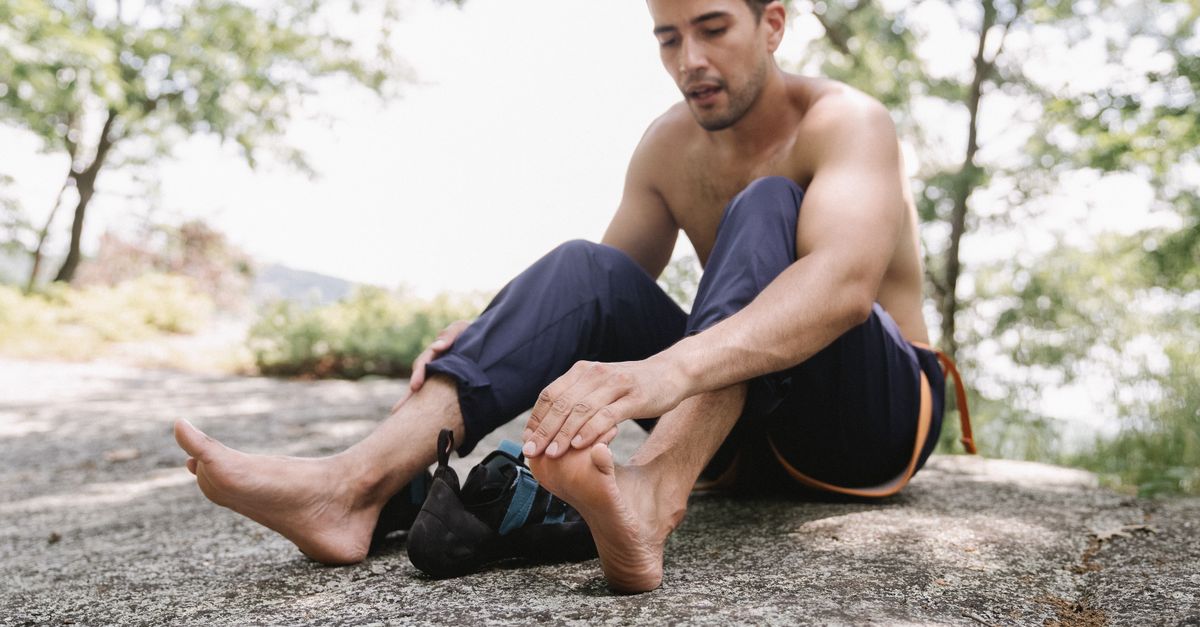
Relieving acute morning heel pain caused by plantar fasciitis requires performing sustained calf and fascial stretches strictly before bearing any weight, utilizing supportive footwear immediately upon rising, and applying localized cold therapy. This intense, stabbing pain results from the microscopic tearing and severe inflammation of the plantar fascia, a thick, fibrous band of connective tissue on the sole of the foot that inherently tightens and contracts during overnight rest. The primary clinical objective is to mechanically elongate this contracted tissue prior to subjecting it to the massive compressive load of standing.
Plantar fasciitis is the most prevalent cause of inferior heel pain worldwide, afflicting individuals across a broad spectrum of age and activity levels. The defining hallmark of this condition is the exquisite, sharp pain experienced during the very first steps out of bed in the morning, which typically lessens into a dull ache as the foot warms up and the tissue slowly stretches out during the day.
Clinical management of plantar fasciitis eschews temporary chemical painkillers in favor of structural realignment and biomechanical rehabilitation. By understanding the mechanical linkage between the calf muscles, the Achilles tendon, and the plantar fascia, patients can deploy targeted physical interventions that halt the daily cycle of micro-tearing, allowing the dense connective tissue the requisite time to heal and remodel permanently.
2. Anatomy of the Plantar Fascia
The plantar fascia is an incredibly dense, broad band of inelastic connective tissue that runs along the bottom of the foot. It originates at the medial tubercle of the calcaneus, the large heel bone, and fans out forward to attach to the base of the toes.
This structure is a critical biomechanical component of the human gait cycle. It functions much like the string of an archer’s bow, acting as a biological tie-rod that maintains the structural integrity of the medial longitudinal arch of the foot. During walking or running, as the body’s full weight comes down on the foot, the arch attempts to flatten.
The plantar fascia fiercely resists this flattening, absorbing immense shock and storing kinetic energy. As the foot pushes off the ground, the fascia recoils, providing a powerful, energy-efficient spring to propel the body forward. Because it bears the entire weight of the body under immense tension, the origin point at the heel is exceptionally vulnerable to mechanical stress.
3. Pathophysiology of Fascial Degeneration
Historically, plantar fasciitis was believed to be a primary inflammatory condition, hence the “-itis” suffix. However, modern histological examinations of surgical tissue samples reveal a completely different pathology. The condition is more accurately classified as fasciosis, representing chronic, non-inflammatory degeneration of the collagen fibers.
Continuous mechanical overloading causes microscopic tears in the fascial tissue right at its insertion point on the heel bone. In a healthy scenario, the body repairs these micro-tears during rest. However, if the mechanical stress is relentless, the rate of damage exceeds the rate of cellular repair.
The tissue becomes structurally disorganized, thickened, and weakened. The local blood supply, which is naturally poor in dense connective tissue, becomes further compromised. This chronic state of structural breakdown and failed healing creates an incredibly sensitive, painful lesion at the base of the heel.
4. The Morning Step Phenomenon
The agonizing pain of the first morning step is the defining clinical feature of this disorder. During sleep, the body naturally rests with the feet pointed slightly downward, a position known as plantar flexion. In this relaxed posture, all tension is removed from the sole of the foot.
Without tension, the damaged plantar fascia attempts to heal by knitting together the microscopic tears. However, because the foot is pointed down, the fascia heals in a shortened, contracted state. Over eight hours of sleep, the tissue becomes stiff and tightly bound.
When the patient wakes up, swings their legs out of bed, and places their full body weight onto the floor, the foot is violently forced into a ninety-degree angle. This massive, sudden mechanical load instantly rips apart the fragile healing tissue that formed overnight. This acute, mechanical tearing of the fascia is what generates the blinding, sharp pain of the first step.
5. Pre-Weight-Bearing Stretching Protocols
To break the cycle of morning tearing, the contracted fascia must be gently and systematically elongated before it is subjected to the weight of the body. The patient must not allow their bare feet to touch the floor until specific stretches have been executed directly in bed.
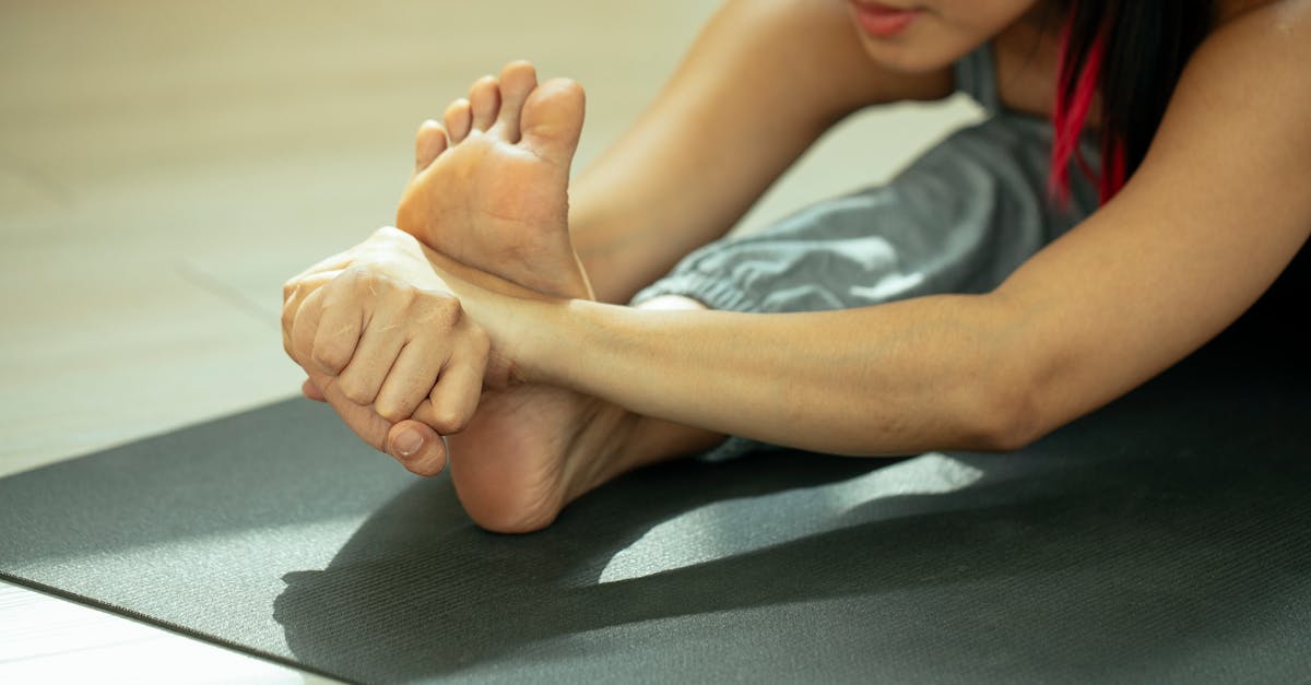
The most effective maneuver is the seated fascial stretch. While sitting on the edge of the bed, the patient crosses the affected leg over the opposite knee. Grasping the base of the toes, the patient slowly pulls the toes backward toward the shin until a firm stretch is felt in the arch of the foot.
Using the opposite hand, the patient should massage the tight band of the fascia while holding the stretch for thirty to sixty seconds. This gentle, sustained tension safely elongates the collagen fibers, preparing them to accept a mechanical load without tearing.
6. The Achilles Tendon Linkage
The plantar fascia does not exist in anatomical isolation; it is directly linked to the massive muscles of the posterior leg. The gastrocnemius and soleus muscles of the calf merge to form the Achilles tendon, which wraps around the back of the heel bone. Dense fascial connections continue under the heel to seamlessly merge with the plantar fascia.
If a patient possesses chronically tight calf muscles or a restricted Achilles tendon, this tension is physically transmitted directly around the heel and into the plantar fascia. A tight calf acts as an unrelenting winch, pulling the fascia taut even while at rest.
Therefore, aggressive rehabilitation of the calf muscles is mandatory for resolving heel pain. Standing wall stretches, where the patient leans forward with a straight back leg and the heel planted firmly on the ground, must be performed multiple times a day to permanently lengthen the posterior kinetic chain and reduce the strain on the bottom of the foot.
7. Immediate Morning Footwear
Walking barefoot is strictly contraindicated for any patient suffering from active plantar fasciitis. Barefoot walking completely removes external support, forcing the inflamed fascia to bear the entirety of the body’s biomechanical shock and arch-flattening forces.
To protect the vulnerable tissue, the patient must place supportive footwear directly next to the bed. The very first step taken after performing the bed stretches must be directly into a rigid, supportive sandal or a structured athletic shoe.
The footwear must possess a firm heel counter and substantial arch support to prevent the arch from collapsing under weight. By mechanically supporting the arch from the outside, the shoe prevents the fascial bowstring from over-stretching, allowing the patient to walk to the bathroom or kitchen without reinjuring the tissue.
8. Cryotherapy and Inflammation Control
By the end of the day, the continuous mechanical stress of walking inevitably causes the damaged tissue to become acutely inflamed and throbbing. Cold therapy is the optimal physical intervention to manage this end-of-day inflammatory spike.
A highly effective clinical technique is the frozen water bottle roll. The patient freezes a standard plastic water bottle, sits in a supportive chair, and places the frozen bottle under the arch of the affected foot. Applying moderate downward pressure, the patient slowly rolls the bottle back and forth from the heel to the base of the toes.
This dual-action therapy provides deep, localized vasoconstriction to flush out inflammatory enzymes and numb the pain receptors, while simultaneously providing a rigorous, mechanical tissue massage that breaks up fascial adhesions and promotes proper collagen alignment.
9. Orthotics and Biomechanical Correction
Many cases of plantar fasciitis are driven by inherent biomechanical faults in the patient’s foot architecture. Individuals with severe pes planus, commonly known as flat feet, inherently over-stretch the fascia because their arch collapses entirely with every step. Conversely, individuals with pes cavus, exceptionally high and rigid arches, possess a fascia that is too tight to absorb shock effectively.
Custom or high-quality over-the-counter orthotic inserts are utilized to mechanically correct these anatomical faults. A proper orthotic physically supports the medial arch and includes a deep heel cup to gather the natural fat pad under the heel, enhancing biological shock absorption.
By optimizing the distribution of weight across the entire surface area of the foot, the orthotic removes the focused, pinpoint stress from the fascial insertion at the heel, providing the structural stability necessary for long-term healing.
10. The Implementation of Night Splints
For patients whose morning pain remains severe despite pre-stretching and supportive footwear, a nocturnal splint is the next clinical intervention. A night splint is a rigid or semi-rigid brace worn while sleeping that physically holds the foot at a strict ninety-degree angle to the leg.
By preventing the foot from dropping into plantar flexion during the night, the splint maintains a gentle, continuous stretch on both the Achilles tendon and the plantar fascia for eight consecutive hours.
Because the tissue is forced to heal in an elongated, stretched position rather than a contracted one, the morning step phenomenon is almost entirely eliminated. The patient can stand up in the morning without instantly ripping the newly repaired collagen fibers.
11. Body Mass and Mechanical Load
The laws of physics dictate the severity of mechanical strain placed on the foot. The plantar fascia must support the entire mass of the human body. Any significant increase in body weight translates to an exponential increase in the sheer physical force applied to the arch of the foot with every single step.
A sudden gain in weight, or the physiological weight gain associated with the third trimester of pregnancy, is a frequent catalyst for the acute onset of plantar fasciitis. The fascial tissue simply fails under the new, unaccustomed load.
While weight loss is a long-term goal and not an immediate acute intervention, reducing total body mass removes the relentless, crushing mechanical force from the heel, heavily reducing the frequency of chronic flare-ups over the patient’s lifetime.
12. Corticosteroid Injections
When severe, blinding pain completely halts a patient’s daily functional mobility, and conservative physical measures have failed, localized pharmacological intervention is considered. A physician may inject a potent corticosteroid directly into the most tender point of the fascial insertion at the heel.
The corticosteroid acts as a massive chemical fire extinguisher, abruptly halting all localized inflammatory and immune responses. This provides profound, rapid pain relief, allowing the patient to participate in physical therapy and stretching programs without agony.
However, repeated corticosteroid injections are strictly avoided. Corticosteroids inherently weaken collagen fibers. Multiple injections dramatically increase the risk of a complete, catastrophic rupture of the plantar fascia, which permanently alters the biomechanics of the foot and causes profound, lifelong disability.
13. Extracorporeal Shockwave Therapy (ESWT)
For chronic, recalcitrant fasciosis that has persisted for more than six months, advanced regenerative therapies are utilized. Extracorporeal shockwave therapy is a non-invasive clinical modality that delivers high-energy acoustic sound waves directly into the fibrotic, damaged heel tissue.
These intense sound waves intentionally cause controlled micro-trauma to the stagnant, non-healing tissue. This acute trauma acts as a biological reset button. It triggers a massive influx of new blood vessels, a process called neovascularization, and recruits stem cells to the area.
By forcing the body to recognize the chronic injury as a new, acute wound, shockwave therapy actively restarts the stalled healing cascade, prompting the body to lay down fresh, structurally sound collagen fibers.
14. Differentiating Heel Spurs and Nerve Entrapment
A common clinical misconception is that heel pain is caused by a bone spur. Radiographs of patients with plantar fasciitis frequently reveal a sharp bony growth, or spur, on the bottom of the heel. However, clinical studies conclusively prove that the spur itself is rarely the source of the pain. The spur is simply a calcified response to the chronic pulling of the tight fascia. Surgical removal of the spur without addressing the fascial tension will not cure the pain.
Furthermore, a clinician must rule out tarsal tunnel syndrome, a condition where the posterior tibial nerve is physically compressed as it wraps around the inside of the ankle.
Nerve entrapment presents with burning, tingling, and electrical pain that radiates into the sole of the foot and the toes, frequently worsening at night or while resting, which is the exact opposite presentation of the mechanical, weight-bearing pain characteristic of true plantar fasciitis.
15. Surgical Fasciotomy Considerations
In the absolute rarest of cases, when all conservative, pharmacological, and regenerative therapies have failed completely after a minimum of twelve months of strict adherence, surgical intervention is cautiously considered.
A plantar fasciotomy involves a surgeon making a tiny incision and physically cutting a portion of the tight fascial band away from its attachment on the heel bone. This surgery instantly releases all structural tension on the tissue.
While highly effective at eliminating the tension pain, severing the biological tie-rod permanently flattens the arch of the foot and alters the entire biomechanical kinetic chain up into the knee and hip. Therefore, it is strictly considered a procedure of last resort.
| Intervention Modality | Physiological Mechanism of Action |
|---|---|
| Pre-Weight-Bearing Stretching | Elongates contracted tissue before mechanical loading prevents tearing. |
| Rigid Arch Support / Orthotics | Prevents arch collapse and removes pinpoint stress from the heel insertion. |
| Frozen Water Bottle Roll | Induces vasoconstriction to reduce pain and mechanically massages tissue. |
| Nocturnal Splinting | Holds the fascia in a stretched position overnight to heal elongated. |
16. Frequently Asked Questions
1. Is it okay to walk barefoot on soft carpet if I have plantar fasciitis?
No. Even on the softest carpet or sand, walking barefoot provides zero structural support to the arch of the foot. Your body weight will continuously force the arch to flatten, aggressively pulling and micro-tearing the inflamed tissue at the heel with every single step.
2. Why do my calves feel so tight when my heel is the part that hurts?
Your calf muscles, Achilles tendon, and plantar fascia are all one continuous mechanical chain. If your calf muscles are chronically tight, they act like a tightened winch, constantly pulling the plantar fascia taut around the bottom of your heel, causing severe friction and pain.
3. Does a heel spur cause the sharp pain?
In the vast majority of cases, no. The bone spur is a symptom of the tight fascia pulling on the bone over many years, causing calcium to build up. The pain originates from the torn, damaged soft tissue of the fascia itself, not the bony spur poking the tissue.
4. How long does it realistically take to cure plantar fasciitis?
Because the plantar fascia has very poor blood flow, it heals exceptionally slowly. Even with strict adherence to daily stretching, supportive footwear, and icing, it typically takes between six to twelve months for the tissue to completely remodel and the pain to resolve permanently.
5. Should I stop exercising completely while my foot heals?
You must stop all high-impact activities like running, jumping, or intense hiking, as these relentlessly pound the damaged tissue. However, you should aggressively transition to low-impact cardiovascular exercises like swimming or cycling, which maintain blood flow and fitness without crushing the heel.
17. Bibliography
Disclaimer: The content is for informational purposes only and does not replace medical advice. Always consult your doctor for personalized treatment.