1. Introduction
Experiencing terrible heel pain during the first steps of the morning is the hallmark clinical symptom of plantar fasciitis. This condition involves the inflammation, irritation, and degenerative microtearing of the plantar fascia, the thick, fibrous band of connective tissue that spans the arch of the foot, connecting the heel bone to the base of the toes. When a person steps out of bed, this injured, tightened tissue is suddenly subjected to the immense mechanical load of the entire body weight, resulting in an acute, sharp, stabbing pain that is universally recognized by clinicians as the defining indicator of this pathology.
The human foot is an engineering marvel designed to absorb shock and propel the body forward. The plantar fascia acts as a crucial biomechanical bowstring, maintaining the structural integrity of the foot’s arch. However, this dense tissue has a remarkably poor blood supply, which severely limits its ability to heal quickly from daily mechanical wear and tear. Prolonged standing, unsupportive footwear, rapid weight gain, or an abrupt increase in physical activity can overwhelm the tissue’s capacity for repair, leading to chronic structural degradation.
Understanding the specific mechanics of morning heel pain requires analyzing what happens to the foot during sleep. The pain is not simply an inflammatory flare-up; it is a direct consequence of nocturnal tissue shortening and abrupt morning elongation. By decoding this biomechanical cycle, individuals can implement targeted interventions to protect the plantar fascia during the night and safely mobilize the foot in the morning, successfully interrupting the cycle of chronic pain.
2. Anatomy of the Plantar Fascia
The plantar fascia is a remarkably dense, aponeurotic band of connective tissue situated on the sole of the foot. It originates from the medial tubercle of the calcaneus (the heel bone), fans out across the arch, and inserts into the ligaments at the base of the five toes.
This structure is integral to the windlass mechanism of the foot. When an individual takes a step and pushes off the ground, the toes bend upward (dorsiflexion). This action winds the plantar fascia tightly around the heads of the metatarsal bones, dynamically lifting the arch and transforming the foot into a rigid lever for efficient forward propulsion.
Because the plantar fascia is constantly stretched and tensioned with every single step, the insertion point at the heel endures immense mechanical stress. This focal point on the calcaneus is precisely where the microtrauma, cellular degeneration, and subsequent excruciating pain of plantar fasciitis occur.
3. Pathophysiology of Plantar Fasciitis
Despite its name ending in “-itis,” which implies acute inflammation, modern histological research demonstrates that plantar fasciitis is more accurately a degenerative condition, or a fasciosis. Chronic mechanical overload leads to repetitive microtears in the collagen fibers of the fascia at its heel attachment.
Because the tissue has poor vascularity, the body’s natural healing response is sluggish and inadequate. Instead of repairing the tears with strong, parallel collagen fibers, the body patches the area with disorganized, fibrotic scar tissue. This scar tissue is stiff, inelastic, and highly sensitive to stretch.
The localized environment becomes flooded with cellular waste and pain-mediating chemicals, leading to a state of chronic, low-grade irritation. The fascia loses its natural resilience, becoming a thick, brittle band that is highly susceptible to acute, stabbing pain whenever a significant mechanical load is applied.
4. The Mechanics of Morning Pain
The severe pain experienced during the first steps of the morning is a direct result of nighttime biomechanics. While a person sleeps, the feet naturally rest in a plantar-flexed position, meaning the toes point slightly downward.
In this relaxed, downward-pointing position, the plantar fascia is completely relieved of tension. Throughout the night, the body attempts to heal the daytime microtears while the tissue is in this shortened, contracted state. The fascia stiffens and essentially heals “short.”
When the alarm rings and the individual steps out of bed, placing their foot flat on the floor, the ankle is forced to ninety degrees, and the full weight of the body flattens the arch. This movement violently and abruptly stretches the stiff, shortened, newly scarred plantar fascia. This rapid, mechanical tearing of the unyielding tissue produces a blinding, sharp pain that can take the breath away.
5. The “Warm-Up” Phenomenon
A characteristic feature of plantar fasciitis is that the intense morning pain usually diminishes after several minutes of walking. This is known clinically as the warm-up phenomenon.
As the individual hobbles around for the first ten to fifteen minutes, the physical movement forces the stiffened plantar fascia to stretch out. The mechanical action also stimulates local blood flow to the sole of the foot, warming the tissue and increasing its pliability.
Once the fascia has stretched and adapted to the elongated position, the acute, stabbing pain subsides into a manageable, dull ache. However, the cycle resets itself if the individual sits at a desk or rests for a prolonged period during the day; the fascia cools and stiffens, and the sharp pain returns upon standing up again.
6. Impact of Foot Arch Biomechanics
The structural architecture of an individual’s foot heavily dictates their susceptibility to plantar fasciitis. Both biomechanical extremes—flat feet (pes planus) and unusually high arches (pes cavus)—place abnormal stress on the plantar fascia.
In individuals with flat feet, the arch collapses completely upon weight-bearing, a process called overpronation. This excessive inward rolling of the foot stretches the plantar fascia far beyond its normal physiological limit, creating severe tensile strain at the heel attachment with every step.
Conversely, individuals with very high, rigid arches possess a foot that is a poor shock absorber. When the heel strikes the ground, the rigid arch does not yield to absorb the kinetic energy. The shockwave is transmitted directly through the heel bone and the origin of the plantar fascia, causing repetitive concussive trauma to the tissue.
7. The Role of the Achilles Tendon and Calf Muscles
The biomechanics of the foot are inextricably linked to the lower leg. The Achilles tendon connects the massive calf muscles (the gastrocnemius and soleus) to the back of the heel bone, while the plantar fascia attaches to the bottom of the same bone.
If the calf muscles are chronically tight, they exert a constant, intense upward pull on the heel bone. This mechanical tension effectively anchors the heel, making it difficult for the ankle to flex normally during walking.
To compensate for this restricted ankle motion, the foot overpronates, and an enormous secondary tensile load is transferred directly to the plantar fascia. Clinicians find that the vast majority of patients suffering from severe plantar fasciitis possess significantly restricted calf and Achilles tendon flexibility.
8. Footwear and Mechanical Support
The external environment, primarily footwear, plays a massive role in the development and exacerbation of heel pain. Shoes with completely flat soles, zero arch support, and thin, unpadded heels offer no mechanical assistance to the plantar fascia.
Walking barefoot on hard surfaces like hardwood or tile floors removes all external shock absorption. The heel bone is forced to endure the full concussive force of the foot strike, which drives the origin of the plantar fascia directly into the rigid floor.
Conversely, footwear with a slight heel raise, a rigid midsole that resists twisting, and a contoured arch provides a mechanical bridge. This support bears a portion of the body weight and reduces the sheer physical tension placed on the fascia, allowing the irritated tissue a vital opportunity to rest and heal.
9. Heel Spurs and Structural Changes
Historically, severe heel pain was frequently attributed to the presence of a heel spur, a sharp, bony growth projecting from the bottom of the calcaneus. However, modern clinical consensus has completely shifted this paradigm.
A heel spur is actually a symptom of the mechanical problem, not the cause of the pain. The body grows the spur in response to the constant, relentless pulling of the tight plantar fascia on the heel bone, a process similar to how a callous forms from friction.
Many individuals have massive heel spurs visible on X-rays without experiencing any pain whatsoever, while others with excruciating plantar fasciitis have perfectly smooth heel bones. The sharp pain is generated by the tearing of the soft tissue (the fascia), not by the spur poking the flesh. Therefore, surgical removal of a spur is rarely the primary treatment objective.
10. Differentiating Heel Pain Causes
While plantar fasciitis is the most common cause of morning heel pain, clinicians must rule out other pathologies that present with similar discomfort.
| Condition | Characteristics of the Pain | Location and Triggers |
|---|---|---|
| Plantar Fasciitis | Sharp, stabbing, worst during first morning steps | Bottom of the heel, radiating into the arch |
| Achilles Tendinitis | Aching, burning stiffness in the back of the ankle | Back of the heel, worsens with running or jumping |
| Tarsal Tunnel Syndrome | Burning, tingling, or electrical shooting pain | Inside of the ankle and heel, worsens with prolonged standing |
| Calcaneal Stress Fracture | Deep, throbbing, unrelenting bone pain | Entire heel bone, hurts even when non-weight-bearing |
Pain that is accompanied by numbness or tingling points toward nerve entrapment, while pain that persists at rest requires evaluation for a potential stress fracture.
11. Clinical Physical Examination
The clinical diagnosis of plantar fasciitis is straightforward and highly reliable through physical examination. The physician will palpate the sole of the foot, applying direct pressure to the medial calcaneal tubercle, the exact spot where the fascia attaches to the inner aspect of the heel. Exquisite tenderness at this precise location confirms the diagnosis.
The physician will also perform the windlass test. They will passively pull the patient’s big toe upward (dorsiflexion) while stabilizing the ankle. This maneuver tightly stretches the plantar fascia across the arch. If this passive stretch reproduces the patient’s sharp heel pain, the fascia is confirmed as the source.
An assessment of ankle range of motion is crucial. The clinician evaluates calf flexibility; an inability to comfortably flex the foot upward past a neutral ninety-degree angle confirms that calf tightness is a primary mechanical driver of the fascial strain.
12. Diagnostic Imaging Capabilities
For classic morning heel pain that aligns perfectly with a plantar fasciitis presentation, imaging is not required for an initial diagnosis. However, if the pain does not respond to weeks of conservative therapy, or if the patient reports a sudden, audible “pop” in the arch, imaging becomes necessary.
Plain X-rays are utilized to rule out calcaneal stress fractures or bone tumors. While they will often reveal a heel spur, the physician will reassure the patient that the spur is an incidental finding.
Diagnostic ultrasound is highly effective for soft tissue evaluation. It allows the physician to measure the exact thickness of the plantar fascia. A fascia thicker than four millimeters, accompanied by dark areas indicating fluid or tissue degeneration, provides objective, visual confirmation of severe chronic fasciosis.
13. Initial Conservative Management
The immediate strategy for managing morning heel pain involves controlling the localized mechanical inflammation and protecting the tissue from further trauma. Rest from high-impact activities, such as running or aerobic jumping, is absolutely mandatory to prevent further tearing.
Ice therapy is a potent analgesic. Rolling a frozen water bottle under the arch of the foot for fifteen minutes every evening reduces the daily accumulation of swelling and provides deep tissue massage. Over-the-counter anti-inflammatory medications offer temporary relief but do not cure the underlying mechanical issue.
The absolute golden rule of plantar fasciitis recovery is avoiding barefoot walking. Patients must wear supportive, highly cushioned footwear from the moment their feet hit the floor in the morning until they return to bed at night. Eliminating the barefoot impact drastically accelerates the healing timeline.
14. Physical Therapy and Stretching Protocols
Fixing the biomechanical flaws requires dedicated physical therapy. The most vital intervention is reversing the tension from the calf muscles. A strict regimen of calf stretching, performed multiple times a day using a wall or a towel, introduces slack into the system, taking the heavy pulling force off the heel bone.
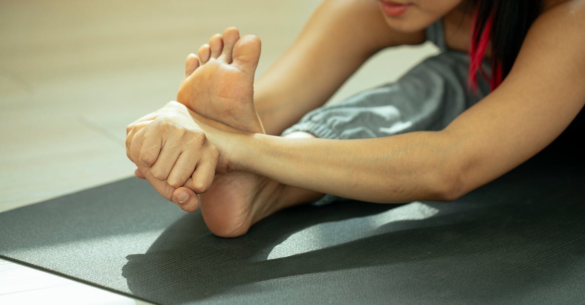
Equally important is stretching the plantar fascia itself before taking the first morning step. While sitting in bed, the patient crosses the affected leg over the opposite knee, grabs the toes, and pulls them backward to stretch the arch. Holding this stretch for a full minute warms up and elongates the fascia gently, completely preventing the violent, painful tearing when stepping onto the floor.
Eccentric loading exercises, such as slow heel drops over the edge of a stair, are introduced as the pain improves. These exercises stimulate the fascia to lay down strong, parallel collagen fibers, permanently repairing the structural integrity of the tissue.
15. Orthotic Devices and Night Splints
Mechanical support often requires external devices. Custom or high-quality over-the-counter orthotic inserts are designed to control overpronation. By providing a firm foundation that prevents the arch from collapsing during the gait cycle, the orthotic mechanically limits how far the plantar fascia can stretch, allowing the microtears to heal undisturbed.
Night splints are a highly effective, specialized tool for eradicating morning pain. A night splint is a rigid boot worn during sleep that holds the foot and ankle in a neutral, ninety-degree angle.
By preventing the foot from pointing downward, the splint forces the plantar fascia and the calf muscles to remain in a gently stretched, elongated position all night. Because the tissue heals in an elongated state rather than a shortened one, the agonizing pain upon the first morning step is entirely eliminated.
16. Pharmacological and Interventional Therapies
If strict stretching, orthotics, and rest fail to resolve the pain after several months, interventional medical therapies are considered. Corticosteroid injections can provide profound, rapid relief of severe acute inflammation. However, they are used sparingly, as multiple steroid injections can weaken the connective tissue, increasing the risk of a full plantar fascia rupture.
Extracorporeal Shockwave Therapy is an advanced, non-invasive option for chronic cases. A device delivers high-energy acoustic sound waves directly into the heel. This micro-trauma stimulates a massive healing response, promoting new blood vessel formation and forcing the chronic, stagnant scar tissue to finally heal.
Surgical intervention, such as a plantar fascia release, is reserved exclusively for the most severe, intractable cases that have failed a full year of conservative management. The surgeon partially cuts the tight fascial band to permanently release the mechanical tension.
17. Frequently Asked Questions (FAQ)
1. Why does my heel only hurt in the morning and then feel better?
While you sleep, your foot points downward, causing the inflamed plantar fascia to relax and tighten up. The tissue attempts to heal in this shortened position. When you stand up in the morning, your body weight violently stretches and re-tears this stiff tissue, causing sharp pain. As you walk, the tissue warms up and stretches, easing the pain temporarily.
2. Does a heel spur cause my plantar fasciitis pain?
No, the bone spur itself rarely causes pain. The sharp, stabbing pain comes from the inflammation and micro-tearing of the soft tissue (the plantar fascia) pulling on the heel. The spur is just the bone’s reaction to being constantly pulled.
3. Is it okay to walk barefoot around the house if I have heel pain?
Absolutely not. Walking barefoot on hard floors provides zero arch support or shock absorption. This places maximum mechanical stress directly on the damaged tissue in your heel, preventing it from healing and guaranteeing the pain will continue.
4. Can tight calf muscles cause pain in my heel?
Yes, they are a primary cause. The calf muscles attach to the back of the heel, while the plantar fascia attaches to the bottom. If your calves are incredibly tight, they constantly pull the heel bone backward, which places enormous, abnormal tension on the plantar fascia every time you take a step.
5. How do I stop the terrible pain when I first get out of bed?
Before you put your feet on the floor, sit on the edge of the bed and stretch. Cross your bad foot over your other knee, grab your toes, and pull them firmly backward to stretch the arch. Massage the arch with your thumb for one minute. This gently stretches the tissue, preventing the violent tearing when you stand.
18. Bibliography
Disclaimer: The content is for informational purposes only and does not replace medical advice. Always consult your doctor for personalized treatment.