1. Introduction
A sensation of instability in the knee when stepping down from a curb is primarily caused by an inability of the quadriceps muscle to adequately perform an eccentric contraction, often coupled with patellofemoral tracking dysfunction or underlying micro-instability of the joint ligaments. Stepping downward shifts the entire body weight onto a single flexed joint, multiplying the mechanical load and requiring precise neuromuscular control. Understanding the biomechanical forces at play and the specific anatomical structures involved is essential for addressing this feeling of giving way and preventing potential falls or structural joint damage.
2. Biomechanics of Stepping Down
The act of stepping down is mechanically more demanding on the knee joint than walking on level ground or climbing stairs. When descending a curb, the lead leg must reach the lower surface while the trailing leg supports the entire body weight on a bending knee.
This action requires the muscles of the trailing leg to lengthen while under tension, a process known as an eccentric contraction. The knee acts as a braking mechanism, absorbing the kinetic energy of the descending body mass to ensure a smooth transition to the lower surface.
During this deceleration phase, the ground reaction forces transmitted through the tibiofemoral joint can reach several times the total body weight. Any weakness or delayed firing in the stabilizing muscles immediately compromises the structural integrity of the joint, manifesting as a sudden feeling of instability.
3. The Role of the Quadriceps Muscle
The quadriceps femoris, the large muscle group on the front of the thigh, is the primary dynamic stabilizer of the knee. It encompasses four distinct muscle bellies that converge into a single tendon attaching to the kneecap and ultimately to the tibia.
During descent from a curb, the quadriceps must execute a highly coordinated eccentric contraction to control the rate of knee flexion. If the quadriceps is weak, fatigued, or inhibited by underlying joint effusion, it cannot generate sufficient braking force.
When the muscle fails to control the descent, the knee joint flexes faster than intended. The nervous system perceives this rapid, uncontrolled flexion as a sudden loss of support, translating into the clinical sensation that the knee is buckling or giving out.
4. Patellofemoral Joint Dynamics
The patellofemoral joint consists of the patella articulating with the trochlear groove of the femur. The patella acts as a fulcrum, increasing the mechanical advantage of the quadriceps muscle.
Optimal function requires the patella to track smoothly within the center of the trochlear groove as the knee bends and straightens. The alignment is dictated by a delicate balance of muscular pull, specifically the vastus medialis obliquus on the inside of the thigh and the vastus lateralis on the outside.
If this muscular balance is disrupted, the patella may track laterally, increasing friction against the femoral condyle. This maltracking disrupts the smooth transmission of force, causing a momentary catch or a feeling that the knee is mechanically unstable during the descent.
5. Patellofemoral Pain Syndrome and Inhibition
Patellofemoral pain syndrome is a prevalent clinical condition characterized by anterior knee pain, particularly during activities that heavily load the patellofemoral joint, such as descending stairs or stepping off curbs.
The pain is often generated by irritation of the highly innervated subchondral bone beneath the patella or the surrounding synovial tissues. Crucially, the presence of pain triggers a reflex neurological response known as arthrogenic muscle inhibition.
This reflex subconsciously prevents the quadriceps muscle from firing at maximum capacity to protect the painful joint from further loading. Consequently, the inhibited muscle fails to provide adequate support during the step-down, leading directly to the sensation of buckling.
6. Ligamentous Laxity and Micro-Instability
The passive stability of the knee is governed by an intricate network of ligaments, including the anterior cruciate ligament, posterior cruciate ligament, medial collateral ligament, and lateral collateral ligament. These structures prevent abnormal translational and rotational movements of the tibia relative to the femur.
Previous sprains, minor tears, or generalized joint hypermobility can result in ligamentous laxity. Even subtle, subclinical laxity, often termed micro-instability, alters the precise kinematics of the joint.
When stepping down, the shear forces applied to the joint may briefly overcome the weakened ligaments. The slight abnormal shifting of the joint surfaces triggers proprioceptive stretch receptors, which signal the brain that the joint is unstable, causing a sudden reflexive giving-way sensation.
7. Meniscal Tears and Joint Degeneration
The menisci are two crescent-shaped wedges of fibrocartilage located between the femur and the tibia. They serve as vital shock absorbers and secondary joint stabilizers, deepening the articular surface of the tibia to cup the rounded femoral condyles.
A tear in the posterior horn of the meniscus can destabilize the joint dynamically. A torn fragment may also become mobile, occasionally impinging between the articulating surfaces during specific degrees of flexion.
When this mechanical impingement occurs while stepping off a curb, it disrupts the smooth gliding motion of the joint. The sudden mechanical block or the sharp pain associated with the pinching of the tissue causes an immediate reflex relaxation of the quadriceps, resulting in acute instability.
8. Proprioception and Neuromuscular Control
Joint stability relies not only on structural strength but also on the nervous system. Proprioception is the unconscious awareness of joint position in space, mediated by specialized mechanoreceptors embedded within the ligaments, joint capsule, and muscles.
These receptors constantly feed information to the central nervous system regarding joint angle, tissue tension, and movement speed. The brain processes this data to orchestrate the precise timing and magnitude of muscle contractions required for stability.
Following an injury or due to natural aging processes, the sensitivity of these mechanoreceptors can decline. A delay in the proprioceptive feedback loop means the stabilizing muscles fire a fraction of a second too late during a step-down, leading to a palpable moment of instability.
9. Hip and Gluteal Muscle Influence
The knee does not function in isolation. It is the middle joint in a kinetic chain that extends from the pelvis to the foot. The stability of the knee is heavily dependent on the alignment dictated by the hip musculature.
The gluteus medius and gluteus minimus are primary hip abductors and external rotators. They stabilize the pelvis and prevent the femur from rotating inward during single-leg weight-bearing tasks.
If these gluteal muscles are weak, stepping down causes the femur to collapse into internal rotation and adduction, a position known as dynamic knee valgus. This poor alignment alters the load distribution across the knee, straining the medial structures and causing a profound sense of mechanical instability.
10. Osteoarthritis and Cartilage Wear
Age-related degeneration of the articular cartilage, known as osteoarthritis, alters the frictionless environment of the knee joint. As the protective cartilage thins, the underlying bone may become exposed, leading to joint space narrowing and the formation of osteophytes.
The altered joint congruency means the femur and tibia no longer articulate smoothly. During the heavy loading of a step-down, the roughened surfaces can cause altered joint mechanics.
Furthermore, osteoarthritic joints frequently develop chronic, low-grade effusions. The presence of excess fluid inside the joint capsule distends the tissue, directly inhibiting quadriceps activation and contributing to the sensation of the knee giving way.
11. Fat Pad Impingement Syndrome
Hoffa’s fat pad is a highly innervated mass of adipose tissue located behind the patellar tendon. It functions as a protective cushion and aids in the distribution of synovial fluid throughout the anterior compartment of the knee.
If the knee undergoes repetitive hyperextension or sustains direct trauma, this fat pad can become inflamed, hypertrophied, and highly sensitive to pressure.
During the descent from a curb, the patella is driven forcefully backward into the trochlear groove, potentially pinching the inflamed fat pad. The acute, sharp pain generated by this impingement elicits a rapid reflex withdrawal, causing the knee to suddenly buckle.
12. Clinical Diagnostic Evaluation
Determining the exact cause of knee instability requires a comprehensive physical examination. A clinician will observe the patient’s gait and perform specific functional tests, such as a single-leg squat, to evaluate neuromuscular control and kinetic chain alignment.
Manual tests assess ligamentous integrity and meniscal health. Palpation of the joint lines and the patellar facets helps isolate areas of acute tenderness.
| Clinical Test | Targeted Structure | Indication of Pathology |
|---|---|---|
| Lachman Test | Anterior Cruciate Ligament | Excessive anterior translation of the tibia. |
| McMurray Test | Menisci | Palpable click or joint line pain during rotation. |
| Patellar Apprehension | Patellofemoral Joint | Patient guarding against lateral patellar translation. |
| Single-Leg Step Down | Neuromuscular Control | Valgus collapse, delayed quadriceps firing, or pain. |
13. Conservative Rehabilitation Strategies
The cornerstone of treating step-down instability is a targeted physical therapy program. Initial phases focus on resolving any joint effusion and reducing pain to minimize arthrogenic muscle inhibition.
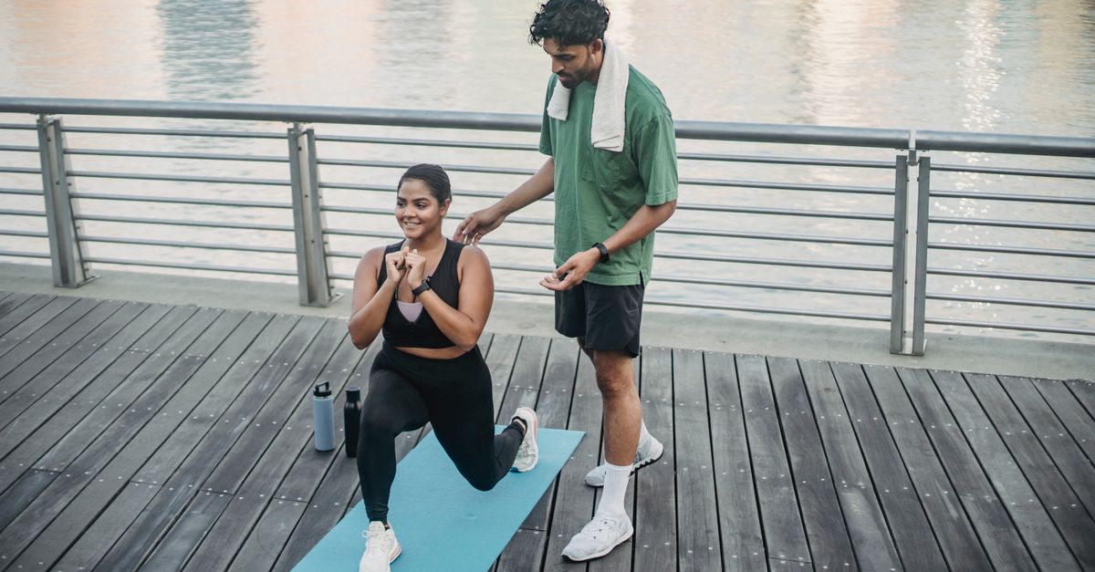
Rehabilitation emphasizes eccentric strengthening of the quadriceps, training the muscle to control the rate of lengthening under load. Closed kinetic chain exercises, such as controlled wall slides and shallow step-downs, safely simulate the mechanical demands of descending a curb.
Strengthening the hip abductors and external rotators is equally critical to prevent dynamic valgus and ensure optimal femoral alignment over the tibia during single-leg weight-bearing.
14. When to Seek Medical Attention
While mild, infrequent feelings of instability might result from temporary muscle fatigue, persistent or severe buckling requires prompt medical evaluation. A knee that completely gives way, resulting in a fall, suggests a significant structural deficit.
Immediate clinical assessment is warranted if the instability is accompanied by recurrent joint swelling, mechanical locking, an inability to fully straighten the knee, or a distinct popping sensation at the time of an initial injury.
Early diagnosis and intervention, potentially including advanced imaging like magnetic resonance imaging, prevent further damage to the articular cartilage and menisci, which can rapidly occur in an unstable joint.
15. Frequently Asked Questions (FAQ)
1. Why does my knee only feel unstable when going down, not up?
Going down requires a demanding eccentric contraction of the quadriceps, where the muscle lengthens under tension to act as a brake. Going up involves a concentric contraction, which is mechanically easier to perform and places less shear stress on the patellofemoral joint.
2. Can wearing a knee brace stop my knee from giving out?
A well-fitted, supportive sleeve can improve joint proprioception by providing continuous sensory feedback to the skin, which enhances muscular control. However, a brace cannot replace the structural support of strong quadriceps and gluteal muscles.
3. Will losing weight help with this sensation?
Yes. The force transmitted through the knee when stepping down is a multiple of your body weight. Reducing total body mass significantly decreases the mechanical load the quadriceps and joint structures must control during the descent.
4. Is a popping sound normal when stepping down?
Painless popping or clicking is often benign, caused by the patella tracking over a slight ridge of cartilage or fluid shifting within the joint. If the popping is accompanied by pain, swelling, or instability, it may indicate a meniscal tear or cartilage defect.
5. How long does it take for physical therapy to improve knee stability?
Improvements in neuromuscular control and proprioception can occur within a few weeks. However, building significant structural strength in the quadriceps and hip muscles typically requires six to twelve weeks of consistent, progressive rehabilitation.
16. Bibliography
Disclaimer: The content is for informational purposes only and does not replace medical advice. Always consult your doctor for personalized treatment.