1. Introduction
Early signs of a brain tumor causing severe morning headaches include a dull, persistent ache that is notably worse upon waking and frequently accompanied by unexplained nausea or vomiting. You should seek an immediate neurological evaluation if this morning head pain is paired with new-onset seizures, progressive weakness on one side of the body, or sudden visual disturbances. Understanding the mechanical relationship between a growing intracranial mass and pressure dynamics during sleep is vital for distinguishing a pathological tumor headache from benign headache disorders.
2. Cranial Anatomy and Intracranial Pressure
The human brain is encased within the rigid, unyielding structure of the skull. This enclosed space contains three primary components: the brain tissue itself, the cerebrospinal fluid that cushions it, and the blood circulating through the cerebral vascular network.
According to the Monro-Kellie doctrine, an increase in the volume of any one of these components must result in a decrease in the volume of the others, or the overall pressure inside the skull will rise. When a tumor begins to grow within this closed system, it introduces a pathological fourth component, progressively exhausting the natural compensatory mechanisms and leading to elevated intracranial pressure.
3. The Pathophysiology of Tumor Growth
Brain tumors can be primary, originating from the brain cells or the supporting glial cells, or secondary, representing metastatic spread from cancers elsewhere in the body. As the mass expands, it inflicts damage through two primary mechanisms.
First, the tumor directly invades and destroys adjacent healthy neural tissue, disrupting local electrical circuits. Second, the growing mass exerts a mechanical compression, creating a mass effect that pushes against surrounding structures. Furthermore, tumors often induce surrounding cerebral edema, a significant accumulation of fluid in the brain tissue, which rapidly accelerates the rise in intracranial pressure and stretches the sensitive meninges.
4. Characteristics of the Morning Headache
Headaches are common, but the specific pattern of a tumor-related headache is distinct. The classic early sign is a headache that is most severe immediately upon waking in the morning.
This occurs because of nocturnal hypoventilation. During deep sleep, breathing naturally slows, causing a mild accumulation of carbon dioxide in the blood. Carbon dioxide is a potent vasodilator; it causes the blood vessels in the brain to widen. In a healthy skull, this slight increase in blood volume is harmless. However, in a skull already crowded by a tumor, this nocturnal vasodilation causes a critical spike in intracranial pressure, manifesting as severe pain upon waking.
5. Exertional Exacerbation of Head Pain
The headache associated with a brain tumor is typically a dull, continuous, non-throbbing ache. A defining clinical feature is its tendency to worsen dramatically with certain physical actions.
Activities such as coughing, sneezing, bending over to tie a shoe, or straining during a bowel movement transiently increase pressure inside the chest and abdomen. This pressure transmits upward through the venous system, temporarily preventing blood from draining out of the head. This brief venous backup causes a sudden, agonizing exacerbation of the head pain, a red flag pointing directly to compromised intracranial dynamics.
6. Nausea and Projectile Vomiting
Elevated intracranial pressure directly stimulates the chemoreceptor trigger zone and the vomiting center located in the medulla oblongata at the base of the brain.
Consequently, the morning headache is frequently accompanied by profound nausea and vomiting. Unlike the nausea caused by gastrointestinal infections, tumor-related vomiting can sometimes occur suddenly and forcefully without any prior feeling of sickness, a phenomenon known as projectile vomiting. Often, patients report that their severe head pain is temporarily relieved immediately after vomiting, as the physical act slightly alters the pressure gradients in the head.
7. Seizures as an Early Indicator
For many patients, the first overt sign of a brain tumor is not a headache, but a new-onset seizure. Seizures occur when the tumor mass physically irritates the surrounding cerebral cortex, triggering a chaotic burst of abnormal electrical activity.
These seizures can take many forms. A generalized tonic-clonic seizure involves a loss of consciousness and full-body convulsions. However, tumors often cause focal seizures, where the electrical storm remains confined to one area of the brain. A patient might experience repetitive twitching in one hand, sudden sensory changes, or a brief period of unresponsive staring, demanding immediate neurological investigation.
8. Focal Neurological Deficits
Because different regions of the brain control specific functions, the location of the tumor dictates the accompanying neurological deficits. As the mass compresses functional areas, patients develop progressive, unilateral symptoms.
A tumor in the frontal lobe may cause progressive weakness or paralysis in the contralateral arm or leg. A mass located in the language centers of the left hemisphere will produce expressive aphasia, where the patient struggles to find words or articulate speech. These focal deficits rarely appear suddenly; they typically worsen gradually over weeks or months as the tumor steadily expands its footprint.
9. Cognitive and Personality Changes
Tumors situated in the frontal or temporal lobes frequently alter a patient’s executive function and emotional regulation long before they cause significant motor weakness.
Family members are often the first to notice these subtle signs. The patient may exhibit uncharacteristic apathy, a severe loss of short-term memory, or inappropriate social behavior. They may become easily confused by simple tasks they previously performed flawlessly, such as balancing a checkbook or navigating a familiar driving route. These personality shifts are often misattributed to stress, depression, or early dementia, delaying a proper oncological diagnosis.
10. Differentiating Primary Headaches from Tumors
Clinical distinction is essential to identify the warning signs hiding within a common symptom.
| Headache Characteristic | Primary Headache (e.g., Tension, Migraine) | Suspicious for Brain Tumor |
|---|---|---|
| Timing and Pattern | Varies, often triggered by stress or late day. | Consistently worst upon waking in the morning. |
| Response to Exertion | May throb with exercise. | Severe, sharp pain when coughing or bending over. |
| Accompanying Symptoms | Light sensitivity, neck tension. | Morning vomiting, new seizures, unilateral weakness. |
| Progression | Episodic, remains relatively stable over years. | Progressively worsens in frequency and intensity. |
11. Visual Disturbances and Papilledema
The optic nerves are directly exposed to the pressure environment of the brain. When intracranial pressure rises significantly, it compresses the optic nerves.
This compression causes swelling of the optic disc at the back of the eye, a clinical sign known as papilledema. While papilledema itself is painless, it leads to distinct visual symptoms. Patients may experience transient visual obscurations, which are brief moments where vision completely blacks out or grays out, especially when standing up quickly. Unexplained double vision or a progressive loss of peripheral vision are also profound warning signs.
12. Warning Signs Requiring Urgent Assessment
While chronic morning headaches warrant a scheduled neurological workup, specific events dictate immediate action. Urgent emergency transport is required if the patient experiences their very first seizure, sudden unresponsiveness, or a precipitous drop in their level of consciousness.
Additionally, if a headache develops with explosive suddenness and unparalleled severity, known as a thunderclap headache, it suggests an acute hemorrhage into the tumor or a ruptured aneurysm. Immediate intervention is required to manage the swelling and prevent catastrophic brain herniation, guiding subsequent chronic headache treatment.
13. Neurological Examination and Fundoscopy
The initial clinical evaluation involves a comprehensive neurological examination. The physician will test cranial nerve function, motor strength, reflexes, and coordination to identify any asymmetrical deficits.
A crucial component is fundoscopy, where the physician uses an ophthalmoscope to look into the patient’s eyes. Visualizing the swollen margins of the optic disc confirms the presence of elevated intracranial pressure, shifting the diagnostic pathway immediately toward urgent neuroimaging to locate the offending mass.
14. Diagnostic Neuroimaging with MRI
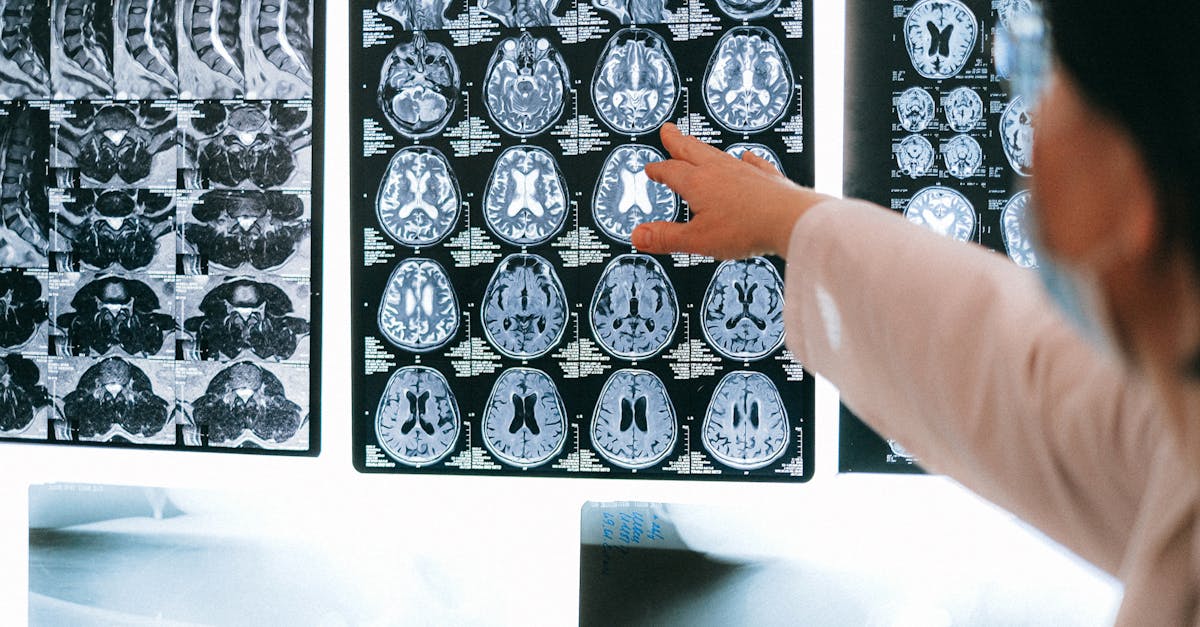
The gold standard for evaluating a suspected brain tumor is magnetic resonance imaging with gadolinium contrast. This advanced imaging modality provides exceptional detail of the brain tissue.
The contrast dye highlights areas where the blood-brain barrier has been compromised by a malignant growth, allowing the radiologist to determine the exact size, location, and vascularity of the tumor. Computed tomography scans may be used in an emergency setting to quickly rule out a massive hemorrhage, but they lack the superior soft-tissue resolution of magnetic resonance imaging required for precise tumor characterization.
15. Surgical Biopsy and Treatment Modalities
Once a mass is identified, a neurosurgeon will typically perform a biopsy, often removing as much of the tumor as safely possible during a craniotomy. The excised tissue is analyzed by a neuropathologist to determine the exact cellular grade and genetic profile of the tumor.
Treatment is highly individualized. It frequently involves a combination of maximal safe surgical resection, followed by targeted radiation therapy to eliminate residual cells. Chemotherapy, designed to cross the blood-brain barrier, may also be administered. For managing the acute symptoms, high-dose corticosteroids like dexamethasone are utilized to rapidly decrease cerebral edema and provide immediate relief from the debilitating morning headaches.
16. Frequently Asked Questions (FAQ)
1. Do all brain tumors cause morning headaches?
No, the presence of a headache depends heavily on the tumor’s size, location, and rate of growth. Small, slow-growing tumors may cause no pain at all, presenting instead with focal symptoms like a hand tremor.
2. Can a tension headache be worse in the morning?
Yes, poor sleep posture or severe teeth grinding during the night can cause muscle tension that results in a morning headache, but this pain is typically described as a tight band rather than the deep, pressure-like ache of a tumor.
3. Will pain relievers help a tumor-related headache?
Standard over-the-counter pain medications may take the edge off in the very early stages, but as the intracranial pressure rises, these medications become entirely ineffective against the mechanical pressure causing the pain.
4. Is a brain tumor the most likely cause of my headache?
Statistically, primary headache disorders like migraines or tension-type headaches are vastly more common. A brain tumor is a rare diagnosis, but the presence of new, progressive neurological signs necessitates ruling it out.
5. How quickly do tumor symptoms progress?
Benign, slow-growing tumors like meningiomas can grow for years before causing symptoms. Aggressive malignancies like glioblastomas can double in size rapidly, causing symptoms to escalate severely over a matter of weeks.
17. Bibliography
Disclaimer: The content is for informational purposes only and does not replace medical advice. Always consult your doctor for personalized treatment.