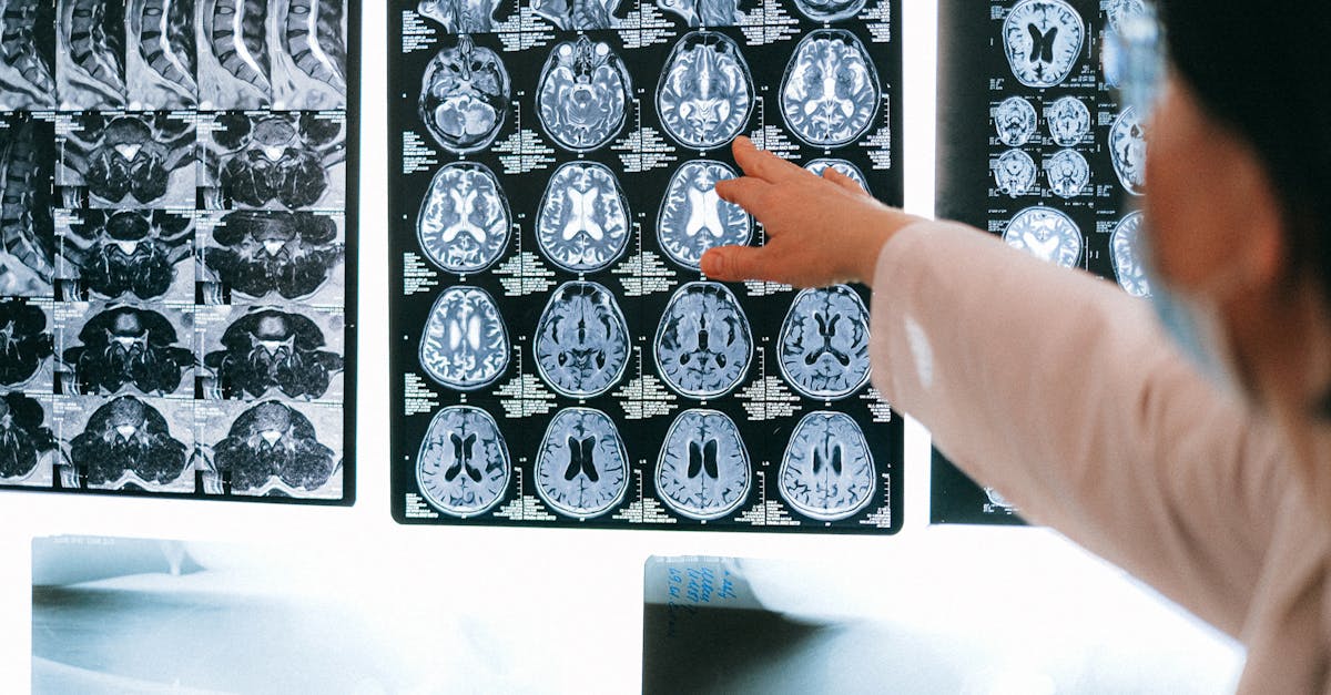
1. Introduction to Intracranial Neoplasms
An intracranial neoplasm, commonly referred to as a brain tumor, represents an abnormal, uncontrolled proliferation of cells within the rigid confines of the skull. This broad medical classification encompasses a highly diverse group of tumors, ranging from slow-growing, benign masses to aggressive, rapidly fatal malignancies. Because the skull forms an enclosed, non-expandable vault, any abnormal cellular growth—regardless of its benign or malignant histological classification—acts as a space-occupying lesion that inevitably compromises the vital neurological structures sharing that limited space.
The clinical approach to an intracranial neoplasm requires a fundamental distinction between two primary categories. Primary intracranial neoplasms originate directly from the tissues of the central nervous system, including the brain parenchyma, the meningeal coverings, or the cranial nerves. Secondary, or metastatic, neoplasms originate as cancer in another organ (such as the lung or breast) and subsequently spread through the bloodstream to seed within the brain tissue.
Managing these complex lesions demands a highly coordinated, multidisciplinary effort involving neurosurgery, neuro-oncology, and radiation oncology. The primary goals of treatment are to secure a precise histological diagnosis, relieve the mechanical pressure exerted by the tumor on the healthy brain, and deploy targeted therapies to halt or eradicate the aberrant cellular growth while preserving the patient’s neurological function and quality of life.
2. Anatomy and the Blood-Brain Barrier
To understand the unique challenges presented by intracranial neoplasms, one must appreciate the specialized anatomy of the central nervous system. The brain is composed of two major categories of cells: neurons, which transmit electrical signals, and glial cells, which provide vital structural, metabolic, and immunological support to the neurons. Interestingly, primary brain tumors almost never arise from mature neurons, which do not actively divide, but rather from the supporting glial cells or the meningeal membranes.
A critical anatomical feature complicating the treatment of these tumors is the blood-brain barrier. The capillaries supplying the brain are constructed with specialized, extremely tight junctions between their endothelial cells. This barrier acts as a rigorous biological filter, protecting the delicate neural tissue from toxins and pathogens circulating in the systemic bloodstream.
However, this highly protective mechanism becomes a profound obstacle in neuro-oncology. The blood-brain barrier effectively blocks the vast majority of traditional, systemic chemotherapy drugs from entering the brain parenchyma in high enough concentrations to destroy tumor cells. Developing therapies capable of bypassing or penetrating this barrier remains one of the most significant challenges in modern cancer research.
3. Primary versus Secondary Neoplasms
The initial diagnostic categorization of a brain mass heavily dictates the subsequent treatment strategy.
Primary intracranial neoplasms originate within the skull. They are classified based on the specific type of central nervous system cell from which they mutated. A defining characteristic of primary malignant brain tumors is that, while they can aggressively invade and destroy adjacent brain tissue, they almost never metastasize (spread) outside the central nervous system to other organs in the body.
Secondary, or metastatic, intracranial neoplasms are far more common than primary brain tumors in adults. These occur when malignant cells break away from a primary cancer located elsewhere—most frequently lung cancer, breast cancer, melanoma, renal cell carcinoma, or colon cancer—and travel through the arterial bloodstream to lodge in the microscopic capillary beds of the brain. Patients with secondary neoplasms frequently present with multiple, distinct tumors scattered throughout the brain hemispheres.
4. Pathophysiology of Tumor Growth and Damage
An expanding intracranial neoplasm inflicts damage upon the central nervous system through two distinct pathophysiological mechanisms: direct invasion and mechanical mass effect.
Malignant primary tumors, such as high-grade gliomas, do not possess a distinct border. They grow by aggressively infiltrating the normal brain parenchyma. The tumor cells migrate along white matter tracts and blood vessels, physically destroying healthy neurons and glial cells as they advance. This infiltrative nature makes complete surgical removal of the tumor on a microscopic level virtually impossible.
Conversely, benign tumors (like meningiomas) and metastatic lesions typically grow as cohesive, well-defined masses. They inflict damage through mechanical mass effect. As the tumor expands, it compresses and displaces adjacent healthy brain tissue, disrupting normal neural circuitry. Furthermore, the tumor triggers severe localized inflammation, causing the surrounding brain tissue to swell with fluid (vasogenic edema). This combined volume of tumor and edema rapidly increases overall intracranial pressure.
5. Common Primary Tumors: The Gliomas
Gliomas represent the most common and clinically challenging category of primary malignant brain tumors. They originate from the glial cells that support the neurons. Gliomas are classified by the World Health Organization (WHO) into grades I through IV, based on their cellular characteristics and biological aggressiveness.
Astrocytomas arise from star-shaped glial cells called astrocytes. A WHO Grade IV astrocytoma, historically known as a Glioblastoma Multiforme, is the most common and devastating primary malignant brain tumor in adults. It is characterized by rapid, aggressive growth, profound invasion into surrounding tissue, and areas of dense necrosis within the tumor core.
Oligodendrogliomas arise from the cells responsible for producing the myelin sheath that insulates nerve fibers. Ependymomas originate from the cells lining the ventricles—the fluid-filled cavities deep within the brain. While low-grade gliomas grow slowly over many years, they inherently possess the biological tendency to eventually mutate into higher-grade, aggressive malignancies.
6. Non-Glial Primary Tumors
Not all primary intracranial neoplasms arise from the brain tissue itself. Several common tumors originate from other structures within the cranial vault.
Meningiomas are the most common primary brain tumor overall, arising from the meningeal membranes covering the brain. The vast majority are benign (WHO Grade I) and grow slowly, causing symptoms solely through the mechanical compression of adjacent brain tissue rather than invasion.
Vestibular schwannomas, also known as acoustic neuromas, are benign, slow-growing tumors that originate from the Schwann cells forming the insulation around the vestibular nerve. They grow within the internal auditory canal, classically causing progressive, unilateral hearing loss, tinnitus, and balance disturbances. Pituitary adenomas are benign tumors arising from the pituitary gland at the base of the brain, frequently causing visual field deficits due to compression of the optic chiasm, alongside complex endocrine imbalances.
7. Clinical Presentation: Increased Intracranial Pressure
The symptoms generated by an intracranial neoplasm depend entirely on the tumor’s size, its growth rate, and its precise anatomical location. However, a universal presentation involves symptoms related to increased global intracranial pressure.
A new-onset, persistent headache is a frequent early indicator. A tumor-related headache is characteristically worse in the morning upon waking, and is frequently exacerbated by maneuvers that momentarily raise intracranial pressure, such as coughing, sneezing, or bending over.
As the pressure within the skull rises further, it irritates the vomiting centers in the brainstem, leading to profound nausea and sudden, unprovoked episodes of vomiting. High pressure also compresses the optic nerves at the back of the eyes, a condition called papilledema, which results in transient visual obscurations, blurred vision, and eventually, permanent vision loss if the pressure is not relieved.
8. Focal Neurological Deficits and Seizures
Beyond the generalized symptoms of pressure, an expanding neoplasm produces focal neurological deficits corresponding directly to the specific area of the brain it is compressing or destroying.
A tumor located in the motor cortex of the frontal lobe will cause progressive weakness or paralysis (hemiparesis) on the opposite side of the body. Tumors in the language centers (typically the left hemisphere) produce aphasia, characterized by profound difficulty producing or comprehending speech. A tumor in the cerebellum leads to severe ataxia, resulting in an uncoordinated, stumbling gait and clumsiness in the extremities.
Seizures are a highly prominent presenting symptom, particularly for slow-growing tumors located near the cerebral cortex. The physical compression of the tumor, combined with the chemical irritation from the surrounding edema, alters the electrical stability of the cortical neurons, triggering focal or generalized seizure activity in adults with no prior history of epilepsy.
9. Diagnostic Neuroimaging Protocols
The definitive diagnosis and precise anatomical mapping of an intracranial neoplasm rely entirely on advanced neuroimaging. Magnetic Resonance Imaging (MRI) with intravenous gadolinium contrast is the absolute gold standard diagnostic modality in neuro-oncology.
The MRI provides exquisite, high-resolution detail of the brain parenchyma. The administration of contrast dye is crucial. Because malignant tumors disrupt the blood-brain barrier and induce the formation of abnormal, leaky blood vessels, the contrast dye rapidly pools within the active tumor tissue, making it appear intensely bright on the scan.
The radiologist evaluates the MRI to determine the exact size, location, and number of lesions. Furthermore, advanced MRI sequences, such as MR spectroscopy, can analyze the chemical composition of the mass, helping to differentiate a high-grade tumor from an infectious abscess or an area of stroke-induced tissue death prior to surgical biopsy.
10. Classification of Common Neoplasms
Understanding the broad categories of intracranial tumors guides the subsequent multidisciplinary treatment plan. The table below outlines common neoplasms.
| Tumor Classification | Common Examples | Typical Biological Behavior |
|---|---|---|
| Primary Benign | Meningioma, Vestibular Schwannoma | Slow-growing, compresses tissue, typically curable with complete resection. |
| Primary Malignant (Glial) | Glioblastoma, Anaplastic Astrocytoma | Rapidly infiltrates brain tissue, highly aggressive, challenging to cure. |
| Primary Low-Grade (Glial) | Diffuse Astrocytoma (Grade II) | Indolent growth, prone to eventual malignant transformation over time. |
| Secondary (Metastatic) | Lung, Breast, Melanoma metastases | Multiple distinct lesions, signifies advanced systemic cancer, behavior depends on primary origin. |
11. Tissue Biopsy and Neuropathology
While MRI characteristics strongly suggest the type of tumor, a definitive diagnosis is only established through the direct microscopic and molecular examination of the tumor tissue by a neuropathologist. This requires a surgical procedure to obtain a tissue sample.
If the tumor is located in an accessible area and surgery is planned, the biopsy is performed during the primary resection. If the tumor is located deep within the brain, in an eloquent (highly functional) area where surgical removal is too dangerous, a stereotactic needle biopsy is performed. The neurosurgeon uses a 3D coordinate system to guide a thin needle precisely into the tumor to extract a core of tissue.
Modern neuropathology relies heavily on molecular and genetic profiling of the tumor tissue. Identifying specific genetic mutations, such as the IDH mutation or MGMT promoter methylation in gliomas, is now mandatory. These molecular markers dictate the final WHO classification, provide critical prognostic information, and determine the tumor’s likely response to specific chemotherapeutic agents.
12. Neurosurgical Intervention
Maximal safe surgical resection is the foundational treatment for the majority of accessible intracranial neoplasms. The primary goals of neurosurgery are to obtain diagnostic tissue, relieve the mechanical mass effect and intracranial pressure, and drastically reduce the total volume of the tumor cells (cytoreduction) to enhance the efficacy of subsequent radiation and chemotherapy.
Craniotomy is the standard approach, utilizing advanced technology. Neurosurgeons employ intraoperative MRI, 3D neuronavigation systems, and specialized fluorescent dyes (like 5-ALA, which causes malignant cells to glow under specific light) to maximize tumor removal while strictly preserving adjacent healthy neural tracts.
For tumors located near speech or motor centers, awake craniotomy is frequently utilized. The patient is awakened during the critical portion of the surgery and asked to perform specific tasks while the surgeon electrically stimulates the brain surface, allowing for real-time mapping to ensure critical functional areas are not damaged during resection.
13. Radiation and Chemotherapy Protocols
For malignant primary tumors and metastatic lesions, surgery is rarely curative on its own due to microscopic cellular infiltration. Adjuvant therapies are essential to target the remaining disease.
Radiation therapy uses high-energy beams to destroy the DNA of tumor cells, preventing them from dividing. Fractionated external beam radiotherapy is delivered daily over several weeks. For small, distinct lesions—particularly brain metastases or small meningiomas—stereotactic radiosurgery (such as the Gamma Knife) delivers a single, massive, highly focused dose of radiation to ablate the tumor while sparing surrounding tissue.
Chemotherapy for primary brain tumors is challenging due to the blood-brain barrier. Temozolomide is an oral alkylating agent specifically designed to penetrate this barrier and is the standard of care for high-grade gliomas, usually administered concurrently with radiation therapy and continued for months afterward.
14. Symptom Management and Corticosteroids
Managing the symptoms caused by the tumor and its treatments is a vital component of neuro-oncological care. The intense inflammatory response and resulting cerebral edema (swelling) surrounding the tumor frequently cause more severe acute neurological deficits than the tumor mass itself.
High-dose corticosteroids, predominantly dexamethasone, are the most effective agents for rapidly reducing tumor-associated vasogenic edema. Administering dexamethasone can profoundly and rapidly improve a patient’s neurological function, relieving headaches and restoring lost motor strength within hours.
However, long-term steroid use carries substantial side effects, including severe muscle weakness, elevated blood glucose, immune suppression, and psychiatric alterations. Therefore, the medical team continuously attempts to taper the steroid dose to the lowest effective level. Concurrently, prophylactic or therapeutic anticonvulsant medications are strictly managed to prevent tumor-associated seizure activity.
15. Multidisciplinary Care and Palliative Support
The treatment of malignant intracranial neoplasms requires a highly coordinated, multidisciplinary approach. The neuro-oncology tumor board—comprising neurosurgeons, medical oncologists, radiation oncologists, neuropathologists, and specialized nurses—collaboratively develops and adjusts the individualized treatment plan for each patient based on ongoing imaging and clinical response.
Because the prognosis for high-grade primary malignancies and extensive metastatic disease remains poor despite aggressive therapy, the integration of palliative care early in the disease process is essential.
Palliative care specialists focus not on curing the disease, but on aggressively managing debilitating symptoms, treating pain, addressing the profound psychological and existential distress of the patient and family, and facilitating complex decisions regarding goals of care and end-of-life transitions when tumor-directed therapies are no longer effective.
16. When to Consult a Neurologist
The sudden onset or progressive worsening of neurological symptoms mandates immediate evaluation. While tension headaches and migraines are common, a headache that is entirely new in character, steadily worsens over weeks, or wakes an individual from sleep requires medical investigation.
Immediate consultation is required if a patient experiences their first seizure, regardless of age, as this is a prominent red flag for a structural brain lesion.
Furthermore, the gradual, unexplained development of focal deficits—such as progressive weakness in a limb, noticeable changes in speech or language comprehension, persistent clumsiness or balance issues, or profound changes in personality, memory, and behavior—warrants a comprehensive neurological examination and prompt neuroimaging to rule out the presence of an expanding intracranial neoplasm.
17. Frequently Asked Questions (FAQ)
1. Are all brain tumors cancerous?
No. Brain tumors are broadly categorized as benign (non-cancerous) or malignant (cancerous). Benign tumors, like most meningiomas, grow slowly and do not invade surrounding tissue, though they can still cause severe problems by pressing on the brain. Malignant tumors grow rapidly and aggressively invade brain tissue.
2. What causes a primary brain tumor?
For the vast majority of primary brain tumors, the exact cause is unknown. Exposure to high doses of ionizing radiation (such as previous radiation therapy) is a known risk factor, as are certain rare inherited genetic syndromes. There is no conclusive evidence linking mobile phone use or specific diets to brain tumors.
3. Can brain cancer spread to the rest of the body?
It is extremely rare. While cancers from the lung or breast very commonly spread (metastasize) to the brain, primary malignant brain tumors (like glioblastoma) almost never spread outside the brain and spinal cord to other organs.
4. Why is brain surgery done while the patient is awake?
An “awake craniotomy” is performed when the tumor is located very close to the areas of the brain that control speech or movement. By keeping the patient awake and talking during the tumor removal, the surgeon can ensure they are not damaging these critical functions, allowing for a safer, more aggressive resection.
18. Bibliography
Disclaimer: The content is for informational purposes only and does not replace medical advice. Always consult your doctor for personalized treatment.