1. Introduction
A pituitary mass is an abnormal growth occurring within the pituitary gland, a small, pea-sized structure situated at the base of the brain. Often referred to as the “master gland,” the pituitary plays a central role in regulating the body’s entire endocrine system. It secretes essential hormones that control growth, metabolism, reproduction, and the body’s response to stress.
Because the pituitary gland governs so many critical physiological functions, a mass developing within this delicate tissue can cause profound systemic disruptions. The vast majority of these masses are benign, non-cancerous tumors known as pituitary adenomas. While they do not spread to distant organs, their physical presence in a confined space and their potential to secrete excess hormones make them significant medical concerns.
The clinical presentation of a pituitary mass is generally divided into two categories: symptoms caused by hormonal imbalances and symptoms caused by the physical size of the tumor compressing nearby structures, such as the optic nerves. Managing these lesions requires a multidisciplinary approach involving endocrinologists, neurologists, and neurosurgeons to accurately restore hormonal balance and preserve neurological function.
2. Anatomy and Location of the Pituitary Gland
The pituitary gland rests securely within a bony depression at the base of the skull known as the sella turcica, directly behind the bridge of the nose. It is connected to the hypothalamus, a region of the brain that serves as the command center, continuously sending chemical signals to the pituitary to release or halt hormone production.
Anatomically, the gland is divided into the anterior lobe and the posterior lobe. The anterior lobe produces prolactin, growth hormone, adrenocorticotropic hormone, thyroid-stimulating hormone, and gonadotropins. The posterior lobe stores and releases oxytocin and antidiuretic hormone. A mass can originate in either lobe, though anterior lobe adenomas are far more common.
Crucially, the pituitary gland sits immediately below the optic chiasm, the anatomical junction where the optic nerves from each eye cross over one another. Furthermore, it is flanked by the cavernous sinuses, which contain major arteries supplying blood to the brain and nerves controlling eye movement. This tightly packed anatomy explains why expanding tumors cause specific neurological deficits.
3. Functioning Versus Non-Functioning Adenomas
Pituitary adenomas are broadly classified based on their biological activity. Functioning adenomas, also called secretory tumors, are composed of cells that actively produce and release excessive amounts of a specific hormone. The symptoms a patient experiences are directly related to the specific hormone being overproduced, leading to distinct clinical syndromes.
Non-functioning adenomas do not secrete active hormones. Because they do not cause obvious hormonal imbalances, they often grow silently for many years. They are typically only discovered when they become large enough to press on surrounding brain structures, causing headaches or vision loss, or they are found incidentally when a patient undergoes a brain scan for an unrelated reason.
Furthermore, adenomas are classified by size. A microadenoma is a tumor measuring less than one centimeter in diameter, while a macroadenoma is a tumor measuring one centimeter or larger. Macroadenomas, whether functioning or non-functioning, pose a significant risk of compressing the optic chiasm and normal pituitary tissue.
4. Prolactinomas: The Most Common Secretory Tumor
A prolactinoma is a functioning adenoma that secretes excessive amounts of prolactin, the hormone responsible for stimulating breast milk production. Prolactinomas are the most frequently diagnosed type of hormone-secreting pituitary mass.
In premenopausal women, elevated prolactin levels disrupt the normal menstrual cycle, leading to irregular periods or a complete cessation of menstruation. It can also cause galactorrhea, which is the inappropriate production of breast milk in women who are not pregnant or nursing. These clear symptoms often lead to early diagnosis while the tumor is still a microadenoma.
In men and postmenopausal women, the symptoms are more subtle. Men may experience decreased libido, erectile dysfunction, and reduced body hair due to prolactin suppressing testosterone production. Because these symptoms are often attributed to aging or stress, prolactinomas in men are frequently diagnosed later, often presenting as large macroadenomas.
5. Growth Hormone Secreting Tumors and Acromegaly
Adenomas that secrete excessive amounts of growth hormone cause profound physical changes. If the tumor develops during childhood before the growth plates in the bones have fused, it results in gigantism, characterized by exceptional linear height.
If the tumor develops in adulthood, after bone growth is complete, it causes a condition known as acromegaly. Adults with acromegaly experience a gradual, progressive thickening of the bones in the hands, feet, and face. Patients often notice that their rings no longer fit, their shoe size increases, and their facial features become noticeably more pronounced and coarse over time.
Beyond physical appearance, excess growth hormone causes severe systemic complications. It leads to significant joint pain, sleep apnea, hypertension, and a profoundly increased risk of developing cardiovascular disease and diabetes, making prompt medical or surgical intervention absolutely essential.
6. ACTH Secreting Tumors and Cushing Disease
An adenoma that overproduces adrenocorticotropic hormone stimulates the adrenal glands to release massive amounts of cortisol, the body’s primary stress hormone. This specific condition is known as Cushing disease.
Chronic exposure to severe cortisol excess dramatically alters the patient’s metabolism and physical appearance. Classic signs include rapid, unexplained weight gain focused centrally on the abdomen and face, resulting in a rounded appearance. The skin becomes thin, bruising easily, and wide, purple stretch marks often develop on the abdomen.
Cushing disease also causes severe muscle weakness, osteoporosis, and difficult-to-control high blood pressure. Diagnosing this condition is complex, requiring careful endocrine testing to prove that the excess cortisol is specifically driven by a pituitary mass and not an underlying retroperitoneal mass involving the adrenal gland itself.
7. Neurological Symptoms: The Mass Effect
As a pituitary macroadenoma expands upward out of the sella turcica, it exerts physical pressure on the brain, creating a phenomenon known as mass effect. The most common neurological symptom is a chronic, dull headache, resulting from the stretching of the fibrous membrane covering the gland.
More concerning is the compression of the optic chiasm. When the tumor presses on the crossing optic nerve fibers, it disrupts the visual pathways. This classically results in bitemporal hemianopsia, which is the progressive loss of peripheral vision in both eyes. Patients often describe feeling as though they are looking through a tunnel.
If the tumor expands laterally into the cavernous sinus, it can compress the cranial nerves responsible for eye movement. This results in double vision, drooping of the eyelid, or a loss of sensation on portions of the face. Any new, unexplained visual field defect requires an immediate neurological evaluation.
8. Pituitary Apoplexy: A Neurological Emergency
Pituitary apoplexy is a rare, life-threatening complication that occurs when a pituitary mass suddenly bleeds internally or when the tumor outgrows its blood supply, causing rapid tissue death. This results in a sudden, dramatic swelling of the mass within the confined bony space of the skull base.
The clinical presentation is acute and dramatic. Patients experience the sudden onset of a severe, explosive headache, often described as the worst headache of their life. This is rapidly followed by double vision, acute vision loss, and sometimes cardiovascular collapse due to the sudden failure of the pituitary gland to produce stress hormones.
Pituitary apoplexy is a critical emergency. It requires immediate administration of high-dose intravenous corticosteroids to stabilize the patient’s blood pressure and frequently necessitates urgent neurosurgery to decompress the optic nerves and remove the dying tumor tissue.
9. Comprehensive Endocrine Laboratory Testing
The diagnostic workup for a suspected pituitary mass begins with a comprehensive panel of blood tests to evaluate the entire endocrine axis. The endocrinologist measures the basal levels of all pituitary hormones and their target organ hormones to detect any pattern of hypersecretion or deficiency.
Tests include measuring serum prolactin, insulin-like growth factor 1 for assessing growth hormone, cortisol levels, thyroid hormones, and testosterone or estrogen. If basal levels are ambiguous, specialized dynamic stimulation or suppression tests are performed.
For example, a glucose tolerance test is used to confirm acromegaly. Normally, consuming a large amount of sugar suppresses growth hormone. In a patient with a growth hormone-secreting adenoma, the hormone levels remain inappropriately elevated despite the sugar load.
10. Magnetic Resonance Imaging Protocols
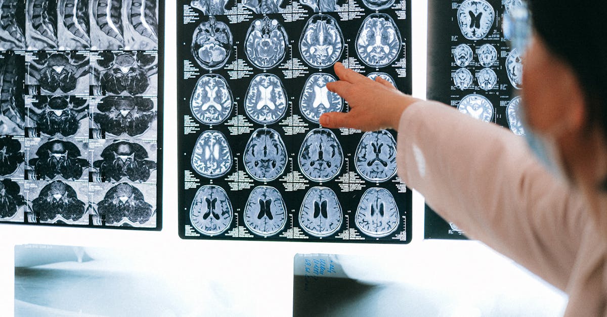
To visualize the pituitary gland and confirm the presence of a mass, Magnetic Resonance Imaging is the gold standard diagnostic tool. A specialized “pituitary protocol” MRI is performed, utilizing thin slices and intravenous contrast material to provide exquisite detail of the small structures at the base of the brain.
The MRI clearly defines the exact size and borders of the adenoma. It demonstrates whether the mass is a microadenoma confined within the sella turcica, or a macroadenoma extending upward to compress the optic chiasm or invading the adjacent cavernous sinuses.
Computed tomography scans are generally inferior for viewing pituitary soft tissues but may be used in emergency settings to quickly rule out acute bleeding (apoplexy) or if the patient has a medical device that prohibits them from undergoing an MRI.
11. Visual Field Testing
Because pituitary masses frequently threaten vision, formal visual field testing by an ophthalmologist or neuro-ophthalmologist is a mandatory component of the evaluation for any macroadenoma.
This non-invasive test maps the complete field of vision for each eye independently. It detects subtle losses in peripheral vision that the patient may not yet have noticed in their daily life.
Documenting a visual field defect provides objective evidence that the tumor is actively compressing the optic chiasm. This finding is a primary indication that surgical removal of the tumor is required to decompress the nerves and prevent permanent blindness.
12. Data Summary: Functioning Pituitary Adenomas
The following table summarizes the most common types of functioning pituitary adenomas, the hormones they overproduce, and their resulting clinical syndromes.
| Tumor Type | Hormone Overproduced | Clinical Syndrome & Key Symptoms |
|---|---|---|
| Prolactinoma | Prolactin | Irregular menses, galactorrhea, decreased libido. |
| Somatotroph Adenoma | Growth Hormone | Acromegaly: enlarged hands/feet, coarse facial features. |
| Corticotroph Adenoma | ACTH (Adrenocorticotropic Hormone) | Cushing Disease: central weight gain, thin skin, hypertension. |
| Thyrotroph Adenoma (Rare) | Thyroid Stimulating Hormone | Hyperthyroidism: rapid heart rate, weight loss, anxiety. |
This hormonal profile dictates the initial approach, differentiating tumors that require immediate surgery from those that can be treated with medication.
13. Pharmacological Therapy: Medical Management
Unlike most brain tumors, the primary treatment for a prolactinoma is medical, not surgical. Dopamine agonist medications, such as cabergoline or bromocriptine, are highly effective at suppressing prolactin production and actively shrinking the tumor.
Many patients with prolactinomas experience a complete normalization of their hormone levels and a significant reduction in tumor size with medication alone, avoiding the need for brain surgery. These medications must often be taken for several years.
Medical therapy is also utilized for acromegaly and Cushing disease, typically as a secondary option if surgery is not fully successful, or to control hormone levels prior to an operation. Somatostatin analogs and specific receptor blockers help mitigate the severe systemic effects of these hormone excesses.
14. Transsphenoidal Neurosurgery
For non-functioning macroadenomas causing vision loss, and for most functioning tumors other than prolactinomas, surgical removal is the definitive treatment. The standard surgical approach is transsphenoidal endoscopic surgery.
This is a minimally invasive technique where the neurosurgeon accesses the pituitary gland by navigating specialized cameras and long, thin instruments through the nasal cavity and the sphenoid sinus, entirely avoiding the need to open the skull or manipulate the main lobes of the brain.
The surgeon carefully removes the adenoma, prioritizing the decompression of the optic nerves and the preservation of the surrounding normal pituitary tissue. Recovery from transsphenoidal surgery is generally rapid, with most patients returning home within a few days.
15. Radiation Therapy and Stereotactic Radiosurgery
If a pituitary mass cannot be completely removed through surgery, or if a functioning tumor continues to secrete excess hormones despite medical and surgical interventions, radiation therapy is employed.
Modern techniques, such as stereotactic radiosurgery (often referred to as Gamma Knife or CyberKnife), deliver a highly focused, intense beam of radiation directly to the residual tumor tissue while sparing the surrounding healthy brain and optic nerves.
Radiation therapy stops the tumor from growing and slowly reduces hormone production over several years. Because the effects are gradual, patients typically continue pharmacological therapy while waiting for the full benefit of the radiation to take effect.
16. Long-Term Endocrine Follow-Up
The presence of a pituitary mass, or the surgical and radiation treatments used to eradicate it, can damage the normal, healthy cells of the pituitary gland. This results in hypopituitarism, a condition where the gland fails to produce sufficient essential hormones.
Patients require lifelong surveillance by an endocrinologist. If hormone deficiencies are identified, the patient must begin hormone replacement therapy. This may include daily thyroid medication, testosterone or estrogen replacement, and crucial corticosteroids to ensure the body can safely respond to stress and illness.
Regular MRI scans and visual field tests are also scheduled to ensure the adenoma does not recur, maintaining the patient’s neurological health and metabolic stability for the rest of their life.
17. Frequently Asked Questions FAQ
1. Is a pituitary mass considered brain cancer?
No. The vast majority of pituitary masses are benign adenomas. They are not brain cancer. While they can cause serious problems by pressing on nerves or releasing too many hormones, they do not spread to other parts of the body.
2. Can a pituitary tumor cause me to go blind?
If a pituitary mass grows large enough, it can press on the optic nerves located right above the gland. This usually causes a slow loss of your peripheral vision. Removing the tumor usually stops the damage and can restore vision.
3. Why am I producing breast milk if I am not pregnant?
This is a classic sign of a prolactinoma, a type of pituitary tumor that produces too much prolactin. Prolactin is the hormone that commands the body to make milk. Medication usually stops this symptom very quickly.
4. How does a surgeon reach the pituitary gland without opening the skull?
Surgeons use a transsphenoidal approach. They pass tiny cameras and tools through your nostrils, going through the natural air spaces in your nasal cavity to reach the base of the skull where the pituitary gland sits.
5. Will I need to take hormones for the rest of my life after surgery?
It depends on how much normal pituitary tissue was damaged by the tumor or the surgery. Some patients require no medication, while others may need to take a daily pill to replace thyroid or stress hormones to keep their body functioning normally.
18. Bibliography
Disclaimer: The content is for informational purposes only and does not replace medical advice. Always consult your doctor for personalized treatment.