1. Introduction
The early signs of a brain tumor often involve a new pattern of severe morning headaches, unexplained nausea, subtle focal neurological deficits, and the sudden onset of seizures. Because the rigid skull cannot accommodate expanding tissue, even a small, slow-growing mass creates localized pressure that disrupts normal cerebral function and demands prompt neurological investigation. A brain tumor is an abnormal proliferation of cells within the central nervous system, and its clinical presentation depends entirely on its size, growth rate, and precise anatomical location.
The human brain is a highly mapped organ where specific regions control distinct cognitive, sensory, and motor functions. A tumor located in the frontal lobe will produce entirely different early symptoms than a tumor situated in the cerebellum. Consequently, the warning signs are not uniform across all patients. Some tumors remain silent for months, while others announce their presence rapidly through a dramatic neurological event.
Differentiating the symptoms of a brain mass from benign, everyday ailments like tension headaches or routine fatigue is a complex clinical task. Recognizing the subtle red flags—such as a headache that wakes a patient from sleep or a progressive, unilateral weakness—is vital. Early detection via advanced neuroimaging significantly broadens the therapeutic avenues available to neurosurgeons and oncologists.
2. Cranial Anatomy and Intracranial Pressure
To understand how a brain tumor produces generalized symptoms, one must consider the fixed volume of the cranial vault. The adult skull is a rigid bone structure containing three elements: brain tissue, cerebrospinal fluid, and blood. According to the Monro-Kellie hypothesis, an increase in the volume of one component must be compensated by a decrease in another, or the overall pressure will rise.
When a tumor begins to grow, it acts as a space-occupying lesion. The brain initially compensates by displacing cerebrospinal fluid into the spinal space and squeezing venous blood out of the skull. However, this compensatory mechanism is quickly exhausted.
Once the reserve is depleted, the growing mass causes a sharp, dangerous rise in intracranial pressure. Furthermore, tumors often lack a normal blood-brain barrier, leading to leaky blood vessels that cause significant edema (swelling) in the surrounding healthy brain tissue. This combination of mass effect and edema drives the generalized early symptoms of a brain tumor.
3. The Characteristics of Tumor Headaches
Headaches are the most frequently reported early symptom, occurring in approximately half of all brain tumor patients. However, a tumor-related headache possesses distinct clinical characteristics that differentiate it from common migraines or tension headaches.
Because intracranial pressure naturally rises when a person lies flat during sleep, a classic tumor headache is typically most severe early in the morning upon waking. Patients often report that the headache actually wakes them up from a deep sleep, a significant clinical red flag.
The pain is usually described as a continuous, dull, aching pressure rather than a throbbing sensation. It frequently worsens with activities that transiently increase cranial pressure, such as coughing, sneezing, bearing down during a bowel movement, or bending over. A new-onset headache in a patient over the age of fifty, or a fundamental change in the pattern of a patient’s historical headaches, mandates immediate neuroimaging.
4. Nausea and Morning Vomiting
Closely linked to the morning headaches is the sudden onset of unexplained nausea and vomiting. The increased intracranial pressure directly stimulates the area postrema, the vomiting center located in the medulla oblongata at the base of the brainstem.
Tumor-related vomiting is highly characteristic. It often occurs early in the morning, immediately upon rising, and is frequently projectile in nature, meaning it occurs suddenly and forcefully without any preceding nausea or gastrointestinal distress.
Once the patient vomits, the resulting rapid exhalation and hyperventilation slightly reduce the intracranial pressure, often providing temporary, paradoxical relief from the morning headache. Unexplained morning vomiting that persists for more than a few days requires swift clinical evaluation to rule out a central nervous system lesion.
5. New-Onset Seizure Activity
For approximately one-third of patients, a sudden, unprovoked seizure is the very first clinical sign of a brain tumor. Tumors act as physical irritants to the surrounding cerebral cortex. As the mass pushes against healthy neurons, it alters their electrical stability, triggering a chaotic burst of synchronized electrical activity.
Tumors in the frontal, temporal, and parietal lobes are highly epileptogenic. The seizure may be a generalized tonic-clonic event, involving a complete loss of consciousness and violent muscle contractions. Alternatively, it may present as a focal seizure, where the electrical storm remains confined to one area of the brain.
Focal seizures present in varied ways, such as uncontrollable twitching in one hand, a sudden, intense sensation of a strange smell (olfactory hallucination), or a brief period of speech arrest. The onset of any seizure in an adult with no prior history of epilepsy is presumed to be caused by a structural brain lesion until an MRI proves otherwise.
6. Focal Motor Weakness
When a tumor invades or compresses the motor cortex located in the posterior frontal lobe, or the nerve tracts descending from it, the patient will experience focal motor weakness. This weakness is typically unilateral, affecting the arm, leg, or face on the opposite side of the body from the tumor.
In its earliest stages, this weakness is exceedingly subtle. A patient might notice they are suddenly dropping their coffee cup, dragging one foot slightly while walking, or struggling to turn a doorknob with their dominant hand.
Unlike a stroke, where weakness occurs instantly, tumor-related hemiparesis develops progressively over days or weeks as the tumor expands. A neurological exam will reveal not only weakness but also hyperreflexia (exaggerated reflexes) and increased muscle tone on the affected side, confirming damage to the upper motor neurons within the central nervous system.
7. Cognitive and Personality Changes
Tumors situated in the frontal lobe, the region responsible for executive function, planning, and social inhibition, often present with profound personality alterations long before any physical symptoms emerge.
Family members frequently are the first to notice these changes. A patient who is typically organized and energetic may become profoundly apathetic, losing all interest in their occupation and hobbies. Conversely, a mild-mannered individual might suddenly exhibit uncharacteristic aggression, inappropriate social behavior, or a complete lack of impulse control.
Cognitive blunting is also prevalent. The patient struggles with severe deficits in short-term memory, an inability to focus, and a generalized slowing of mental processing. Because these symptoms mirror primary psychiatric disorders or early dementia, older adults with frontal lobe tumors are occasionally misdiagnosed initially.
8. Visual Disturbances and Optic Nerve Pressure
The visual pathways traverse a large portion of the brain, making them highly vulnerable to compression from various tumor locations. A tumor in the occipital lobe directly impairs the brain’s ability to process visual information, resulting in partial or complete visual field deficits (hemianopia), where the patient loses vision in one half of their visual field in both eyes.
If a tumor is located near the pituitary gland, it frequently presses against the optic chiasm, the point where the optic nerves cross. This specific compression leads to bitemporal hemianopsia, a loss of peripheral vision in both eyes, creating a “tunnel vision” effect.
Furthermore, a generalized increase in intracranial pressure from a mass anywhere in the brain pushes cerebrospinal fluid down the optic nerve sheath. This compresses the optic disc at the back of the eye, a clinical condition known as papilledema, which causes transient blurring, double vision, and eventual permanent visual loss if not decompressed.
9. Speech and Language Difficulties
Language processing is heavily lateralized in the human brain, almost exclusively located in the left hemisphere for the vast majority of the population. A tumor encroaching on the left frontal or temporal lobes rapidly disrupts the complex neural networks required for speech.
Patients may develop expressive aphasia, where they retain full comprehension but struggle intensely to find the right words or form complete sentences, speaking in slow, halted fragments. Alternatively, they may develop receptive aphasia, where their speech is fluent but devoid of meaning, and they completely lose the ability to understand spoken or written language.
Even subtle difficulties, such as consistently substituting incorrect words or suddenly struggling to name familiar objects (dysnomia), serve as critical early localizing signs that direct the neurologist toward a left-hemisphere lesion.
10. Balance and Coordination Issues
The cerebellum, located at the lower back of the brain, coordinates all voluntary muscle movements, maintaining posture, balance, and spatial orientation. Tumors growing in or near the cerebellum present with highly specific coordination deficits known as ataxia.
Patients will experience a sudden, unexplainable clumsiness. They may exhibit a wide-based, staggering gait, frequently veering to one side and heavily prone to falling. Fine motor coordination is severely disrupted; tasks requiring precision, such as buttoning a shirt or touching a finger to the nose, become erratic and impossible to execute smoothly.
In pediatric populations, cerebellar tumors are the most common type of central nervous system malignancy. Early signs in children often include a sudden deterioration in athletic ability, frequent tripping, or an inability to walk in a straight line.
11. Distinguishing Primary from Metastatic Tumors
When evaluating early signs, clinicians must distinguish between a primary brain tumor and a metastatic brain tumor. Primary tumors, such as glioblastomas or meningiomas, originate directly from the brain tissue or its surrounding membranes.
Metastatic tumors, however, are far more common. These are cancers that have originated elsewhere in the body—most frequently the lungs, breasts, colon, or melanoma—and have traveled through the bloodstream to seed in the brain.
Patients with metastatic brain tumors often present with multiple neurological symptoms simultaneously, as these cancers frequently seed in several different areas of the brain at once. A patient presenting with new neurological signs who has a known history of systemic cancer is immediately evaluated for brain metastasis.
12. Diagnostic Imaging Protocols
The definitive diagnosis of a brain tumor relies entirely on advanced neuroimaging. A computerized tomography (CT) scan is often the first test performed in an emergency setting. It quickly identifies large masses, acute hemorrhage, and life-threatening swelling, but it lacks the resolution to detect small or low-grade tumors.
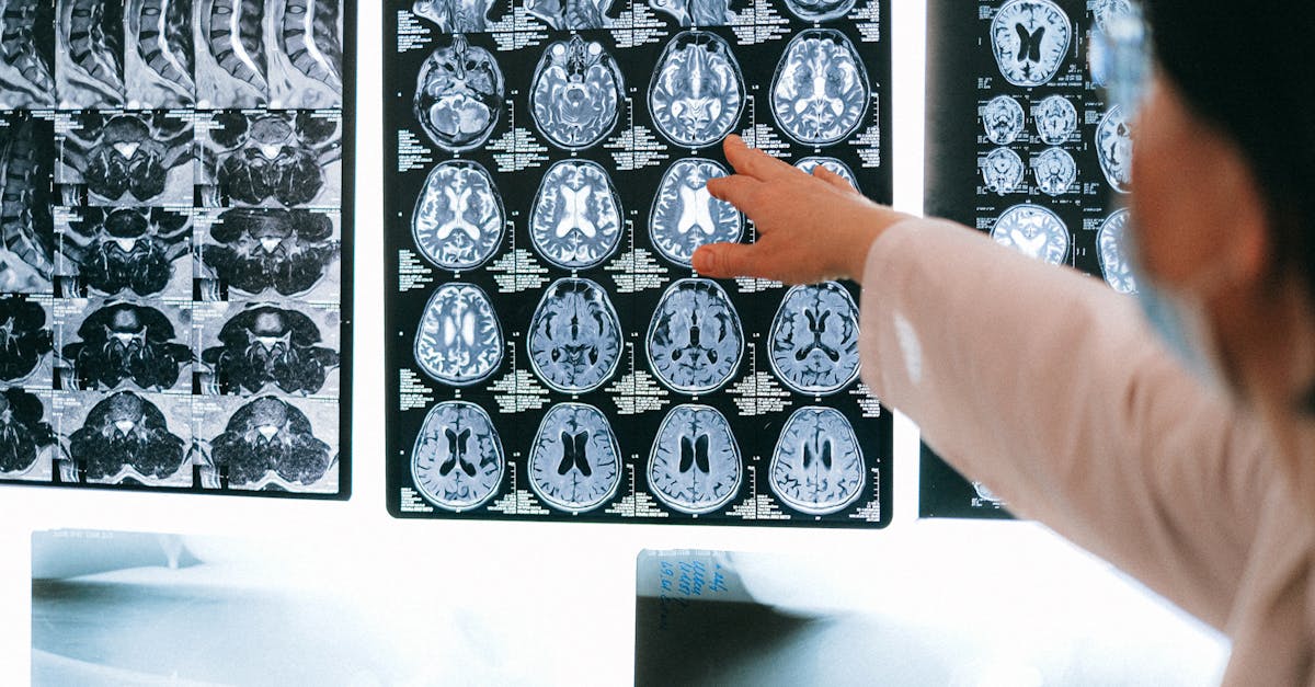
The gold standard for diagnosing a brain tumor is a brain Magnetic Resonance Imaging (MRI) scan with and without intravenous gadolinium contrast. The contrast dye leaks out of the abnormal tumor blood vessels, causing the tumor tissue to light up brightly on the scan, clearly defining its borders, exact location, and proximity to eloquent brain regions.
Advanced MRI techniques, such as functional MRI and diffusion tensor imaging, allow neurosurgeons to map the exact location of motor and speech pathways in relation to the tumor, providing a crucial roadmap for safe surgical resection.
| Tumor Location | Dominant Early Clinical Sign | Neurological Deficit |
|---|---|---|
| Frontal Lobe | Personality shifts, apathy | Loss of executive function and impulse control. |
| Motor Cortex | Progressive hemiparesis | Unilateral weakness in arm, leg, or face. |
| Left Temporal/Parietal | Aphasia, dysnomia | Inability to speak fluently or comprehend language. |
| Cerebellum | Ataxia, severe clumsiness | Staggering gait, loss of fine motor coordination. |
| Any Location (Mass Effect) | Morning headaches, projectile vomiting | Increased intracranial pressure, papilledema. |
13. Therapeutic Avenues
The treatment of a brain tumor requires a highly coordinated multidisciplinary team consisting of neurosurgeons, neuro-oncologists, and radiation oncologists. The initial and most critical step is typically a maximal safe surgical resection. The goal is to remove as much of the tumor as possible to relieve the intracranial pressure without damaging critical healthy brain tissue.
The tissue removed during surgery is sent to a neuropathologist for molecular and genetic profiling. This detailed analysis dictates the follow-up therapy. High-grade, malignant tumors require aggressive postoperative intervention, combining precisely targeted focal radiation therapy with systemic oral chemotherapy agents that cross the blood-brain barrier.
For slow-growing, benign tumors located in highly sensitive areas where surgery is too dangerous, clinicians may opt for stereotactic radiosurgery (such as a Gamma Knife), which delivers a massive, focused dose of radiation directly to the tumor to halt its growth without requiring an open cranial incision.
14. When to Seek Urgent Neurological Care
Neurological symptoms should never be ignored, as they indicate direct compromise of the central nervous system. Early evaluation provides the highest chance of preserving cognitive and motor function.
Seek prompt emergency care if you experience a sudden, unprovoked seizure for the first time, or if you develop rapidly progressive weakness or numbness on one side of your body. An inability to speak, sudden confusion, or profound visual loss are absolute medical emergencies demanding an immediate brain scan.
Furthermore, if you develop a new pattern of intense morning pain resembling a severe headache that is entirely different from anything you have experienced before, especially if it is accompanied by morning vomiting or wakes you from sleep, consult a neurologist or proceed to an emergency department for a comprehensive evaluation.
15. Frequently Asked Questions FAQ
1. Do all brain tumors cause headaches?
No, nearly half of all patients with a brain tumor never experience a headache. Tumors growing in areas with ample space or growing very slowly may only present with focal symptoms, such as weakness or speech difficulty, without causing pressure-related pain.
2. Is a brain tumor a death sentence?
Absolutely not. There are over one hundred distinct types of brain tumors. Many, such as meningiomas and acoustic neuromas, are completely benign, slow-growing, and curable with surgery. Malignant tumors vary widely in their aggressiveness and response to treatment.
3. Why do brain tumors cause seizures?
The tumor mass acts as a physical irritant, pushing against the healthy neurons of the cerebral cortex. This pressure and local inflammation disrupt the delicate chemical balance required for electrical signaling, triggering a chaotic electrical storm known as a seizure.
4. Can a blood test detect a brain tumor?
Currently, there is no standard blood test that can diagnose a brain tumor. Diagnosis relies exclusively on neurological examinations and high-resolution neuroimaging, specifically a contrast-enhanced MRI.
5. Does using a cell phone cause brain tumors?
Decades of extensive, large-scale epidemiological studies have consistently found no conclusive evidence linking the non-ionizing radiofrequency radiation emitted by cell phones to the development of brain tumors.
16. Bibliography
Disclaimer: The content is for informational purposes only and does not replace medical advice. Always consult your doctor for personalized treatment.