1. Introduction to Fascial Fibroma
A fascial fibroma is a highly specific, benign soft tissue proliferation originating from the deep connective tissue fascia. In clinical practice and formal pathology, this entity is overwhelmingly recognized and classified under the diagnostic term nodular fasciitis. It represents a rapid, localized, and self-limiting overgrowth of myofibroblasts, the specialized cells responsible for tissue repair and structural support.
The biological behavior of this lesion is fascinating and frequently alarming. Unlike typical slow-growing benign tumors, a fascial fibroma is characterized by a sudden, explosive growth trajectory. The patient often discovers a rapidly expanding, tender nodule that achieves its maximum size within a matter of mere weeks. This exceptionally rapid clinical presentation routinely raises severe clinical suspicion for a highly aggressive, high-grade soft tissue sarcoma.
Clinical management is entirely centered on securing an accurate histological diagnosis to rule out malignancy. Because the cellular pathology can mimic aggressive cancer under the microscope, evaluation by an expert soft tissue pathologist is mandatory. Once definitively diagnosed, the standard of care emphasizes profound reassurance and conservative observation, as these unique lesions frequently undergo spontaneous, complete regression without the need for extensive surgical intervention.
2. Cellular Origin and Pathophysiology
The fundamental pathophysiology of a fascial fibroma involves the abnormal, localized proliferation of myofibroblasts. Myofibroblasts are specialized connective tissue cells that share characteristics of both structural fibroblasts and contractile smooth muscle cells. They are the primary cellular engines utilized by the body to execute normal wound healing and tissue contraction.
Historically, nodular fasciitis was categorized exclusively as a reactive, inflammatory process triggered by unrecognized localized trauma or infection. However, modern molecular pathology has revolutionized our understanding of this condition. Research demonstrates that the vast majority of these lesions harbor a highly specific, recurrent genetic alteration: the MYH9-USP6 gene fusion.
The presence of this specific gene fusion classifies the fascial fibroma as a true, albeit transient, neoplastic process rather than a simple reactive scar. Despite harboring a distinct genetic mutation, the biological growth program is strictly self-limiting. The myofibroblasts proliferate explosively for a short duration, but the process spontaneously arrests, and the tissue eventually degenerates and resolves naturally.
3. Microscopic Cellular Architecture
The definitive diagnosis of a fascial fibroma requires meticulous microscopic evaluation. The primary diagnostic challenge is that the rapid growth velocity of the tumor is directly mirrored by highly active, alarming cellular features under the microscope, frequently leading to misdiagnoses of aggressive sarcomas by inexperienced pathologists.
The tissue matrix is composed of plump, actively dividing spindle-shaped myofibroblasts. These cells are arranged in loose, intersecting fascicles, frequently described as having a “tissue culture-like” appearance because the cells look identical to rapidly growing fibroblasts in a laboratory petri dish.
The background stroma is highly variable, often appearing myxoid (mucus-like) or edematous, giving the tissue a feathery appearance. The pathologist routinely observes abundant mitotic figures, confirming rapid cellular division. However, crucially, these mitoses are structurally normal. The strict absence of bizarre, atypical mitotic figures, severe nuclear pleomorphism, and large areas of spontaneous tissue necrosis confirms the benign nature of the proliferation.
4. Anatomical Distribution
Fascial fibromas can theoretically develop in any anatomical region containing deep fascial connective tissue. However, they exhibit a distinct predilection for the superficial soft tissues of the upper extremities, particularly the volar aspect of the forearm.
The trunk and the head and neck region are also frequently affected, particularly in pediatric and adolescent patients. Depending on their exact relationship with the deep connective tissue planes, pathologists classify these lesions into three specific anatomical subtypes: subcutaneous, fascial, and intramuscular.
The subcutaneous variant is the most common, presenting as a highly mobile nodule directly beneath the skin. The fascial variant originates directly from the superficial fascia and frequently exhibits a stellate, irregular growth pattern extending outward along the fibrous septa. The intramuscular variant is the deepest and typically the largest, developing entirely within the belly of a skeletal muscle, creating a profound diagnostic challenge.
5. Clinical Presentation and Growth Velocity
The clinical presentation of a fascial fibroma is defined by its alarming temporal timeline. Patients typically present to the clinic in a state of high anxiety, reporting the sudden discovery of a solid lump that has noticeably enlarged over the past two to four weeks.
Upon physical examination, the clinician palpates a firm to rubbery, well-defined nodule. The size is generally small, usually arresting between one and three centimeters in maximum diameter; lesions exceeding five centimeters are exceptionally rare. The mass may be mildly tender to direct palpation, reflecting the rapid tissue distension and localized inflammatory response.
Depending on the anatomical subtype, the nodule may be freely mobile or appear firmly fixed to the underlying deep fascia. Despite the rapid growth, the overlying skin remains completely normal, devoid of any erythema, warmth, or ulceration. You can explore how generalized superficial tissue abnormalities are evaluated in our comprehensive clinical guide regarding skin anomalies.
6. Diagnostic Imaging Strategies
Following the clinical discovery of a rapidly growing soft tissue mass, radiological imaging is mandatory to evaluate the internal characteristics and confirm the anatomical depth of the lesion. Magnetic resonance imaging is the gold standard diagnostic modality for all complex soft tissue tumors.
On magnetic resonance imaging, a fascial fibroma typically appears as a relatively small, well-circumscribed, ovoid mass. Because the internal stroma is frequently edematous or myxoid, the tumor typically exhibits high signal intensity (appearing distinctly bright) on T2-weighted sequences.
Following the administration of intravenous contrast, the mass generally demonstrates prominent, uniform enhancement due to the rich capillary network supporting the rapid cellular growth. While the magnetic resonance imaging clearly defines the size and boundaries of the lesion, it cannot reliably differentiate this rapidly growing benign nodule from a highly aggressive, small soft tissue sarcoma. Therefore, a tissue biopsy is absolutely required.
7. The Crucial Role of Core Needle Biopsy
Securing a definitive histological diagnosis prior to undertaking any major surgical intervention is the cornerstone of safe oncological practice. For a rapidly growing soft tissue mass, an ultrasound-guided core needle biopsy is the mandatory procedure utilized to secure diagnostic tissue.
The radiologist extracts several intact, cylindrical tissue cores from the center of the nodule. This procedure preserves the crucial architectural arrangement of the myofibroblasts and the background stroma, providing the pathologist with the spatial context required to identify the characteristic “tissue culture” appearance.
Fine-needle aspiration is strictly contraindicated for these specific lesions. Extracting loose, individual, rapidly dividing myofibroblasts via aspiration will almost certainly lead to an alarming, false-positive diagnosis of a malignant sarcoma, potentially resulting in a disastrously unnecessary radical surgery.
8. Differentiating from Soft Tissue Sarcomas
The absolute most critical diagnostic task for the pathologist is differentiating the benign fascial fibroma from high-grade soft tissue sarcomas, specifically fibrosarcoma, myxofibrosarcoma, and spindle cell variants of melanoma or carcinoma.
The diagnostic overlap is severe because all these entities present clinically as rapidly enlarging masses and exhibit high cellularity and abundant mitotic division under the microscope. The pathologist must meticulously evaluate the cells at high magnification.
In a fascial fibroma, the cells, despite dividing rapidly, remain uniform, bland, and perfectly regular in shape. There is no atypical, bizarre cellular mutation. Furthermore, if the diagnosis remains ambiguous, modern molecular pathology laboratories can perform advanced genetic testing on the biopsy tissue. Confirming the presence of the specific MYH9-USP6 gene fusion definitively diagnoses a benign fascial fibroma and completely rules out any malignant sarcoma.
9. Data Summary: Differentiating Rapid Soft Tissue Masses
To clarify the critical distinctions guiding surgical planning, the following table summarizes the differences between benign fascial fibromas and aggressive sarcomas.
| Diagnostic Feature | Fascial Fibroma (Nodular Fasciitis) | Soft Tissue Sarcoma |
|---|---|---|
| Growth Timeline | Explosive growth, arrests at 2-3 cm | Continuous, relentless enlargement |
| Cellular Atypia | Strictly Absent, bland uniform cells | Severe, profound nuclear pleomorphism |
| Mitotic Figures | Abundant, but structurally normal | Abundant, frequently abnormal/bizarre |
| Gene Fusion | MYH9-USP6 frequently positive | Negative for this specific fusion |
10. The Paradigm of Spontaneous Regression
Once the core needle biopsy definitively confirms the diagnosis of a fascial fibroma, the clinical management paradigm shifts dramatically. Historically, these lesions were routinely excised surgically. However, extensive clinical research has conclusively demonstrated that these specific tumors are entirely self-limiting.
Following the initial explosive growth phase lasting a few weeks, the myofibroblastic proliferation spontaneously arrests. Subsequently, the cellular matrix begins a process of natural degeneration. Over a period of several weeks to months, the nodule shrinks and frequently disappears entirely without any medical or surgical intervention.
Understanding this biological life cycle is crucial. The primary medical intervention is providing profound clinical reassurance to the highly anxious patient, educating them that the alarming mass is a temporary, harmless condition that the body will resolve naturally.
11. Conservative Observation Strategy
The absolute gold standard of clinical care for a confirmed fascial fibroma is active, conservative observation. Because the lesion possesses zero potential for malignant transformation and will likely resolve spontaneously, surgical removal is deemed medically unnecessary.
The standard surveillance protocol involves returning to the clinic for a physical examination and targeted ultrasound imaging every four to six weeks to monitor the dimensions of the nodule.
During this observation period, the clinician documents the cessation of growth and the initiation of regression. Once the nodule demonstrates a clear pattern of decreasing in size, the clinical team and the patient are fully reassured, and active medical surveillance is typically discontinued.
12. Indications for Surgical Excision
Although spontaneous regression is the expected outcome, specific clinical scenarios justify the surgical removal of a fascial fibroma. The primary indication for surgical intervention is diagnostic uncertainty. If the core needle biopsy yields ambiguous results or if the pathologist cannot definitively rule out a low-grade sarcoma, complete surgical removal is absolutely necessary for an exhaustive, whole-tumor evaluation.
Surgery is also indicated if the tumor violates the typical biological timeline. If the mass continues to grow aggressively beyond a few weeks, or if it exceeds five centimeters in diameter, the diagnosis of a benign fascial fibroma must be severely questioned, mandating surgical excision.
Finally, elective excision is frequently performed if the nodule causes the patient significant, persistent pain due to nerve compression, or if the psychological burden of waiting for a palpable lump to disappear naturally causes the patient severe and unmanageable anxiety.
13. Surgical Techniques and Marginal Excision
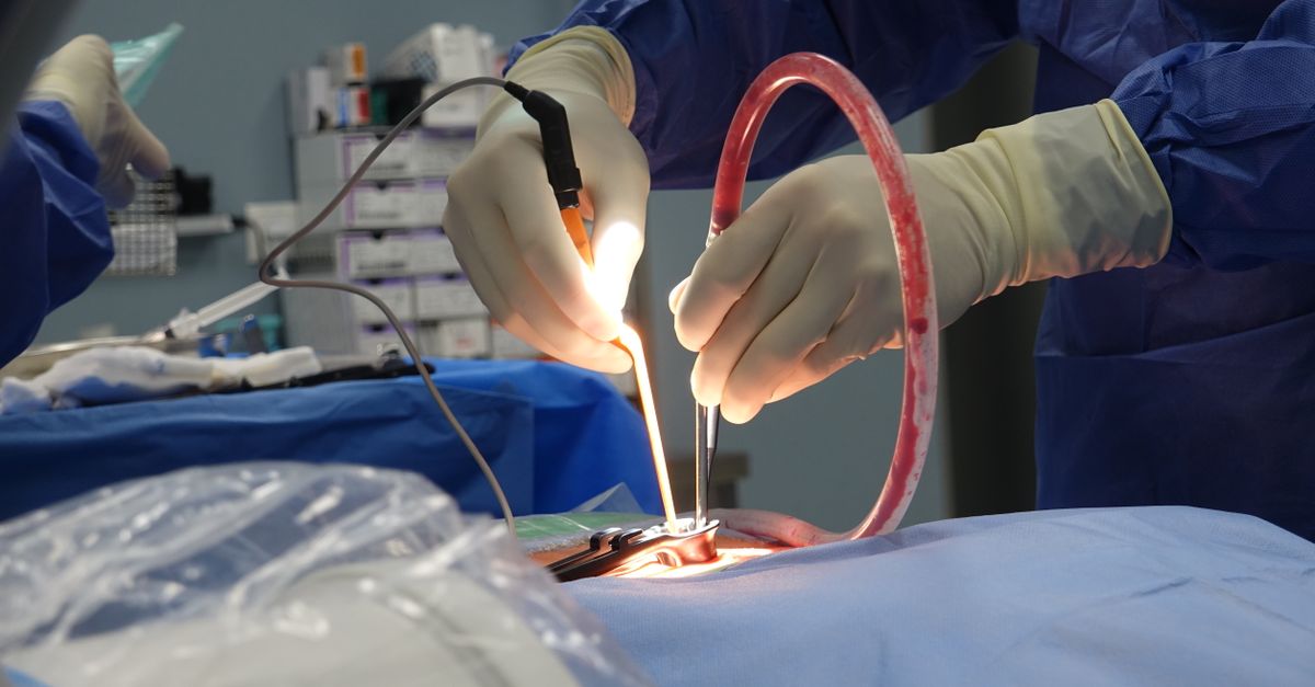
When surgical removal is indicated, the procedure is generally straightforward. The excision is typically performed on an outpatient basis under local or regional anesthesia, depending on the anatomical depth and location of the tumor.
Because a fascial fibroma is strictly non-malignant, wide, radical surgical margins are completely unnecessary. The surgeon performs a marginal excision, carefully dissecting close to the visible boundary of the nodule, ensuring complete removal of the gross mass while preserving all surrounding healthy muscle fibers, nerves, and blood vessels.
This precise, tissue-sparing approach minimizes surgical trauma, limits postoperative pain, and ensures an excellent functional and cosmetic outcome for the patient. The resulting tissue defect heals naturally without the need for complex reconstructive flap closures.
14. Adjuvant Therapy Contraindications
Given the strictly benign biological nature of a fascial fibroma, adjuvant systemic therapies or localized radiation treatments are absolutely contraindicated under any clinical circumstance.
Standard sarcoma treatments, including aggressive cytotoxic chemotherapy and targeted radiation fields, provide zero clinical benefit for these self-limiting lesions. Employing these therapies would unnecessarily expose the patient to severe systemic toxicities and long-term morbidity without improving their already excellent prognosis.
The medical consensus confirms that simple observation or marginal surgical extirpation of the mass provides optimal and sufficient local control, rendering any adjuvant oncological interventions entirely obsolete.
15. Prognosis and Clinical Follow-up
The long-term prognosis for a patient diagnosed with a fascial fibroma is outstanding. For patients managed observationally, the lesion reliably undergoes complete spontaneous regression, leaving behind normal, healthy tissue.
For patients who undergo a marginal surgical excision, the procedure is entirely curative. The risk of local recurrence at the exact surgical site is exceptionally low, estimated at less than two percent. If a recurrence does occur, it almost universally represents an incomplete initial excision rather than aggressive tumor behavior, and simply requires a minor secondary excision.
A diagnosis of a fascial fibroma does not elevate a patient lifetime statistical risk of developing soft tissue sarcomas or any other malignancies in the future. Once the mass has regressed or been successfully excised, the patient is officially discharged from active surgical care with profound clinical reassurance.
16. Frequently Asked Questions (FAQ)
1. Is a fascial fibroma a type of fast-growing cancer?
No. Despite its alarming, rapid growth, it is a strictly benign, temporary overgrowth of normal tissue repair cells. It completely lacks the ability to spread to other organs and poses no threat to your overall health.
2. Why did this lump appear so suddenly?
The specific cells involved in this condition, called myofibroblasts, are designed to multiply rapidly to heal wounds. In this condition, a minor genetic glitch causes them to activate and multiply quickly, forming a lump within a few weeks before shutting down again.
3. Will the lump eventually go away without surgery?
Yes, in the vast majority of cases. Once the rapid growth phase stops, the body naturally breaks down the extra tissue, and the lump typically shrinks and disappears completely over a few months.
4. Why did the doctor insist on a biopsy if it is benign?
Because this benign lump grows so rapidly, it looks remarkably similar to a very dangerous, aggressive cancer called a sarcoma. A biopsy is the absolute only way to examine the cells under a microscope and prove definitively that it is harmless.
5. If I choose to have surgery, will it damage my muscles?
No. Surgeons use precise, tissue-sparing techniques. Because the tumor is not a destructive cancer, the surgeon only needs to remove the lump itself, safely peeling it away from your healthy muscle fibers and nerves.
17. Bibliography
Disclaimer: The content is for informational purposes only and does not replace medical advice. Always consult your doctor for personalized treatment.