1. Introduction to Soft Tissue Giant Cell Tumors
Giant cell tumor of soft tissue is an exceptionally rare primary neoplasm that arises outside of the skeletal system. Histologically, it closely mimics the cellular architecture of its more common counterpart found in the bone. Despite this microscopic resemblance, the soft tissue variant represents a distinct clinical entity with specific diagnostic criteria and behavioral patterns.
These tumors typically manifest as painless, enlarging masses located in either the superficial or deep soft tissues of the extremities. Because soft tissue masses can represent a wide spectrum of pathologies ranging from harmless cysts to aggressive sarcomas, precise medical evaluation is paramount to ensure accurate categorization and proper therapeutic intervention.
Clinical management requires careful differentiation from other giant cell-rich soft tissue anomalies. Providing an accurate diagnosis ensures that patients receive the appropriate surgical care without undergoing unnecessary, overly radical procedures or, conversely, inadequate excisions that lead to rapid disease recurrence.
2. Pathophysiological Foundations
The fundamental pathophysiology of a soft tissue giant cell tumor mirrors the complex cellular dynamics seen in bone tumors. The mass is composed of a biphasic cellular population. The primary neoplastic element consists of mononuclear stromal cells that drive the abnormal tissue growth within the fascial or muscular planes.
These specialized stromal cells secrete specific cytokine signals that attract circulating monocytes directly into the surrounding soft tissue matrix. Under the influence of the tumor microenvironment, these recruited monocytes fuse to form multinucleated osteoclast-like giant cells.
Unlike bone tumors where these giant cells dissolve mineralized matrix, in the soft tissues, they form a dense, nodular mass rich in vascular networks. The tumor expands by displacing rather than directly invading adjacent muscle fibers and subcutaneous fat, creating a well-circumscribed but highly cellular mass.
3. Superficial Versus Deep Tumors
Giant cell tumors of soft tissue are anatomically classified into superficial and deep categories. Superficial lesions develop in the subcutaneous tissues, just beneath the dermal layers. These are more easily palpable, often recognized early by the patient as a discrete, mobile lump just under the skin.
Deep lesions arise within the intramuscular spaces or along deep fascial planes. Because they are constrained by thick muscle groups, they may grow significantly larger before becoming visibly apparent or causing noticeable mechanical symptoms.
The anatomical depth influences the surgical approach. Superficial masses are generally amenable to straightforward excision. Deep lesions require more complex surgical navigation to protect adjacent major nerves and blood vessels while ensuring complete removal of the tumor capsule.
4. Typical Patient Presentation
Patients typically present to the clinic noting a slow-growing, solid mass. The upper and lower extremities, particularly the thighs and forearms, are the most common anatomical sites. Less frequently, these tumors may present on the trunk or the head and neck region.
Pain is generally not a primary symptom in the early stages. The mass is usually firm to the touch and moves slightly when palpated, indicating it is not tethered to the underlying bone. Rapid growth is unusual and should immediately raise clinical suspicion for a more aggressive form of soft tissue abnormalities.
When deep tumors reach a significant volume, they can cause pressure-related symptoms. Patients may experience localized aching, muscle fatigue, or radiating nerve pain if the mass impinges upon a peripheral nerve pathway traversing the muscle compartment.
5. Diagnostic Imaging Modalities
Initial evaluation of any unidentified soft tissue mass usually begins with ultrasonography. Ultrasound provides real-time, dynamic imaging to confirm the solid nature of the tumor and assess its internal vascularity, effectively distinguishing it from simple fluid-filled cysts or localized hematomas.
Magnetic resonance imaging remains the gold standard for comprehensive soft tissue evaluation. It offers precise anatomical localization, defining the relationship of the tumor to deep fascial layers, muscles, and vascular bundles. The tumor typically demonstrates specific signal intensities that, while not entirely diagnostic, help guide the surgical approach.
Computed tomography is less frequently utilized for the primary mass but is an essential component of systemic staging. A computed tomography scan of the chest is standard protocol to screen for the presence of asymptomatic pulmonary metastases, a known although rare complication of this specific tumor type.
6. Histological Hallmarks
A definitive diagnosis rests entirely on expert histopathological examination of biopsy tissue. Core needle biopsy provides sufficient material for the pathologist to analyze the architectural patterns and cellular constituents of the lesion.
Microscopically, the hallmark is a highly cellular proliferation of mononuclear cells intimately admixed with numerous, large, multinucleated osteoclast-like giant cells. The tissue is highly vascular, often containing areas of micro-hemorrhage and deposits of hemosiderin, a byproduct of broken-down red blood cells.
| Microscopic Component | Description in Soft Tissue GCT |
|---|---|
| Mononuclear Cells | Round to spindle-shaped, uniform nuclei |
| Giant Cells | Osteoclast-like, evenly dispersed |
| Vascularity | Prominent thin-walled blood vessels |
| Matrix Formation | Focal areas of metaplastic bone occasionally seen |
7. Differential Diagnosis
Pathologists face the complex task of distinguishing a primary soft tissue giant cell tumor from other lesions that contain high numbers of giant cells. This differential diagnosis is critical because the required treatments vary significantly between different conditions.
A primary differential includes the giant cell tumor of tendon sheath, which typically occurs in the hands and feet and displays different cellular ratios and structural collagen deposits. Extraskeletal osteosarcoma with giant cells must also be rigorously excluded, as it is a high-grade malignancy requiring aggressive chemotherapy.
Advanced immunohistochemical staining is employed to clarify ambiguous cases. Soft tissue giant cell tumors lack certain specific protein markers found in distinct sarcomas, allowing pathologists to rule out more aggressive mimickers and secure the correct benign classification.
8. Benign and Malignant Subtypes
The vast majority of soft tissue giant cell tumors follow a benign clinical course. They exhibit localized growth and do not possess the capacity to widely disseminate throughout the body. However, their biological behavior remains unpredictable regarding local site recurrence.
A very small subset of these tumors can present with malignant histological features from the outset, or undergo malignant transformation over time. Malignant variants demonstrate high mitotic activity, profound cellular atypia, and extensive areas of spontaneous cellular necrosis.
The distinction between benign and malignant behavior dictates the urgency and extent of surgical intervention. A diagnosis of malignant giant cell tumor of soft tissue shifts the treatment paradigm from a simple excision to a comprehensive oncological protocol often involving radiation and expanded surgical margins.
9. Principles of Surgical Excision
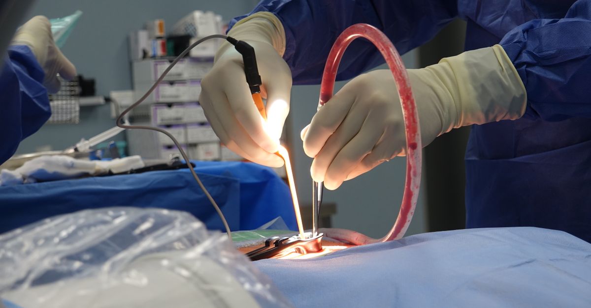
The cornerstone of treatment for a benign soft tissue giant cell tumor is complete surgical excision. The objective is to remove the entire tumor mass along with an intact pseudo-capsule, ensuring that no microscopic clusters of neoplastic cells are left behind in the tumor bed.
Surgeons aim for marginal or wide excision depending on the proximity to vital structures. If a tumor is intimately wrapped around a major nerve or blood vessel, a careful marginal dissection is performed to peel the mass away while preserving limb function and sensation.
Meticulous hemostasis is required during the procedure due to the inherent vascularity of these tumors. Postoperative hematomas can complicate wound healing and obscure the interpretation of future follow-up magnetic resonance imaging scans.
10. Radiotherapy Considerations
Adjuvant radiotherapy is generally not indicated for standard benign soft tissue giant cell tumors that have been completely excised. The risks of radiation-induced complications usually outweigh the benefits in fully resected, non-malignant disease.
However, in specific clinical scenarios where the tumor location prevents complete surgical removal without causing severe functional impairment, radiation therapy may be considered as an adjunctive measure to control residual microscopic disease.
In the rare instances of definitively malignant soft tissue giant cell tumors, radiotherapy plays a central role. It is often administered prior to surgery to shrink the tumor borders, or postoperatively to sterilize the surgical bed and reduce the likelihood of aggressive local recurrence.
11. Recovery and Wound Care
Postoperative recovery depends largely on the anatomical depth and size of the excised tumor. Superficial resections usually require minimal downtime. Patients are advised to keep the incision clean and avoid strenuous mechanical stretching of the overlying skin until the wound is fully consolidated.
Deep intramuscular resections require more structured rehabilitation. Removing mass from within a muscle belly causes localized tissue trauma and internal scarring. Patients often participate in directed physical therapy to restore flexibility, break up scar tissue adhesions, and regain full muscular strength.
Surgeons monitor the wound closely for signs of seroma fluid accumulation or delayed healing, addressing any minor complications early to ensure an optimal cosmetic and functional outcome.
12. Local Recurrence Rates
The local recurrence rate for giant cell tumors of soft tissue is recognized in the oncological literature, though it is generally lower than that of its bony counterpart. Recurrences usually occur when microscopic disease escapes surgical detection during the primary excision.
When a recurrence develops, it presents as a new nodular growth within or directly adjacent to the original surgical scar. Re-excision is the standard clinical response. Recurrent tumors are carefully analyzed by pathologists to ensure the cellular biology has not evolved into a more aggressive or malignant phenotype.
13. Clinical Surveillance Protocols
Because of the recognized risk of local return, patients enter a formal surveillance program following surgical recovery. Regular clinical physical examinations of the surgical site are paramount.
Baseline magnetic resonance imaging of the operative bed is usually obtained a few months post-surgery to establish a new normal for the surgical scar. Subsequent imaging is performed periodically over the next several years to detect any non-palpable early recurrences deep within the tissue planes.
Patients are counseled on self-examination techniques and are instructed to promptly report any new palpable nodules, localized swelling, or changes in sensation near the primary surgical site.
14. Frequently Asked Questions
1. Is a soft tissue giant cell tumor the same as a bone giant cell tumor?
They share an almost identical microscopic appearance but are distinct clinical entities. The soft tissue variant forms in muscles or fat instead of bone and generally has a slightly different pattern of growth and recurrence.
2. Does this mass mean I have soft tissue sarcoma?
No. The vast majority of these giant cell tumors are classified as benign. They require surgical removal to prevent local growth but are not considered high-grade sarcomas.
3. Will a needle biopsy spread the tumor cells?
Core needle biopsies are highly safe and routine. Surgeons carefully plan the biopsy pathway so that the entire needle tract is safely removed during the main surgery, preventing any cellular spread.
4. How large can these tumors grow?
If left untreated, deep tumors can grow to several centimeters in diameter, eventually causing pressure on nearby muscles and nerves, which highlights the need for elective surgical removal.
5. Will I need physical therapy after the removal?
If the tumor was located deep within a major muscle group, physical therapy is often recommended to help stretch the healing scar tissue and restore full strength to the affected limb.
15. Bibliography
Disclaimer: The content is for informational purposes only and does not replace medical advice. Always consult your doctor for personalized treatment.