1. Introduction
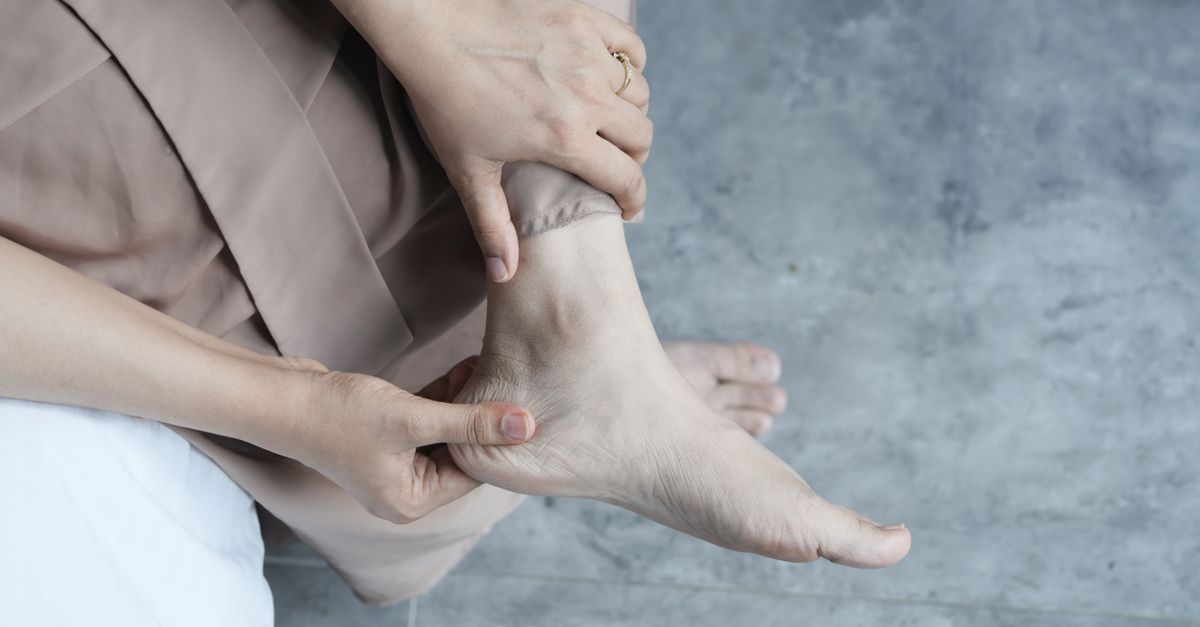
Aching pain under the heel after walking five thousand steps is primarily caused by mechanical stress and inflammation of the plantar fascia, a thick band of tissue connecting the heel bone to the toes. Repetitive impact from walking can create microtears in this tissue, leading to localized inflammation and discomfort. Addressing this pain requires understanding the biomechanical forces placed on the foot and identifying any underlying structural vulnerabilities.
2. Anatomy of the Plantar Fascia
The plantar fascia is a robust aponeurosis that runs along the sole of the foot. It originates at the medial tubercle of the calcaneus, which is the heel bone, and extends forward to attach to the base of the proximal phalanges. This tissue is crucial for maintaining the longitudinal arch of the foot.
During the gait cycle, the plantar fascia acts as a shock absorber and a dynamic tension bridge. It distributes the weight of the body across the foot and assists in propulsion during the push-off phase of walking.
When structural integrity is compromised, the ability of the fascia to absorb mechanical loads diminishes. This localized tissue failure is a frequent starting point for chronic heel pain syndromes.
3. The Biomechanics of Walking
Walking places a significant amount of ground reaction force on the feet. With every step, the heel strikes the ground first, absorbing a force that can equal or exceed total body weight. This impact is transmitted upward through the skeletal system.
As the body moves forward over the planted foot, the arch flattens slightly. This flattening stretches the plantar fascia. During a walk of five thousand steps, this stretching and contracting cycle repeats thousands of times, generating immense repetitive tensile strain on the tissue attachment at the heel.
Any deviation in optimal biomechanics, such as overpronation where the foot rolls inward excessively, amplifies this tensile strain. Over time, the structural limits of the soft tissue are exceeded, triggering an inflammatory cascade.
4. Understanding Plantar Fasciitis
Plantar fasciitis is the most prevalent clinical diagnosis for inferior heel pain. It is characterized by acute or chronic pain at the bottom of the heel, especially notable after periods of rest or prolonged weight-bearing activities. The term implies inflammation, though chronic cases often demonstrate collagen degeneration rather than active inflammatory cells.
The primary site of tissue damage is the enthesis, the area where the fascia inserts into the heel bone. Repetitive microtrauma from walking outpaces the natural healing capacity of the body.
Patients typically describe a sharp or bruised sensation directly under the heel. The pain is often proportional to the volume of activity, explaining why discomfort peaks after reaching a specific threshold like five thousand steps.
5. Heel Spurs and Bone Stress
A calcaneal spur is a bony outgrowth that develops on the underside of the heel bone. Historically, heel spurs were incorrectly identified as the primary source of heel pain. Current clinical understanding indicates that spurs are a secondary response to chronic tension applied by the plantar fascia.
The body attempts to reinforce the stressed attachment site by depositing calcium, forming the spur. While the spur itself is rarely the direct source of pain, its presence confirms long-standing mechanical stress and chronic inflammation in the region.
In some cases, the pain originates from bone marrow edema within the calcaneus itself. This stress reaction occurs when the bone is subjected to impact forces that exceed its remodeling capacity.
6. Achilles Tendinopathy Considerations
The Achilles tendon and the plantar fascia function synergistically. They are connected via a continuous fascial plane around the heel bone. Tightness or dysfunction in the Achilles tendon can directly influence the tension applied to the plantar fascia.
When calf muscles are tight, ankle dorsiflexion is restricted. This limitation forces the midfoot to compensate during the gait cycle, increasing the load on the plantar arch.
Managing heel pain often requires addressing the entire posterior kinetic chain, including the calf muscles and the Achilles tendon, to ensure equitable distribution of mechanical forces.
7. Fat Pad Atrophy in the Heel
The heel is protected by a specialized layer of adipose tissue known as the plantar fat pad. This structure acts as a primary shock absorber during heel strike. It features unique fibrous septa that lock the fat in place under pressure.
With advancing age or after repetitive high-impact activities, this fat pad can thin out or lose its elasticity, a condition known as fat pad atrophy. When the protective cushioning is diminished, the underlying heel bone is exposed to greater impact forces.
Pain originating from fat pad atrophy feels like a deep, dull ache directly in the center of the heel pad. Unlike plantar fasciitis, this discomfort tends to worsen uniformly with prolonged standing or walking on hard surfaces.
8. Nerve Entrapment and Tarsal Tunnel Syndrome
Inferior heel pain can occasionally stem from a neurological origin. Tarsal tunnel syndrome involves the compression of the posterior tibial nerve as it passes through a narrow anatomical tunnel on the inside of the ankle.
Branches of this nerve, specifically the medial calcaneal nerve, supply sensation to the bottom of the heel. Compression or irritation of these nerve branches can radiate pain, tingling, or a burning sensation into the heel region.
This type of pain might worsen with increased physical activity due to swelling in the tissues surrounding the nerve. Differentiating nerve entrapment from mechanical tissue strain requires a careful clinical evaluation.
9. Systemic Inflammatory Conditions
While mechanical overload is the most common culprit, systemic inflammatory conditions must be considered in the differential diagnosis. Conditions belonging to the spondyloarthropathy family can present with persistent heel pain.
These systemic conditions often cause enthesitis, which is inflammation at the sites where tendons and ligaments attach to bone. The attachment of the plantar fascia is a classic site for such systemic inflammation.
If heel pain is bilateral, accompanied by morning stiffness in the lower back, or unresponsive to standard mechanical treatments, a clinical investigation into autoimmune or systemic inflammatory markers is warranted.
10. The Role of Footwear and Arch Support
Footwear choices play a critical role in the mechanical loading of the foot. Shoes that lack adequate arch support or shock absorption force the intrinsic muscles and the plantar fascia to absorb the entirety of the ground reaction forces.
Walking thousands of steps in flat, unsupportive shoes, or shoes with worn-out outsoles, rapidly accelerates tissue fatigue. The heel counter of the shoe is also vital for stabilizing the calcaneus upon impact.
Transitioning to footwear designed with structural integrity, proper cushioning, and an appropriate heel-to-toe drop can immediately reduce the mechanical strain on the inferior heel.
11. Diagnostic Approaches and Clinical Evaluation
Diagnosing the exact cause of heel pain relies on a comprehensive clinical history and a focused physical examination. Palpation of specific anatomical landmarks helps isolate the involved tissues. Pain elicited by pressing the medial calcaneal tubercle strongly points to plantar fasciitis.
Clinicians assess joint mobility, muscle strength, and gait mechanics to identify contributing biomechanical faults. A visual inspection of the foot architecture helps determine if a high arch or a flat foot is contributing to the problem.
Imaging modalities are utilized when conservative measures fail or to rule out specific pathologies.
| Diagnostic Imaging | Clinical Utility in Heel Pain |
|---|---|
| Plain Radiography | Identifies fractures, bone tumors, and calcaneal spurs. |
| Diagnostic Ultrasound | Evaluates the thickness of the plantar fascia and identifies tears. |
| Magnetic Resonance Imaging | Assesses bone marrow edema, soft tissue inflammation, and nerve compression. |
12. Conservative Treatment Modalities
Initial management of mechanical heel pain focuses on reducing tissue strain and modulating the inflammatory response. Rest or activity modification is the cornerstone of conservative care, allowing the overwhelmed tissues time to heal.
Stretching protocols targeting the plantar fascia and the calf musculature are highly effective. These exercises improve tissue compliance and reduce tension at the insertion point. Icing the area after walking can provide temporary symptomatic relief by causing vasoconstriction.
Over-the-counter nonsteroidal anti-inflammatory medications can assist in managing acute pain flares, though they do not alter the underlying biomechanical issues.
13. Preventive Strategies and Foot Care
Preventing the recurrence of heel pain requires ongoing management of foot health. Gradual progression in walking distance allows the soft tissues to adapt to increased loads safely. Spikes in activity volume are a primary trigger for tissue failure.
Maintaining a healthy body weight is a direct way to reduce the total mechanical load applied to the feet during walking. Every step taken multiplies the body weight force, meaning even a modest weight reduction yields significant benefits for the lower extremities.
Consistent use of supportive footwear, even inside the home, prevents the arches from collapsing and maintains optimal tissue resting length.
14. When to Seek Medical Attention
While many cases of post-activity heel pain resolve with rest and basic interventions, specific symptoms warrant prompt medical evaluation. Pain that is severe, unrelenting, or present during non-weight-bearing periods is a cause for concern.
If the heel becomes visibly swollen, red, or warm to the touch, this may indicate an acute inflammatory process or infection. Furthermore, an inability to bear any weight on the affected foot could signal a stress fracture of the calcaneus.
Early consultation with a specialist ensures an accurate diagnosis and prevents the progression of a minor strain into a chronic, debilitating condition.
15. Frequently Asked Questions (FAQ)
1. Why does the pain start only after a certain distance?
Tissue failure often occurs once a specific mechanical threshold is crossed. The muscles and fascia can absorb shock adequately for a certain period, but sustained repetitive loading eventually leads to fatigue, microtears, and subsequent pain.
2. Should I continue walking if my heel aches?
Pushing through sharp pain often exacerbates tissue damage and prolongs recovery. It is advisable to rest the foot and modify activity levels until the acute discomfort subsides.
3. Can stretching really help mechanical heel pain?
Yes. Stretching the calf muscles and the plantar fascia directly reduces the tension placed on the heel bone insertion point. Improved flexibility allows for better force distribution during the gait cycle.
4. Are custom orthotics necessary for this type of pain?
Not always. Many individuals find adequate relief with supportive, off-the-shelf footwear or basic over-the-counter arch supports. Custom orthotics are typically reserved for complex biomechanical abnormalities or cases resistant to initial conservative therapy.
5. How long does this type of heel discomfort take to heal?
Recovery timelines vary based on the extent of tissue damage. Mild strains may resolve within a few weeks with rest, whereas chronic degenerative conditions like severe plantar fasciitis can require several months of consistent rehabilitation.
16. Bibliography
Disclaimer: The content is for informational purposes only and does not replace medical advice. Always consult your doctor for personalized treatment.