1. Introduction to Intracranial Meningioma
An intracranial meningioma is a primary tumor of the central nervous system that originates not from the brain tissue itself, but from the meninges—the three protective membranous layers enveloping the brain and spinal cord. Meningiomas are the most frequently diagnosed primary intracranial tumors in adults. The vast majority of these tumors are benign, characterized by a slow, indolent growth pattern that frequently allows them to remain clinically silent for many years before discovery.
Because they grow inward from the coverings of the brain, meningiomas typically compress adjacent neural structures rather than invading and destroying them. However, within the rigid confines of the skull, even a slow-growing, benign mass eventually exerts significant pressure on delicate cortical tissues, cranial nerves, or vital blood vessels. This mechanical compression is responsible for the diverse array of neurological symptoms associated with the disease.
Medical management of an intracranial meningioma is highly individualized. While many small, asymptomatic tumors require only careful observation, expanding tumors that provoke symptoms necessitate intervention. Modern neurosurgery and advanced stereotactic radiosurgery provide highly effective, curative treatment options for most patients, though precise anatomical location dictates the complexity and risk of these procedures.
2. Anatomy of the Meninges
To comprehend the origin and behavior of a meningioma, one must understand the anatomy of the cranial meninges. The brain is protected by three distinct layers of tissue situated directly beneath the skull. The outermost layer is the dura mater, a tough, thick, inelastic fibrous membrane closely attached to the inner table of the cranial vault.
Beneath the dura lies the arachnoid mater, a delicate, web-like membrane. The innermost layer is the pia mater, which adheres tightly to the complex contours and folds of the brain’s surface. Between the arachnoid and pia mater is the subarachnoid space, which contains the circulating cerebrospinal fluid.
Meningiomas arise specifically from arachnoid cap cells. These specialized cells are most densely clustered along the arachnoid granulations—the structures responsible for reabsorbing cerebrospinal fluid into the venous system. Because arachnoid cap cells are distributed throughout the entire meningeal system, a meningioma can theoretically develop anywhere within the cranial vault or along the spinal canal.
3. Tumor Origin and Biology
The transformation of normal arachnoid cap cells into a neoplastic meningioma is driven by specific genetic alterations. These tumors exhibit a remarkably uniform, slow-growing biology. As they proliferate, they typically form a well-circumscribed, firm, and heavily vascularized mass that adheres broadly to the overlying dura mater.
A defining characteristic of a typical benign meningioma is its sharp demarcation from the underlying brain parenchyma. The tumor pushes against the brain, creating a distinct indentation, but a thin layer of the pia mater and a plane of cerebrospinal fluid almost always separate the tumor from the functional brain tissue. This distinct cleavage plane is a critical feature that allows neurosurgeons to safely dissect and remove the tumor without resecting actual brain matter.
However, as the tumor expands, it requires a robust blood supply. Meningiomas frequently parasitize their blood supply from the dural arteries, most notably branches of the external carotid artery. This intense vascularity makes these tumors highly prone to bleeding during surgical resection, requiring meticulous preoperative planning and intraoperative hemostasis.
4. The World Health Organization Grading System
Pathologists classify meningiomas based on their microscopic cellular features and their propensity for aggressive behavior, utilizing a standardized grading system established by the World Health Organization (WHO). This grading is paramount for determining the patient’s long-term prognosis and the need for adjuvant therapies.
WHO Grade I meningiomas account for approximately 80 percent of all cases. These are classically benign, slow-growing tumors with well-differentiated cells and low proliferative activity. If a Grade I tumor is completely surgically removed, the risk of recurrence is exceptionally low.
WHO Grade II, or atypical meningiomas, comprise about 15 to 20 percent of cases. These exhibit increased cellularity, a faster growth rate, and a higher tendency to invade the adjacent dura or bone. They carry a significantly higher rate of recurrence following surgery. WHO Grade III, or anaplastic (malignant) meningiomas, are very rare (1 to 2 percent). They are highly aggressive, rapidly dividing tumors that can frankly invade the brain tissue and possess a high likelihood of recurrence and metastasis.
5. Genetic and Environmental Risk Factors
The precise etiology of meningiomas remains the subject of ongoing research, but specific genetic and environmental risk factors have been definitively identified. The most established environmental risk factor is prior exposure to ionizing radiation, particularly high-dose radiation administered to the head or neck for childhood malignancies, which can induce meningioma formation decades later.
Genetically, a significant proportion of sporadic meningiomas exhibit mutations in the NF2 gene on chromosome 22. This gene is responsible for producing merlin, a tumor suppressor protein. When this gene is inactivated, arachnoid cap cells lose their normal growth regulation.
Furthermore, individuals with the inherited genetic disorder Neurofibromatosis type 2 possess a profound predisposition to developing multiple meningiomas at a young age, alongside other nervous system tumors like bilateral vestibular schwannomas. Hormonal influences are also strongly suspected, given that meningiomas are approximately twice as common in women than in men, and tumor cells frequently express progesterone and estrogen receptors.
6. Common Anatomical Locations
Because the meninges cover the entire central nervous system, meningiomas can arise in highly diverse locations, dictating their specific clinical presentation and surgical complexity.
Parasagittal and falcine meningiomas are among the most common. They grow along the falx cerebri, the thick fold of dura mater separating the left and right hemispheres of the brain. Convexity meningiomas grow on the outer surface of the brain directly beneath the skull, making them generally the most accessible for surgical removal.
Skull base meningiomas present significant clinical challenges. Tumors located on the sphenoid wing, the olfactory groove, or the petroclival region grow intimately among the critical cranial nerves and major cerebral arteries entering the base of the skull. Surgically resecting tumors in these locations carries a much higher risk of inducing permanent neurological deficits, requiring highly specialized skull base neurosurgical techniques.
7. Neurological Symptoms and Compression
The clinical presentation of an intracranial meningioma is largely dependent on the tumor’s size, its specific location, and the rate at which it grows. Because Grade I tumors grow very slowly, the brain has an astonishing capacity to adapt to the increasing pressure, often delaying the onset of symptoms until the tumor reaches a substantial size.
When symptoms do occur, they are the direct result of localized compression of adjacent neural tissue or increased global intracranial pressure. A new onset, persistent headache is a frequent early symptom, particularly one that is worse in the morning or exacerbated by coughing or straining.
Focal neurological deficits correspond directly to the compressed area of the brain. A tumor compressing the motor cortex will cause progressive weakness or paralysis in the opposite arm or leg. Tumors near the optic nerves or chiasm produce progressive visual field deficits or frank vision loss. Meningiomas in the frontal lobes frequently cause subtle but progressive changes in personality, decision-making, and emotional regulation.
8. Seizures and Cognitive Changes
A very common and defining presentation for a convexity or parasagittal meningioma is the sudden onset of a focal or generalized seizure in an adult with no prior history of epilepsy.
As the tumor compresses the cerebral cortex, it physically irritates the highly sensitive neurons on the brain’s surface. Furthermore, the tumor can induce localized swelling (vasogenic edema) in the adjacent white matter. This mechanical compression and chemical irritation lower the electrical threshold of the neurons, triggering abnormal electrical discharges that manifest as seizures.
In the elderly population, slow-growing frontal lobe meningiomas are frequently misdiagnosed initially as primary dementia or depression. Because these tumors cause gradual memory impairment, apathy, and cognitive slowing without obvious physical weakness, the underlying structural cause may remain hidden until neuroimaging is finally performed.
9. Diagnostic Neuroimaging Protocols
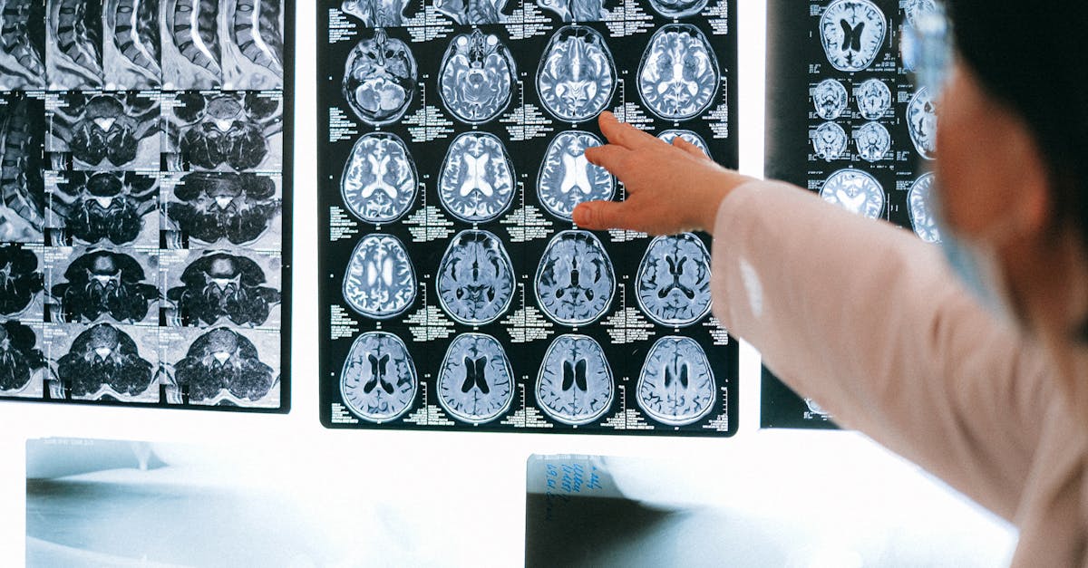
The definitive diagnosis and precise anatomical mapping of an intracranial meningioma rely entirely on advanced neuroimaging. Magnetic Resonance Imaging is the absolute gold standard diagnostic modality.
On an MRI utilizing gadolinium contrast, a meningioma typically displays a highly characteristic appearance. It presents as a well-circumscribed, extra-axial mass (meaning it is clearly outside the brain tissue) that enhances intensely and uniformly when the contrast dye is administered. A classic radiological feature is the “dural tail”—a tapering enhancement of the dura mater extending away from the base of the tumor, signifying reactive thickening of the meninges.
A Computed Tomography scan of the head is often utilized as a complementary study. CT is superior to MRI in identifying calcifications within the tumor, which are common in meningiomas, and for assessing whether the tumor has caused hyperostosis—an abnormal thickening and reaction of the adjacent skull bone, which is a hallmark sign of this specific neoplasm.
10. Differential Diagnosis
While the radiological appearance of a meningioma is often classic, the neuroradiologist must consider a differential diagnosis for any extra-axial mass to ensure accurate treatment planning.
| Tumor Type | Distinguishing Features on Imaging |
|---|---|
| Meningioma | Dural tail present, broad-based dural attachment, often calcified. |
| Dural Metastasis | Multiple lesions, known primary cancer (e.g., breast, prostate), aggressive bone destruction. |
| Hemangiopericytoma | Lacks calcification, narrower dural attachment, frequently invades bone aggressively rather than thickening it. |
| Vestibular Schwannoma | Grows specifically within the internal auditory canal; rare dural tail. |
11. Conservative Monitoring: The “Watch and Wait” Approach
Not all diagnosed meningiomas require immediate intervention. For small, asymptomatic meningiomas discovered incidentally, particularly in elderly patients or those with significant medical comorbidities, the standard of care is often conservative management, frequently termed “watch and wait.”
Because the majority of these tumors are benign and exhibit remarkably slow growth rates (often growing less than one millimeter per year), the risks of major cranial surgery may far outweigh the potential benefits.
The conservative approach involves strict, serial MRI surveillance. The patient typically undergoes an MRI scan every six to twelve months initially, followed by less frequent scans if the lesion proves to be stable. Intervention is only considered if the serial imaging demonstrates significant tumor growth, or if the patient develops new neurological symptoms attributable to the mass.
12. Neurosurgical Resection
When a meningioma is symptomatic, rapidly enlarging, or causing significant brain swelling, definitive treatment is required. Open microsurgical resection remains the primary and most effective treatment modality. The goal of surgery is the complete removal of the tumor, along with the segment of dura mater from which it originated, to minimize the risk of recurrence.
The procedure involves a craniotomy to access the tumor. Utilizing a surgical microscope and advanced intraoperative neuronavigation systems, the neurosurgeon meticulously dissects the tumor away from the surrounding healthy brain tissue.
Because meningiomas are highly vascular, the surgeon must carefully identify and coagulate the feeding arteries before attempting to remove the bulk of the tumor. In cases of convexity meningiomas, complete resection is frequently achievable. However, for complex skull base tumors entangled with major cranial nerves or the carotid artery, the surgeon may intentionally leave a small remnant of the tumor behind to avoid causing devastating, permanent neurological damage, prioritizing the patient’s functional quality of life over total tumor eradication.
13. Stereotactic Radiosurgery
Stereotactic radiosurgery is a highly effective, non-invasive alternative or adjunct to open surgery. Systems such as the Gamma Knife or CyberKnife deliver a highly concentrated, precise dose of radiation directly to the tumor volume, sparing the surrounding healthy brain tissue from significant radiation exposure.
Radiosurgery is not designed to physically remove or dissolve the tumor. Instead, the intense radiation damages the DNA of the tumor cells and destroys the microscopic blood vessels feeding the mass. This halts the tumor’s growth and frequently causes it to shrink slightly over a period of months to years.
Radiosurgery is frequently utilized as a primary treatment for small (less than 3 centimeters), deep-seated meningiomas located in surgically inaccessible areas, such as the cavernous sinus. It is also routinely employed as an adjuvant therapy following surgery to treat any small tumor remnants intentionally left behind on critical neurovascular structures.
14. Postoperative Care and Complications
The postoperative recovery following meningioma resection depends largely on the size and location of the tumor. Patients are monitored closely in a neuro-intensive care unit immediately following surgery to watch for complications such as postoperative bleeding or seizures.
A significant challenge in the immediate postoperative period is managing cerebral edema. Brain tissue that has been compressed for years can swell significantly once the pressure of the tumor is removed. Corticosteroids, such as dexamethasone, are frequently prescribed before and after surgery to aggressively control this inflammatory swelling.
Patients who presented with seizures will continue on anticonvulsant medications. Temporary or permanent neurological deficits, such as mild weakness or cranial nerve palsies, can occur, particularly after complex skull base resections, requiring comprehensive physical, occupational, and speech rehabilitation to optimize functional recovery.
15. Long-Term Surveillance and Recurrence
Meningiomas require lifelong medical surveillance. Even following a seemingly complete, aggressive surgical resection, meningiomas possess a notable rate of recurrence. The recurrence risk is heavily influenced by the WHO grade of the tumor and the extent of the initial surgical resection (measured by the Simpson grading scale).
Grade I tumors completely removed along with their dural attachment have a low recurrence rate (under 10 percent at ten years). However, atypical Grade II and malignant Grade III tumors recur with high frequency and often require postoperative fractionated radiation therapy to delay progression.
Patients commit to a schedule of regular follow-up MRI scans, typically annually, for many years. If a recurrence is detected early while it is still small, it can often be effectively treated with stereotactic radiosurgery before it causes new neurological symptoms.
16. When to Consult a Neurologist
While most headaches are benign, certain warning signs mandate a prompt evaluation by a neurologist and subsequent neuroimaging.
A patient experiencing a new onset of seizures, regardless of age, must undergo an immediate MRI of the brain to rule out a structural lesion like a meningioma. A headache that is consistently worse upon waking in the morning, wakes the patient from sleep, or is exacerbated by coughing or bending over is a classic sign of increased intracranial pressure requiring investigation.
Furthermore, the gradual, unexplained onset of focal neurological deficits—such as progressive weakness in a limb, a slow decline in vision, persistent double vision, or subtle but noticeable changes in personality and cognition—warrants a comprehensive neurological examination to ensure an indolent, slow-growing meningioma is not quietly compressing vital brain structures.
17. Frequently Asked Questions (FAQ)
1. Is an intracranial meningioma considered brain cancer?
The vast majority of meningiomas (about 80 percent) are benign, meaning they are not cancer. They grow slowly and do not spread to other organs. Only a very small percentage are atypical or malignant (cancerous) and behave aggressively.
2. Will a meningioma always require brain surgery?
No. If the tumor is small, benign, and not causing any symptoms, doctors often recommend simply monitoring it with regular MRI scans. Many people live their entire lives with a small meningioma without ever needing surgery.
3. Can a meningioma grow back after it is removed?
Yes, there is always a risk of recurrence, even if the surgeon believes they removed the entire tumor. The risk depends on the tumor’s grade and how much tissue was able to be safely removed. This is why lifelong MRI monitoring is essential.
4. What causes a meningioma to form?
In most cases, the exact cause is unknown. We do know that prior high-dose radiation therapy to the head and certain rare genetic conditions (like Neurofibromatosis type 2) significantly increase the risk. Hormones may also play a role, as these tumors are more common in women.
18. Bibliography
Disclaimer: The content is for informational purposes only and does not replace medical advice. Always consult your doctor for personalized treatment.