1. Introduction
The sharp, stabbing pain localized directly under your heel bone when you step down is a classic hallmark of plantar fasciitis. This condition occurs when the thick, fibrous band of connective tissue running along the bottom of your foot, known as the plantar fascia, becomes mechanically overloaded and inflamed at the exact point where it attaches to the calcaneus, or heel bone. The sudden, intense pain experienced upon bearing weight, especially after periods of rest, is the result of applying immediate tensile force to a highly sensitized, irritated, and microscopically damaged area of tissue.
The plantar fascia is designed to support the arch of the foot and absorb the tremendous shock generated by walking and running. Under normal circumstances, it is exceptionally strong and resilient. However, chronic repetitive stress, poor biomechanics, or unsupportive footwear can subject this tissue to forces that exceed its capacity to repair itself.
When the fascia is overloaded, tiny micro-tears develop at the calcaneal insertion site. The body responds to this microtrauma with inflammation, causing the tissue to become swollen, tight, and highly reactive to pressure.
Understanding the structural mechanics of the foot and the physiological healing process is essential for managing this severe heel pain. Appropriate interventions can halt the cycle of damage and facilitate tissue repair, restoring pain-free mobility.
2. Anatomy of the Plantar Fascia and Heel Bone
The plantar fascia is a robust, inelastic aponeurosis—a broad, flat sheet of dense connective tissue. It originates from the medial tubercle of the calcaneus, which is the prominent bony ridge on the underside of the heel bone. From this singular attachment point, it fans out forward across the sole of the foot, dividing into five distinct slips that anchor into the base of the toes.
This anatomical configuration creates a vital structural tie-rod for the foot. It acts to maintain the longitudinal arch, preventing it from collapsing under the weight of the body. The structural integrity of the entire foot relies heavily on the tension maintained by this fibrous band.
The calcaneus itself is the largest bone in the foot and is uniquely designed to bear the initial impact of a heel strike during the gait cycle. The underside of the heel is covered by a specialized, dense layer of fat encased in fibrous septa, known as the plantar fat pad, which serves as a biological shock absorber.
The intersection of these structures—the dense bone, the rigid fascia, and the shock-absorbing fat pad—is a highly complex mechanical zone. Any disruption in this delicate balance, particularly an overload at the fascial attachment site, rapidly results in severe clinical symptoms.
3. The Mechanism of Plantar Fasciitis
Despite its name ending in “-itis,” which implies active inflammation, contemporary histological studies reveal that chronic plantar fasciitis is more accurately described as a tendinosis or fasciosis. This means it is primarily a degenerative condition rather than a purely inflammatory one.
The mechanism begins with repetitive mechanical overload. With each step, as the foot flattens, the plantar fascia is stretched tight. If the tensile load is too great—due to factors like increased body weight, sudden increases in activity, or biomechanical faults—collagen fibers at the heel attachment sustain microscopic tears.
Initially, an inflammatory response does occur to try and heal these microtears. However, because the foot is in constant use, the tissue is repeatedly re-injured before the healing cycle can complete. The repair process becomes stalled.
Over time, the healthy parallel collagen fibers degenerate and are replaced by disorganized, weak scar tissue. The fascia thickens and loses its natural elasticity. It is this disorganized, mechanically compromised tissue at the heel insertion that generates the sharp, stabbing pain when subjected to the load of stepping down.
4. The “First Step” Phenomenon Explained
The most defining and frequently reported symptom of plantar fasciitis is severe, stabbing pain with the very first steps taken out of bed in the morning, or after sitting at a desk for a prolonged period. This specific clinical presentation is known as post-static dyskinesia.
The biological explanation for this phenomenon lies in tissue adaptation during rest. During sleep or extended periods of sitting, the foot naturally relaxes into a slightly plantarflexed, or downward-pointing, position. In this relaxed state, the tension on the plantar fascia is removed, and the injured tissue contracts and shortens.
During the night, the body attempts to heal the microtears by laying down delicate new collagen fibers while the fascia is in this shortened position.
When you stand up and place your foot flat on the floor, the ankle is forced into dorsiflexion, and the arch of the foot lengthens. This sudden mechanical demand violently stretches the shortened fascia, instantly tearing the fragile new healing tissue that formed overnight. The sharp, stabbing pain is the neurological response to this acute micro-tearing event.
5. The Role of Microtears and Inflammation
The pain pathway in plantar fasciitis is deeply connected to the cycle of micro-tearing and failed healing. The plantar fascia is densely innervated with specialized nerve endings designed to monitor tissue tension and detect potential damage.
When a microtear occurs at the calcaneal insertion, these nerves immediately transmit pain signals. Concurrently, local cells release biochemical markers that trigger an inflammatory cascade. While inflammation is a necessary precursor to healing, chronic inflammation leads to tissue sensitization.
The nerve endings in the inflamed area become hyper-reactive. They lower their firing threshold, meaning that even a normal amount of tension—such as simple standing—is interpreted by the brain as an acutely painful, stabbing sensation.
Breaking this cycle requires preventing the daily micro-tearing events, particularly the aggressive first-step tearing in the morning. Only when the mechanical disruption is halted can the chronic inflammation subside and genuine tissue remodeling begin.
6. Heel Spurs: Correlation Versus Causation
Historically, a sharp pain under the heel was almost universally attributed to the presence of a “heel spur.” A heel spur, or calcaneal exostosis, is a bony projection that grows on the underside of the heel bone, exactly where the plantar fascia attaches.
X-rays of patients with heel pain frequently reveal these spurs. However, modern medical understanding has fundamentally shifted regarding their role. Extensive clinical data shows that a significant percentage of the population has substantial heel spurs but experiences absolutely no pain. Conversely, many patients with excruciating plantar fasciitis have perfectly smooth heel bones on X-ray.
The heel spur is generally considered a secondary symptom of the problem, not the root cause. It is a biological reaction to the chronic tension pulling on the bone. Over years, the body lays down calcium deposits at the attachment site to reinforce the bone against the constant pulling force of the tight fascia.
Therefore, the sharp pain is generated by the inflamed and degenerating fascial tissue pulling on the bone, not by the bony spur itself piercing downward into the flesh. Surgical removal of the spur is rarely indicated; treating the tension in the fascia is the primary clinical objective.
7. Biomechanical Risk Factors
Certain mechanical realities of the human body can predispose an individual to developing severe heel pain. Foot architecture plays a critical role. Individuals with pes planus, or flat feet, lack inherent skeletal arch support, meaning the plantar fascia must bear an abnormally high tensile load to keep the foot from completely collapsing inward.
Conversely, individuals with pes cavus, or excessively high and rigid arches, possess a foot structure that is very poor at absorbing shock. Because the arch does not yield upon impact, the concussive force of every step is transferred directly to the heel bone and the fascial attachment, leading to rapid tissue breakdown.
Tightness in the posterior muscular chain is a nearly universal finding in patients with heel pain. If the gastrocnemius or soleus muscles in the calf are tight, they limit the upward flexibility of the ankle joint. To compensate for this lack of motion, the foot over-pronates, placing extreme stretch on the plantar fascia.
Addressing these biomechanical imbalances through specific stretching or targeted orthotic support is often necessary to achieve a permanent resolution of the sharp heel pain.
8. The Impact of Footwear and Surface Hardness
The environment in which the foot operates heavily influences the health of the plantar fascia. Modern, flat, unyielding surfaces—such as concrete floors, tile, and hardwood—provide zero shock absorption. Walking barefoot or in unsupportive shoes on these hard surfaces dramatically increases the mechanical impact transferred directly to the heel.
Footwear choices are often the primary catalyst for an acute flare-up. Worn-out athletic shoes that have lost their midsole cushioning fail to absorb concussive forces. Flat-soled shoes, such as ballet flats or minimal sandals, offer no arch support, forcing the fascia to bear the full burden of dynamic stabilization.
Occupations that demand prolonged standing on hard floors—such as teaching, nursing, or retail work—exponentially increase the time under tension for the connective tissues. The sustained isometric load without adequate rest or supportive footwear is a recipe for chronic fascial irritation.
Modifying the environment by introducing highly cushioned, supportive footwear with a slight heel elevation is one of the most effective initial interventions to reduce the immediate strain on the heel attachment.
9. Fat Pad Atrophy as a Differential Diagnosis
While plantar fasciitis is the most common cause of under-heel pain, it is not the only one. A highly relevant differential diagnosis, particularly in older adults, is plantar fat pad atrophy.
The thick pad of specialized fat under the heel bone is designed to buffer the impact of walking. With age, or following multiple corticosteroid injections into the heel, this fat pad can thin out and degenerate, losing its shock-absorbing capacity.
When the fat pad atrophies, the calcaneus bone essentially strikes the ground with minimal cushioning. The pain associated with this condition is typically described as a deep, bruising ache right in the center of the heel pad. It worsens with prolonged standing and walking barefoot on hard floors.
Crucially, fat pad atrophy does not typically present with the classic “first step” sharp, stabbing pain characteristic of plantar fasciitis. Recognizing this distinction is vital, as treating fat pad atrophy requires maximizing external cushioning, whereas treating the fascia involves modifying tension and biomechanics.
10. Nerve Entrapment and Tarsal Tunnel Syndrome
Another condition that can mimic or co-exist with heel pain is nerve entrapment. The tibial nerve travels down the inside of the ankle and branches out to supply the bottom of the foot. One of these branches, known as Baxter’s nerve, can become compressed or pinched as it passes under the heel bone.
Baxter’s nerve entrapment causes a sharp, burning, or shooting pain that radiates across the bottom of the heel. Unlike the mechanical pain of plantar fasciitis, nerve pain often worsens as the day progresses and may be accompanied by a tingling or numb sensation in the foot.
Similarly, Tarsal Tunnel Syndrome occurs when the main tibial nerve is compressed higher up, behind the inner ankle bone. This can refer pain down into the heel and the arch.
A thorough clinical examination is necessary to differentiate mechanical tissue pain from neurological pain. Tapping on the nerve to see if it reproduces the shooting symptoms is a common diagnostic technique used to identify nerve entrapment.
11. Diagnostic Approaches to Heel Pain
Diagnosing the specific cause of sharp under-heel pain is primarily achieved through a detailed clinical history and a focused physical examination. A physician will identify the exact location of maximum tenderness, which in plantar fasciitis is highly specific to the medial tubercle of the calcaneus.
The physician will also assess the flexibility of the ankle joint and evaluate the patient’s gait to identify any biomechanical abnormalities like overpronation. The classic reproduction of sharp pain upon dorsiflexing the toes, which activates the windlass mechanism and tightens the fascia, is a strong diagnostic indicator.
Imaging studies are not usually required for a preliminary diagnosis. However, if the pain does not respond to initial conservative treatments, X-rays may be ordered to rule out a calcaneal stress fracture or to assess the architecture of the foot.
Ultrasound imaging is becoming increasingly valuable in the clinical setting. A high-resolution ultrasound can visualize the thickness of the plantar fascia, identify areas of disorganized tissue, and confirm the presence of localized inflammation, providing clear evidence of the pathology.
12. Conservative Management and Immediate Relief
The most effective initial management of sharp heel pain involves mechanically unloading the irritated tissue and managing the acute inflammation. The absolute priority is preventing the micro-tearing that occurs with the first morning steps.
Keeping a pair of highly supportive, slip-on shoes immediately next to the bed ensures that the foot is never placed flat on the ground completely unsupported. A supportive shoe provides an artificial arch and slightly elevates the heel, significantly reducing the tensile stretch on the fascia.
Rolling the arch of the foot over a frozen water bottle for ten minutes at the end of the day serves a dual purpose: the cold temperature causes vasoconstriction to manage deep tissue inflammation, while the rolling motion provides a gentle, mobilizing massage to the tight fascial band.
Taping techniques, such as low-dye strapping applied by a physical therapist, can effectively strap the foot to artificially support the arch, mimicking the effect of an orthotic and providing immediate, short-term relief during daily activities.
13. Stretching and Strengthening Protocols
Long-term resolution of plantar fasciitis requires correcting the underlying mechanical tension. The most evidence-based intervention is a specific stretching protocol targeting both the calf complex and the plantar fascia itself.
Because the calf muscles connect to the heel bone via the Achilles tendon, tightness in the calf directly pulls upward on the heel bone, which in turn increases tension on the plantar fascia pulling in the opposite direction. Regular, sustained stretching of the gastrocnemius and soleus muscles is mandatory to relieve this systemic tension.
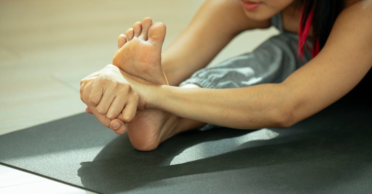
Specific plantar fascia stretching is equally critical. While seated, the patient crosses the affected leg over the opposite knee, grasps the toes, and pulls them backward toward the shin until a stretch is felt in the arch. Massaging the tight band of tissue while it is under tension helps remodel the collagen fibers.
Once the acute pain subsides, strengthening the intrinsic muscles of the foot with exercises like towel scrunches and short-foot activations helps build a dynamic internal support system, reducing the passive load the fascia must bear during activity.
14. Advanced Interventions and Medical Therapies
If conservative measures fail to provide relief after several months of diligent application, advanced medical therapies may be considered. Custom orthotics crafted from a biomechanical mold of the foot can provide highly specific structural correction, particularly for significant overpronation.
Extracorporeal Shockwave Therapy is a non-invasive clinical treatment utilizing high-energy acoustic waves targeted directly at the heel insertion. These waves create controlled microtrauma that re-initiates a stalled healing response and stimulates the formation of new blood vessels in the degenerated tissue.
Corticosteroid injections can provide rapid, potent relief of severe acute inflammation. However, they are used judiciously, as repeated steroid injections can physically weaken the fascial tissue and significantly increase the risk of a complete fascial rupture or accelerate the atrophy of the protective heel fat pad.
In severe, chronic cases that are entirely refractory to all conservative and advanced medical management for over a year, surgical intervention such as a partial plantar fascia release may be discussed, though it is considered a last resort.
15. Frequently Asked Questions (FAQ)
1. Why does my heel pain suddenly stab when I get out of bed?
While you sleep, your foot relaxes and points downward, allowing the inflamed plantar fascia to shorten and attempt to heal. When you stand up, the tissue is violently stretched and re-torn, causing the sharp, stabbing morning pain.
2. Should I walk barefoot if I have sharp heel pain?
Absolutely not. Walking barefoot on hard surfaces offers zero arch support or shock absorption, placing maximum mechanical strain directly on the injured fascial attachment. Always wear supportive shoes, even inside the house.
3. Do heel spurs need to be surgically removed to stop the pain?
In the vast majority of cases, no. The pain is caused by the inflamed soft tissue pulling on the bone, not the bony spur itself. Treating the tension in the plantar fascia almost always resolves the pain without needing to remove the spur.
4. How long does it typically take for this heel pain to go away completely?
Plantar fasciitis is notorious for being a stubborn condition because the tissue is subjected to body weight daily. With consistent stretching, proper footwear, and rest, full resolution can take anywhere from three to nine months.
5. Can tight calf muscles really cause pain under my heel?
Yes. The calf muscles attach to the back of the heel bone, and the plantar fascia attaches to the bottom. Tightness in the calves acts like a tight pulley system, physically hauling on the heel bone and increasing the tension transferred to the fascia.
16. Bibliography
Disclaimer: The content is for informational purposes only and does not replace medical advice. Always consult your doctor for personalized treatment.