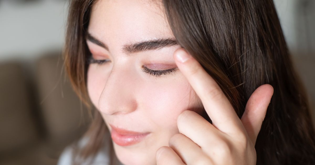
1. Introduction to Myokymia
Myokymia is a neurological phenomenon characterized by spontaneous, fine, fascicular contractions of muscle fibers without causing actual movement of a joint. Clinically, it manifests as a continuous, undulating, or rippling movement beneath the skin, often described by patients as a persistent “twitching” or “fluttering.” While it can theoretically affect any skeletal muscle, it is most frequently observed in the facial musculature, particularly around the eyes.
In the vast majority of cases, localized myokymia is an entirely benign condition. The most common presentation, facial myokymia (specifically eyelid twitching), is a widespread occurrence in the general population. It is usually temporary, self-limiting, and heavily linked to environmental and lifestyle factors such as fatigue, stress, and excessive caffeine consumption.
However, when myokymia is chronic, widespread, or accompanied by other neurological deficits, it necessitates a more thorough clinical evaluation. Understanding the subtle distinctions between benign fasciculations, physiological myokymia, and pathological muscle rippling is essential for an accurate diagnosis and for providing the patient with appropriate reassurance or targeted medical treatment.
2. Pathophysiology and Mechanisms
The exact pathophysiological mechanism underlying myokymia involves the hyperexcitability of the lower motor neurons or their axons. Under normal circumstances, a motor neuron transmits an electrical impulse that results in a coordinated contraction of a motor unit. In myokymia, there is a spontaneous, repetitive discharge of these motor units at an ectopic site along the peripheral nerve.
This ectopic generation of action potentials is often linked to localized alterations in the microenvironment of the nerve. Changes in the concentration of extracellular ions, particularly potassium and calcium, or localized ischemia can lower the threshold for depolarization. This causes the nerve fibers to fire independently and asynchronously, producing the characteristic worm-like rippling effect in the muscle belly.
Unlike conditions involving the central nervous system, benign myokymia represents a strictly peripheral nerve hyperexcitability. The rhythmic bursting discharges can be recorded on an electromyogram (EMG), which shows regular or irregular groups of action potentials firing at specific frequencies, a hallmark that helps differentiate it from other movement disorders.
3. Eyelid Myokymia: The Most Common Form
Superior or inferior oblique eyelid twitching, medically termed eyelid myokymia, is by far the most prevalent manifestation of this condition. It typically involves the lower eyelid of one eye, though the upper eyelid can also be affected. The twitching is usually intermittent, lasting for a few seconds to hours, and may recur over several days or weeks before resolving completely.
This specific form of myokymia is almost exclusively benign. It is considered an episodic phenomenon rather than a chronic disease. Patients often report the sensation as highly distracting and annoying, sometimes believing that the twitching is visible to others, even when the contractions are microscopic and barely detectable to an observer.
The clinical evaluation of isolated eyelid myokymia is generally straightforward. If the twitching is strictly localized to the orbicularis oculi muscle, does not force the eye closed, and there are no other cranial nerve deficits, the diagnosis of benign eyelid myokymia is secure, and aggressive diagnostic testing is not warranted.
4. Common Lifestyle Triggers
Benign eyelid and facial myokymia are heavily influenced by physiological stress and lifestyle habits. Identifying and modifying these triggers is usually the only intervention required. Fatigue and severe sleep deprivation are among the most common culprits. Lack of restorative sleep lowers the overall threshold for nerve excitability, making spontaneous discharges more likely.
Psychological stress and anxiety are also potent triggers. During periods of high stress, the sympathetic nervous system is hyperactive, increasing circulating catecholamines which can heighten peripheral nerve irritability. Many patients notice the onset of a persistent eyelid twitch during periods of intense work deadlines, academic examinations, or personal emotional turmoil.
Dietary stimulants, most notably caffeine, directly stimulate the central and peripheral nervous systems. Excessive consumption of coffee, energy drinks, or certain teas can easily provoke myokymia. Similarly, alcohol use, tobacco smoking, and sudden withdrawal from certain medications can create a physiological imbalance that triggers localized muscle twitching.
5. Facial Myokymia as a Neurological Sign
While eyelid myokymia is benign, myokymia affecting broader areas of the face (facial myokymia) can sometimes serve as an important localizing sign in neurology. Pathological facial myokymia involves continuous, unilateral rippling of the facial muscles, and unlike the benign eyelid form, it typically persists during sleep and is not relieved by rest.
This continuous undulating movement indicates a structural or inflammatory lesion affecting the facial nerve (Cranial Nerve VII) pathway within the brainstem (pons). One of the classic neurological conditions associated with continuous facial myokymia is Multiple Sclerosis (MS). In MS, demyelinating plaques in the brainstem alter nerve conduction and promote hyperexcitability of the facial nerve nucleus.
Other central nervous system pathologies that can present with facial myokymia include brainstem gliomas, pontine strokes, and syringobulbia. Additionally, it may appear during the recovery phase of a severe acute inflammatory demyelinating polyneuropathy, such as Guillain-Barré syndrome, or following radiation therapy directed at the head and neck.
6. Systemic and Limb Myokymia
Although rare, myokymia can manifest systematically or in the limbs. Limb myokymia presents as continuous muscle rippling in the arms or legs, often accompanied by muscle stiffness, cramps, and delayed relaxation after voluntary contraction (pseudomyotonia). This presentation requires a comprehensive neurological workup.
Systemic myokymia is heavily associated with a group of rare autoimmune nerve hyperexcitability disorders, the most notable being Isaacs syndrome (neuromyotonia). In Isaacs syndrome, autoantibodies target voltage-gated potassium channels on the peripheral nerves. The failure of these channels to properly regulate potassium flow leads to continuous, spontaneous nerve firing, resulting in profound muscle twitching, stiffness, and hyperhidrosis (excessive sweating).
Limb myokymia can also result from localized peripheral nerve damage, such as a severe radiculopathy or plexopathy. In these instances, the nerve hyperexcitability is secondary to chronic compression or trauma, and the myokymia is restricted to the specific muscles innervated by the damaged nerve segment.
7. Differentiating Myokymia from Other Movement Disorders
Accurate clinical diagnosis requires distinguishing myokymia from other conditions that cause involuntary muscle movements. Fasciculations are brief, isolated twitches of a single motor unit. While myokymia is a continuous, rippling sequence of fasciculations, isolated benign fasciculations (commonly felt in the calves) are usually scattered and irregular. Pathological fasciculations are a hallmark of lower motor neuron diseases like Amyotrophic Lateral Sclerosis (ALS), but these are accompanied by muscle atrophy and weakness.
Hemifacial spasm is a critical differential diagnosis for facial myokymia. Hemifacial spasm involves forceful, synchronized contractions of an entire side of the face, often causing the eye to clamp shut and the corner of the mouth to pull upward. In contrast, myokymia causes fine rippling that does not usually result in gross joint movement or forced eye closure.
Blepharospasm is another distinct condition characterized by bilateral, forceful, involuntary closure of the eyelids. It is a form of focal dystonia originating in the basal ganglia. Unlike myokymia, blepharospasm affects both eyes simultaneously and is characterized by sustained muscle spasms rather than a continuous subtle flutter.
8. Clinical Diagnostic Evaluation
When a patient presents with muscle twitching, the clinical evaluation begins with a meticulous medical history and neurological examination. The physician assesses the distribution of the twitching (is it confined to one eyelid, or does it involve the lower face and limbs?), the duration, and any alleviating or aggravating factors.
During the physical examination, the clinician will observe the affected area closely at rest and during voluntary movement. Assessing cranial nerve function, muscle strength, deep tendon reflexes, and sensory pathways is vital to rule out broader neurological involvement. If the exam is entirely normal aside from isolated eyelid twitching, further testing is generally unnecessary.
If the presentation is atypical—such as continuous facial rippling, limb involvement, muscle weakness, or concurrent neurological symptoms—the clinician will order specific diagnostic studies to investigate potential central nervous system or autoimmune pathologies.
9. Electromyography (EMG) and Neuroimaging
Electromyography (EMG) is the gold standard diagnostic tool for confirming myokymia and differentiating it from other forms of nerve hyperexcitability. A fine needle electrode is inserted into the affected muscle to record its electrical activity at rest.
In classic myokymia, the EMG reveals distinctive “myokymic discharges.” These are rhythmic or semi-rhythmic bursts of normal-appearing motor unit potentials that fire in groups (doublets, triplets, or multiplets), followed by periods of silence. The firing rate within a burst is high (up to 60 Hz), producing a sound on the EMG audio monitor often likened to “marching soldiers.”
If pathological facial myokymia is suspected, an MRI of the brain, specifically focusing on the brainstem and the cerebellopontine angle, is mandatory. The MRI aims to detect demyelinating lesions (as seen in Multiple Sclerosis), tumors, or vascular loops that might be compressing the facial nerve. Blood tests may also be ordered to screen for voltage-gated potassium channel antibodies if a systemic syndrome is suspected.
10. Data: Differentiating Facial Muscle Spasms
The following table summarizes the key clinical differences between common conditions that cause abnormal facial movements, assisting in correct clinical identification.
| Condition | Clinical Presentation | Area Affected | Typical Cause |
|---|---|---|---|
| Benign Myokymia | Fine, undulating, rippling twitch; no forced closure. | Usually one eyelid (upper or lower). | Fatigue, stress, caffeine. |
| Hemifacial Spasm | Forceful, synchronized spasms; pulls face. | Unilateral, involves entire half of face. | Vascular compression of facial nerve. |
| Blepharospasm | Sustained, forceful eye closure; blinking spasms. | Bilateral (both eyes). | Focal dystonia (basal ganglia dysfunction). |
| Pathological Myokymia | Continuous undulating rippling, persists in sleep. | Unilateral face. | Multiple Sclerosis, brainstem lesion. |
11. Management of Benign Myokymia
For the vast majority of patients experiencing isolated eyelid myokymia, conservative management and lifestyle modification are the only required treatments. Education and reassurance are paramount; once patients understand that the condition is benign and self-limiting, their anxiety—which often exacerbates the twitching—frequently subsides.
Patients are advised to optimize their sleep hygiene, aiming for 7-9 hours of restful sleep per night. Drastically reducing or entirely eliminating the intake of caffeine, energy drinks, and alcohol is often highly effective in resolving the symptoms within a few days. Applying warm compresses to the affected eyelid and gently massaging the area can also provide temporary mechanical relief and relax the orbicularis oculi muscle.
If the patient relies on digital screens for prolonged periods, implementing the “20-20-20 rule” (looking at an object 20 feet away for 20 seconds every 20 minutes) helps reduce visual fatigue, which is a known trigger for periorbital muscle spasms.
12. Pharmacological and Interventional Treatments
In cases where myokymia is chronic, widespread (such as Isaacs syndrome), or secondary to a central nervous system lesion, specific medical therapy is required. The goal is to stabilize the neuronal membrane and reduce the ectopic firing of the peripheral nerves.
Membrane-stabilizing antiepileptic medications are the first line of treatment for symptomatic myokymia. Drugs such as carbamazepine, phenytoin, and gabapentin alter sodium or calcium channel function, effectively suppressing the abnormal nerve discharges. For patients with autoimmune-mediated neuromyotonia, immunosuppressive therapy (such as corticosteroids, plasmapheresis, or intravenous immunoglobulin) is necessary to target the underlying antibody production.
In severe, refractory cases of facial myokymia that cause significant psychological distress or functional impairment, botulinum toxin (Botox) injections provide excellent localized relief. The toxin temporarily paralyzes the hyperactive muscle fibers by blocking the release of acetylcholine at the neuromuscular junction, providing relief for several months at a time.
13. When to See a Doctor
While an occasional twitching eyelid is not a cause for medical concern, certain specific signs indicate the need for a comprehensive neurological evaluation. Patients should consult a doctor if the twitching persists continuously for more than a few weeks despite lifestyle modifications like reducing caffeine and improving sleep.
Immediate medical attention is required if the twitching spreads to other parts of the face or body, or if it is accompanied by forced closure of the eye (preventing normal vision). Furthermore, if the myokymia presents alongside red flag symptoms such as facial weakness, drooping, numbness, visual disturbances, or difficulty swallowing, it may indicate an underlying brainstem pathology requiring urgent investigation.
Early clinical assessment is essential to distinguish between a benign, stress-induced flutter and a subtle sign of a more complex neurological condition, ensuring that the patient receives appropriate reassurance or timely intervention.
14. Frequently Asked Questions (FAQ)
1. Is an eyelid twitch a sign of a stroke?
No. An isolated eyelid twitch (myokymia) is a local muscle spasm usually caused by fatigue or caffeine, not a vascular event. A stroke typically presents with sudden weakness, paralysis, numbness, or difficulty speaking, not a fine rippling muscle twitch.
2. Can staring at a computer screen cause myokymia?
Yes. Prolonged screen time causes eye strain and visual fatigue, which are common triggers for benign eyelid myokymia. Taking regular breaks to rest the eyes can help prevent it.
3. Will myokymia go away on its own?
In most cases, benign eyelid myokymia resolves spontaneously within a few days to a few weeks, especially if the underlying triggers (stress, caffeine, lack of sleep) are addressed.
4. When are Botox injections used for this condition?
Botulinum toxin is rarely used for typical, brief eyelid twitching. It is reserved for severe, chronic, and unremitting cases that do not respond to conservative measures and cause significant distress to the patient.
5. Can nutritional deficiencies cause myokymia?
While severe imbalances in electrolytes (such as magnesium, potassium, or calcium) can lower the threshold for muscle spasms and nerve excitability, they are rarely the sole cause of isolated myokymia without other systemic symptoms.
Bibliography
Disclaimer: The content is for informational purposes only and does not replace medical advice. Always consult your doctor for personalized treatment.