1. Introduction to Nodular Fasciitis
Nodular fasciitis is a benign, rapidly growing soft tissue lesion that originates from the superficial fascia, the layer of connective tissue situated just beneath the skin. This condition often provokes significant anxiety for patients and clinicians alike because its rapid enlargement clinically mimics malignant soft tissue sarcomas. However, despite its alarming growth rate, nodular fasciitis is entirely non-cancerous and poses no threat of spreading to distant organs.
The primary objective during clinical evaluation is to differentiate this benign reactive process from true malignancies. Because the clinical presentation is so striking, a careful combination of patient history, physical examination, and pathological analysis is required. Healthcare providers strive to deliver a precise diagnosis promptly to alleviate patient distress and prevent unnecessarily aggressive surgical interventions.
Understanding the nature of nodular fasciitis allows patients to approach their diagnosis with reassurance. While the mass can become tender or cosmetically bothersome, treatment options are straightforward, and the prognosis is uniformly excellent. In many cases, the condition is self-limiting and may even resolve without extensive surgical treatment.
2. Pathophysiology and Rapid Growth
The pathophysiology of nodular fasciitis is characterized by the sudden and robust proliferation of fibroblasts and myofibroblasts. These are the specialized connective tissue cells responsible for wound healing and structural support within the body. In this condition, these cells multiply rapidly within the fascial layer, creating a firm, well-defined nodule.
This proliferation is considered a reactive or inflammatory process rather than a true neoplastic cancer. The cellular behavior resembles an exaggerated healing response that has occurred without a proportional initial injury. The rapid cellular division is the primary reason the nodule can double in size within a matter of weeks, reaching its maximum dimension quite abruptly before halting its growth.
Once the mass reaches its peak size, the cellular proliferation ceases. The lesion then enters a stationary phase where it no longer expands. This self-limiting biological behavior is a hallmark of nodular fasciitis and a critical differentiator from malignant tumors, which grow relentlessly and invasively without ceasing.
3. The USP6 Gene Mutation
Recent advancements in molecular pathology have significantly clarified the biological origins of nodular fasciitis. Scientific research has identified that a large majority of these lesions harbor a specific genetic alteration involving the USP6 gene. This involves a chromosomal rearrangement where the USP6 gene fuses with a promoter gene, most commonly MYH9.
This genetic fusion leads to an overexpression of the USP6 protein, which acts as a catalyst for the rapid proliferation of fibroblasts. The discovery of this recurrent genetic mutation has prompted some medical authorities to reclassify nodular fasciitis as a transient, benign neoplasm rather than a purely reactive inflammatory condition.
Despite this genetic driver, the mutation does not confer any malignant potential to the cells. The presence of the USP6 fusion is strictly associated with benign behavior. In challenging diagnostic cases, molecular testing for this specific genetic alteration can provide absolute certainty, distinguishing nodular fasciitis from other aggressive spindle cell tumors.
4. Microscopic and Histological Features
To secure a definitive diagnosis, a pathologist must examine the tissue under a microscope. Nodular fasciitis displays a highly characteristic cellular landscape that is often described as resembling tissue culture. The cells are plump, immature-appearing fibroblasts and myofibroblasts arranged in short, intersecting bundles or loosely scattered in a feathery pattern.
The background stroma of the tumor is typically myxoid, meaning it contains a gelatinous, mucus-like substance that gives the tissue a bluish tint under standard staining. As the lesion matures over time, this myxoid background often becomes more densely collagenous and fibrous. Extravasated red blood cells, which are red blood cells that have leaked out of small capillaries into the surrounding tissue, are also a classic microscopic finding.
Crucially, while the cells are dividing rapidly and show numerous mitotic figures, they lack the atypical, bizarre nuclear features that characterize cancer cells. The pathologist relies on this combination of rapid cellular division without malignant cellular changes to confirm the benign nature of the lesion.
5. Clinical Presentation and Symptoms
Patients with nodular fasciitis typically report the sudden appearance of a small, firm lump just beneath the skin. The most striking symptom is the timeline; the mass often grows from being unnoticeable to two or three centimeters in diameter within a span of two to four weeks.
The lesion is frequently tender to the touch, and patients may experience mild, localized pain, especially if the mass is pressing against nearby superficial nerves or if it is located in an area subject to frequent friction. The overlying skin usually appears completely normal, without redness, warmth, or ulceration, as the primary process is located deeper in the subcutaneous tissue.
Because the nodule arises from the fascia, it may feel somewhat tethered to the underlying muscle but remains relatively mobile beneath the skin. The firm texture and rapid onset are the primary drivers that prompt patients to seek urgent medical evaluation, fearing a serious underlying condition.
6. Common Anatomical Locations
Nodular fasciitis can theoretically develop in any region of the body containing fascial connective tissue, but it exhibits a strong predilection for certain anatomical areas. The upper extremities, particularly the volar aspect of the forearm, are the most frequently affected sites, accounting for nearly half of all documented cases.
The trunk, including the chest wall and the back, is the second most common location. In pediatric and adolescent populations, the head and neck region is affected more frequently than in adults. When it occurs in the head and neck, it can arise in the subcutaneous tissues of the face or the deeper fascial planes of the neck, requiring careful clinical assessment.
Regardless of the anatomical site, the fundamental clinical behavior and rapid growth trajectory of the lesion remain consistent. Its location primarily influences the degree of mechanical discomfort the patient experiences and dictates the most appropriate surgical approach if excision is pursued.
7. Demographics and Incidence
Nodular fasciitis is a relatively uncommon condition, but it is considered the most frequent benign pseudosarcomatous lesion encountered in clinical practice. It affects individuals of all ages but demonstrates a clear peak incidence in young to middle-aged adults, typically those between twenty and forty years old.
The condition affects both men and women with equal frequency, showing no distinct gender predilection. There are no identified racial or ethnic disparities in the incidence of nodular fasciitis. Furthermore, it is a sporadic condition, meaning it does not run in families and is not associated with any known inherited genetic syndromes.
While it is less common in young children and the elderly, clinicians must maintain a broad diagnostic awareness across all age groups, as the alarming presentation can occur at any stage of life.
8. Potential Causes and Risk Factors
The exact trigger that initiates the development of nodular fasciitis remains a subject of ongoing medical inquiry. Historically, it was widely believed to be a localized reaction to physical trauma, such as a blunt force injury, an insect bite, or a minor penetrating wound.
However, careful clinical histories reveal that fewer than fifteen percent of patients can recall any specific traumatic event at the site of the lesion prior to its rapid growth. Consequently, while trauma may play a role in a minority of cases, it is not considered the primary or necessary causative factor.
Currently, medical science views the condition as an idiopathic process, meaning it arises spontaneously without a clearly defined external cause. There are no known lifestyle factors, dietary influences, or environmental exposures that increase an individual’s risk of developing nodular fasciitis.
9. Diagnostic Imaging Techniques
When a patient presents with a rapidly growing soft tissue mass, diagnostic imaging is an essential component of the initial evaluation. Ultrasound is often the first modality utilized because it is readily available and provides excellent visualization of superficial tissues. On ultrasound, nodular fasciitis typically appears as a well-defined, solid, hypoechoic nodule situated within the subcutaneous fat or abutting the fascia.
For a more comprehensive anatomical assessment, magnetic resonance imaging is the gold standard. Magnetic resonance imaging provides superior soft tissue contrast, allowing clinicians to evaluate the exact depth of the lesion and its relationship to surrounding muscles, nerves, and blood vessels.
On magnetic resonance imaging, the appearance of the lesion varies depending on its cellular composition. Highly cellular and myxoid lesions often show hyperintense, or bright, signals on T2-weighted images. Imaging helps outline the mass but cannot definitively distinguish it from a malignant sarcoma, underscoring the necessity of a tissue biopsy.
10. Core Needle Biopsy and Pathology
Because the clinical and radiological features of nodular fasciitis overlap significantly with malignant soft tissue tumors, obtaining a tissue sample is mandatory for a conclusive diagnosis. A core needle biopsy is the preferred initial diagnostic procedure.
During a core needle biopsy, a physician uses a hollow needle, often guided by ultrasound, to extract small, cylindrical samples of tissue from the mass. This procedure is performed under local anesthesia and carries a minimal risk of complications. The extracted tissue preserves the cellular architecture, allowing the pathologist to evaluate the specific spatial arrangement of the fibroblasts and the surrounding stroma.
Fine needle aspiration, which extracts only individual cells, is generally discouraged for this condition. The isolated cells of nodular fasciitis appear highly active and can easily be misidentified as malignant sarcoma cells without the crucial context of the surrounding tissue architecture.
11. Differential Diagnosis
Accurately distinguishing nodular fasciitis from other soft tissue neoplasms is the most critical step in clinical management.
| Tumor Type | Key Differentiating Features |
|---|---|
| Nodular Fasciitis | Rapid growth followed by stabilization, USP6 gene mutation, myxoid background, lack of nuclear atypia. |
| Fibrosarcoma | Relentless growth, significant nuclear atypia, highly infiltrative margins, absence of USP6 mutation. |
| Desmoid Fibromatosis | Slower growth, deep muscular infiltration, beta-catenin mutation, dense collagenous stroma. |
| Schwannoma | Arises directly from a nerve, frequently painful, strongly positive for S100 protein on immunohistochemistry. |
Pathologists meticulously evaluate the tissue to rule out these alternative conditions. Misdiagnosing nodular fasciitis as a sarcoma can lead to devastating consequences, including unnecessary, radical surgery or radiation therapy. For related considerations regarding persistent localized tissue irritation, patients might explore muscle pain guidelines.
12. Observation and Spontaneous Regression
One of the most remarkable clinical characteristics of nodular fasciitis is its capacity for spontaneous regression. Because the condition is a self-limiting process, the cellular proliferation naturally halts once the lesion reaches a certain size, typically less than three centimeters.
In many confirmed cases, particularly when the diagnosis is securely established via a core needle biopsy and the patient is asymptomatic, active observation is a highly appropriate management strategy. Under a “watch and wait” approach, the physician monitors the lesion periodically.
Over several months, the nodule frequently begins to shrink and may resolve completely without any surgical intervention. This conservative approach is increasingly favored, as it spares the patient from surgical scarring and potential procedural complications, provided the patient is comfortable with careful monitoring.
13. Surgical Excision Procedures
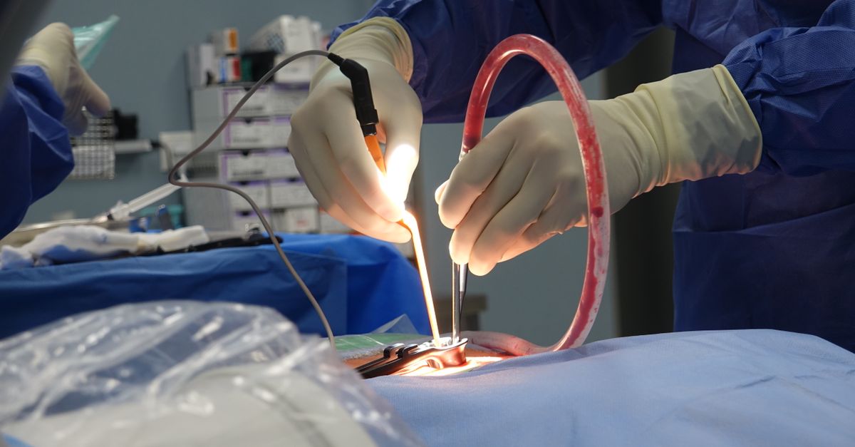
When active observation is not feasible, or if the patient prefers immediate resolution, surgical excision is the standard definitive treatment. Surgery is generally indicated if the mass is causing significant pain, limiting joint mobility, or generating profound cosmetic distress.
The surgical procedure involves a marginal excision, where the surgeon removes the nodule along with a very thin rim of surrounding normal tissue. Because nodular fasciitis does not aggressively invade neighboring structures, wide surgical margins, which are necessary for malignant tumors, are completely unneeded.
The procedure is typically performed on an outpatient basis under local anesthesia, allowing the patient to return home the same day. The surgical team ensures that the excised tissue is sent for final pathological confirmation to verify the initial biopsy results and ensure complete removal of the benign lesion.
14. Post-Surgical Recovery and Prognosis
Recovery following the marginal excision of nodular fasciitis is usually rapid and uncomplicated. Patients are advised to keep the surgical site clean and dry and to avoid strenuous activities that might stretch the incision line during the initial healing phase.
Postoperative pain is generally minimal and can be managed effectively with standard over-the-counter analgesics. Sutures are typically removed within one to two weeks, and patients can resume their normal daily activities shortly thereafter.
The long-term prognosis is exceptionally favorable. Once the lesion is removed, or if it resolves spontaneously, the patient is considered completely cured. The condition does not predispose the individual to any future connective tissue disorders or cancers. Local recurrence after surgical excision is exceedingly rare, occurring in less than two percent of cases, and is usually attributed to incomplete initial removal.
15. When to Seek Medical Evaluation
Discovering a rapidly growing lump anywhere on the body is an alarming experience that warrants immediate medical evaluation. Patients should not attempt to self-diagnose or monitor a new, fast-growing mass without professional guidance.
A prompt consultation with a healthcare provider or a dermatologist is necessary to initiate the appropriate diagnostic pathway. Timely clinical assessment and imaging can quickly differentiate between benign reactive processes and potentially serious neoplasms.
Early medical intervention ensures that a precise diagnosis is established through biopsy, alleviating unwarranted fear and anxiety. Establishing care with a specialized medical team guarantees that the patient receives evidence-based recommendations tailored to their specific clinical presentation.
16. Frequently Asked Questions (FAQ)
1. Is nodular fasciitis a type of cancer?
No, nodular fasciitis is entirely benign. Despite its rapid growth, which can mimic cancer, it does not have the biological ability to spread to other parts of the body or cause systemic illness.
2. Why did the lump grow so fast?
The rapid growth is a hallmark of the condition. It is caused by an exaggerated, reactive proliferation of connective tissue cells, often driven by a specific, benign genetic alteration in the USP6 gene.
3. Does nodular fasciitis go away on its own?
Yes, in many cases, once the nodule stops growing, it may gradually shrink and disappear completely over several months without any surgical treatment.
4. Will I need to have surgery to remove it?
Surgery is not always mandatory. If a biopsy confirms the diagnosis and the lump is not causing pain, your doctor may recommend observing it. Surgery is an option if the lump is painful, restrictive, or cosmetically bothersome.
5. Can the lump come back after it is removed?
The recurrence rate after surgical removal is extremely low, occurring in less than two percent of cases. If a lump returns at the same site, it requires prompt re-evaluation by a physician.
17. Bibliography
Disclaimer: The content is for informational purposes only and does not replace medical advice. Always consult your doctor for personalized treatment.