1. Introduction
Hazy vision is a frequent and significant clinical complaint characterized by a general loss of visual sharpness. Patients typically describe this symptom as looking through a dirty window, a dense fog, or a cloudy film. Unlike a localized blind spot or a specific visual field defect, hazy vision generally affects the entire field of view uniformly.
This symptom is not a disease in itself but rather a primary indicator of an underlying anatomical or physiological abnormality within the visual system. The etiology of hazy vision spans a broad spectrum, ranging from benign, easily correctable refractive errors to progressive, vision-threatening ocular pathologies.
A timely and comprehensive ophthalmological evaluation is essential when a patient presents with this symptom. Determining whether the onset was sudden or gradual, and identifying any accompanying symptoms such as pain or light sensitivity, guides the clinician in diagnosing the precise cause and implementing the appropriate medical or surgical intervention.
2. The Anatomy of Clear Vision
To understand the mechanisms behind hazy vision, one must trace the physiological pathway of light as it enters the eye. Light first passes through the cornea, the clear, dome-shaped outer window of the eye. The cornea provides the majority of the eye’s focusing power.
After passing through the aqueous humor in the anterior chamber, the light travels through the pupil and reaches the crystalline lens. The lens fine-tunes the focus, adjusting its shape to allow for clear vision at varying distances. The light then traverses the vitreous humor, a gel-like substance filling the central cavity of the eye.
Finally, the focused light hits the retina, the light-sensitive tissue lining the back of the eye. The macula, the central portion of the retina, is responsible for sharp, detailed vision. The retina converts the light into electrical signals, which are transmitted via the optic nerve to the brain for visual processing. Any disruption or opacity along this precise anatomical pathway will result in hazy vision.
3. Refractive Errors
The most common cause of hazy or blurry vision is a simple refractive error. This occurs when the physical shape of the eye prevents light from focusing directly on the retina. The primary types of refractive errors are myopia, hyperopia, and astigmatism.
In myopia, or nearsightedness, the eyeball is slightly too long, causing light rays to focus in front of the retina. This results in hazy distance vision while close objects remain clear. Conversely, hyperopia, or farsightedness, occurs when the eyeball is too short, causing light to focus behind the retina, blurring close objects.
Astigmatism is caused by an irregular curvature of the cornea or the lens. Instead of being perfectly spherical like a basketball, the surface resembles the shape of an American football. This irregular shape distorts incoming light rays, leading to hazy vision at all distances. Refractive errors are easily diagnosed and typically corrected with eyeglasses or contact lenses.
4. Cataracts and Lens Opacification
As the human body ages, the crystalline lens inside the eye undergoes structural changes. The proteins within the lens begin to break down and clump together. This slow, progressive process is the formation of a cataract.
A cataract acts as a physical barrier, scattering and blocking the light before it can reach the retina. The hallmark symptom of a developing cataract is a gradual, painless onset of hazy vision. Patients often report that colors appear faded or yellowed, and they may experience significant glare or halos around bright lights, particularly when driving at night.
Cataracts are a normal physiological consequence of aging, though their development can be accelerated by systemic conditions like diabetes, prolonged use of corticosteroid medications, or extensive exposure to ultraviolet radiation. When the hazy vision severely interferes with daily activities, surgical intervention is the definitive treatment.
5. Corneal Edema and Abnormalities
The cornea must remain perfectly clear and relatively dehydrated to function properly. Specialized cells on the inner surface of the cornea, known as the endothelium, continuously pump fluid out of the corneal tissue. If these cells are damaged or fail, fluid accumulates, leading to corneal edema, or swelling.
Corneal edema disrupts the uniform arrangement of the collagen fibers within the cornea, causing incoming light to scatter and resulting in severe hazy vision. Fuchs’ endothelial dystrophy is a common genetic disorder that causes the progressive loss of these vital pump cells, leading to chronic corneal swelling.
Furthermore, infections of the cornea, such as bacterial or viral keratitis, can cause hazy vision by generating a dense, opaque inflammatory ulcer on the corneal surface. These infections are severe medical emergencies that require immediate antimicrobial therapy to prevent permanent scarring.
6. Dry Eye Syndrome
A healthy tear film is critical for maintaining clear vision. The tear film smooths the microscopic irregularities on the surface of the cornea, creating a perfectly refractive surface. In dry eye syndrome, the eye fails to produce an adequate volume of tears, or the tears evaporate too quickly due to a lack of protective lipid layers.
When the tear film becomes unstable, the corneal surface dries out and becomes microscopically rough. Light passing through this irregular surface is scattered, resulting in transient hazy vision.
Patients with dry eye often report that their hazy vision fluctuates throughout the day, frequently worsening after prolonged reading or computer use, and temporarily improving immediately after blinking rapidly or instilling artificial lubricating drops.
7. Glaucoma and Intraocular Pressure
Glaucoma is a group of progressive eye diseases that damage the optic nerve, frequently associated with elevated intraocular pressure. While open-angle glaucoma, the most common form, typically causes a slow, painless loss of peripheral vision without initial haziness, acute angle-closure glaucoma presents very differently.
In acute angle-closure glaucoma, the drainage system of the eye becomes suddenly and completely blocked. The intraocular pressure spikes rapidly to dangerous levels. This massive pressure forces fluid into the cornea, causing acute corneal edema.
The patient experiences a sudden, dramatic onset of severely hazy vision, accompanied by intense eye pain, profound redness, nausea, and the perception of rainbow-colored halos around lights. This is a critical ocular emergency requiring immediate medical intervention to lower the pressure and save the optic nerve.
8. Macular Degeneration
Age-related macular degeneration affects the macula, the small central area of the retina responsible for sharp, detailed, straight-ahead vision. As the macular tissue deteriorates, the patient central vision becomes progressively hazy and distorted.
In the dry form of the disease, small yellow deposits called drusen accumulate beneath the macula, slowly thinning the tissue and blurring central vision. The wet form is less common but more severe, occurring when abnormal blood vessels grow beneath the retina and leak fluid or blood, causing rapid and severe haziness and distortion in the center of the visual field.
Unlike refractive errors or cataracts, macular degeneration does not affect peripheral vision. However, the hazy central vision profoundly impairs the ability to read, recognize faces, or perform intricate tasks.
9. Diabetic Retinopathy
Patients with diabetes are at a significant risk for developing diabetic retinopathy, a condition where chronic high blood sugar levels damage the delicate blood vessels supplying the retina.
In the early stages, these weakened vessels may leak fluid and proteins into the macula, a condition known as diabetic macular edema. This fluid accumulation causes the retinal tissue to swell, resulting in hazy and distorted vision.
In advanced, proliferative diabetic retinopathy, the eye attempts to grow new, fragile blood vessels to replace the damaged ones. These abnormal vessels can easily rupture, bleeding directly into the clear vitreous gel that fills the center of the eye. A vitreous hemorrhage blocks light from reaching the retina, presenting as a sudden, profound haziness or a shower of dark spots obscuring the vision.
10. Inflammatory Conditions of the Eye
Uveitis is the inflammation of the uvea, the vascular middle layer of the eye. This condition can be triggered by autoimmune disorders, systemic infections, or localized trauma.
During an episode of uveitis, inflammatory cells and proteins leak from the blood vessels into the clear fluids of the eye, particularly the anterior chamber and the vitreous cavity. The presence of these cellular debris and inflammatory proteins clouds the clear ocular media.
Patients with uveitis typically present with hazy vision, significant deep eye pain, marked light sensitivity (photophobia), and a severely red eye. Prompt treatment with potent topical or systemic corticosteroids is required to suppress the inflammation and prevent permanent structural damage to the eye.
11. Neurological Causes of Hazy Vision
Occasionally, hazy vision stems not from a problem within the physical structure of the eye itself, but from a neurological disruption along the visual pathway. Optic neuritis is an inflammation of the optic nerve, frequently associated with demyelinating diseases such as multiple sclerosis.
When the optic nerve becomes inflamed, its ability to transmit precise visual signals to the brain is compromised. The patient experiences a sudden onset of hazy or dimmed vision, often accompanied by a distinct loss of color vision and pain that worsens with eye movement.
Similarly, cerebrovascular events, such as a transient ischemic attack or a stroke affecting the visual cortex in the occipital lobe, can manifest as a sudden, hazy loss of vision in specific segments of the visual field, necessitating an immediate neurological evaluation.
12. Clinical Evaluation and Eye Examination
A systematic ophthalmological examination is required to determine the exact cause of hazy vision. The clinician begins by recording a detailed medical history, focusing on the onset, duration, and associated symptoms of the haziness.
Visual acuity testing is performed to quantify the degree of visual impairment. The clinician then performs a refraction test to determine if the haziness can be corrected simply with lenses.
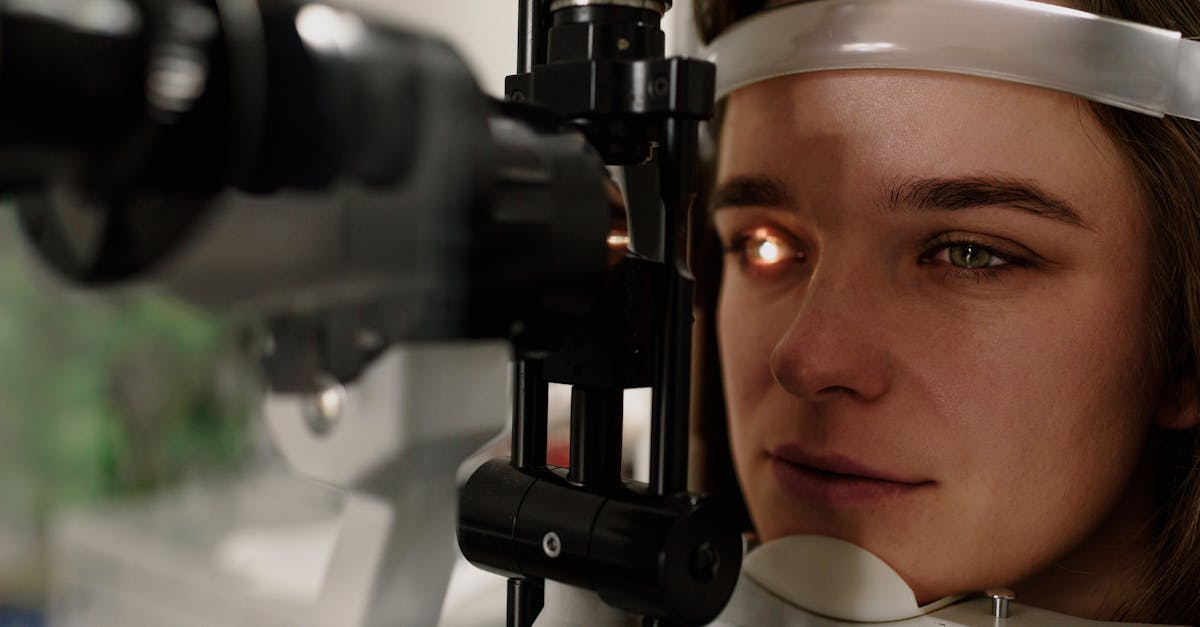
A slit-lamp biomicroscope is utilized to intensely magnify and illuminate the front structures of the eye. This allows the clinician to detect microscopic signs of corneal edema, evaluate the tear film, identify the presence of inflammatory cells in the anterior chamber, and assess the density of a developing cataract.
13. Diagnostic Imaging and Testing
To evaluate the posterior segment of the eye, the clinician dilates the pupils to obtain a comprehensive view of the retina, macula, and optic nerve.
Optical Coherence Tomography is a highly advanced, non-invasive imaging test that uses light waves to take cross-sectional pictures of the retina. This technology allows the clinician to visualize each distinct layer of the retina, making it an invaluable tool for diagnosing and managing macular degeneration and diabetic macular edema.
Tonometry is performed to measure the intraocular pressure, checking for signs of glaucoma. If a neurological cause is suspected, visual field testing and a referral for magnetic resonance imaging of the brain and optic nerves may be indicated.
14. Corrective and Medical Interventions
Treatment strategies are directly targeted at the underlying etiology. Refractive errors are managed easily with the prescription of corrective eyeglasses or contact lenses, which alter the trajectory of incoming light to focus precisely on the retina.
For dry eye syndrome, a comprehensive regimen of preservative-free artificial tears, warm compresses, and potentially prescription immunomodulatory eye drops, such as cyclosporine, is utilized to stabilize the tear film and restore the smooth optical surface of the cornea.
Inflammatory conditions like uveitis or optic neuritis require aggressive medical therapy. High-dose corticosteroids are administered topically, orally, or intravenously to rapidly suppress the immune response and clear the inflammatory debris from the visual axis.
15. Surgical Treatments
When anatomical structural changes cause the hazy vision, surgical intervention is often the only definitive solution. Cataract surgery is one of the most common and successful surgical procedures performed globally. The surgeon removes the cloudy, opacified lens and replaces it with a clear, synthetic intraocular lens implant, instantly restoring visual clarity.
For patients with severe corneal edema due to Fuchs’ dystrophy or corneal scarring from infections, a corneal transplant may be necessary. Modern techniques, such as endothelial keratoplasty, allow the surgeon to replace only the diseased inner layer of the cornea rather than the entire thickness, resulting in faster recovery and better visual outcomes.
In cases of advanced diabetic retinopathy with non-clearing vitreous hemorrhage, a vitrectomy is performed. The surgeon meticulously removes the blood-filled vitreous gel from the center of the eye, clearing the visual pathway, and treats the underlying abnormal blood vessels with a surgical laser.
16. Summary of Diagnostic Data
The clinical approach to evaluating hazy vision relies heavily on categorizing the symptoms and identifying the specific anatomical location of the disruption. The table below outlines common clinical correlations.
| Anatomical Origin | Common Condition | Characteristic Clinical Presentation |
|---|---|---|
| Cornea | Dry Eye Syndrome / Edema | Fluctuating haziness, foreign body sensation, light scatter |
| Lens | Cataract | Gradual, painless clouding, faded colors, increased glare |
| Retina/Macula | Macular Degeneration | Central hazy or distorted vision, peripheral vision preserved |
| Optic Nerve | Optic Neuritis | Sudden hazy vision, pain with eye movement, loss of color saturation |
Thorough diagnostic testing is essential to differentiate between benign, transient conditions and those requiring urgent surgical care.
17. Frequently Asked Questions (FAQ)
1. Is hazy vision the same thing as astigmatism?
Astigmatism is a specific physical shape of the eye that causes light to scatter, which is one of many possible causes of hazy vision. Hazy vision is the symptom; astigmatism is a potential underlying reason.
2. Can high blood pressure cause my vision to become cloudy?
Yes. Extremely high blood pressure can cause the blood vessels in the retina to leak fluid, or it can lead to a stroke in the eye, both of which can present as a sudden, painless onset of hazy or blurred vision.
3. Why is my vision hazy when I wake up, but it clears later?
This is very common with dry eye syndrome. During sleep, your eyes do not produce as many tears. When you open them, the surface is dry and rough, causing light to scatter. Blinking a few times restores the tear film and clears the vision.
4. Will eye drops cure a cataract?
No. A cataract is a structural change where the proteins inside the lens of the eye clump together and turn cloudy. No eye drop can dissolve this cloudiness. The only effective treatment for a cataract is surgical removal.
5. When should I seek emergency medical care for hazy vision?
You should go to an emergency room immediately if your hazy vision occurs suddenly, affects only one eye, is accompanied by severe eye pain, a severe headache, nausea, or a sudden loss of visual field (like a curtain falling over your vision).
18. Bibliography
Disclaimer: The content is for informational purposes only and does not replace medical advice. Always consult your doctor for personalized treatment.