1. Introduction
High blood sugar levels can directly cause blurry vision in one eye by inducing rapid osmotic changes within the crystalline lens or by causing localized, severe damage to the retinal blood vessels. The delicate structures of the eye are exceptionally sensitive to fluctuations in systemic glucose. When the body cannot properly regulate blood sugar, the resulting hyperglycemic environment structurally degrades the ocular microvasculature.
Visual disturbances tied to blood sugar often present bilaterally, affecting both eyes symmetrically. However, it is entirely possible, and clinically significant, for the blurriness to present unilaterally. This asymmetry usually indicates that a specific, localized complication, such as a microscopic hemorrhage or concentrated macular swelling, has occurred in one eye before the other.
Prompt clinical evaluation is necessary when unilateral blurriness arises in a diabetic patient. Ophthalmic assessment differentiates between transient, harmless lens swelling and permanent, vision-threatening retinal damage. Achieving tight glycemic control and utilizing advanced retinal therapies can halt disease progression and frequently restore visual clarity.
2. Blood Glucose and Ocular Health
The human eye requires a constant, highly regulated supply of nutrients and oxygen to maintain clear vision. The retina, the light-sensitive tissue lining the back of the eye, possesses one of the highest metabolic demands in the body. It relies on an intricate, dense network of microscopic capillaries to fulfill this demand.
When systemic blood glucose levels remain chronically elevated, this excess glucose becomes toxic to the vascular network. Hyperglycemia damages the endothelial cells that line the interior of these capillaries and destroys pericytes, the structural support cells that wrap around the vessels and maintain their integrity.
Without pericyte support, the capillary walls weaken, bulge, and become highly permeable. This microvascular degradation is the foundational pathology of diabetic eye disease, establishing an environment where fluid leakage and microscopic bleeding threaten the precise focal points required for clear vision.
3. Osmotic Changes in the Crystalline Lens
Before structural damage occurs to the retina, sudden spikes in blood sugar can cause rapid, temporary blurry vision through an entirely different mechanism involving the crystalline lens. The lens sits behind the pupil and changes shape to focus light accurately onto the retina.
The lens capsule is freely permeable to glucose. When blood sugar levels spike rapidly, the glucose concentration within the fluid of the eye increases. Glucose is an osmotically active molecule, meaning it strongly draws water toward itself.
As glucose floods into the lens, water follows rapidly. The lens swells significantly, altering its precise curvature. This sudden physical swelling changes the refractive index of the eye, making the patient acutely nearsighted or blurring their vision profoundly. This osmotic swelling can occasionally affect one eye more noticeably than the other depending on minute anatomical variations.
4. Transient Hyperglycemic Visual Blurring
Blurry vision caused by osmotic swelling of the lens is characteristically transient. It directly mirrors the patient’s immediate glycemic control. When blood sugar levels are exceptionally high, the vision becomes blurred and distorted.
As the patient administers insulin or hydration to lower their systemic blood glucose, the osmotic gradient reverses. The excess water is slowly drawn back out of the lens, allowing it to return to its original shape and resting curvature.
This recovery process can take several days to a few weeks of stable blood sugar control to completely resolve. Clinicians explicitly advise diabetic patients against receiving new corrective lens prescriptions during periods of uncontrolled blood sugar, as the glasses will be useless once the lens shape normalizes.
5. Diabetic Retinopathy Pathophysiology
If high blood sugar levels remain uncontrolled over years, the damage transitions from temporary lens swelling to permanent structural failure of the retina, a condition known as diabetic retinopathy. This disease progresses through specific stages of vascular degradation.
In the initial stage, non-proliferative diabetic retinopathy, the weakened capillaries develop microscopic bulges called microaneurysms. These weakened vessel walls begin to leak blood plasma and lipid deposits directly into the delicate retinal tissue.
This fluid accumulation disrupts the precise cellular alignment required for sharp visual processing. If this localized leakage occurs primarily in the left eye, for instance, the patient will experience significant unilateral blurry vision, while the right eye may remain temporarily asymptomatic despite possessing similar underlying microvascular damage.
6. Macular Edema and Central Vision
The macula is a highly specialized, central area of the retina responsible for sharp, detailed, straight-ahead vision, essential for reading and facial recognition. The fovea, the very center of the macula, contains the highest concentration of photoreceptor cells.
Diabetic macular edema is the most common cause of vision loss in patients with diabetes. It occurs when the leaking fluid and proteins from damaged capillaries accumulate directly within the macula. This fluid acts like a blister, physically lifting and separating the photoreceptor cells.
When the macula swells, the central visual field becomes profoundly blurred, warped, or washed out. Because microvascular leakage does not always occur symmetrically, profound macular edema can easily strike one eye, causing a sudden and severe unilateral drop in visual acuity.
7. Why Symptoms Can Be Unilateral
While diabetes is a systemic disease affecting blood vessels throughout the entire body, the manifestations of vascular failure are often asymmetric. The capillary networks in the left and right eyes are structurally independent.
A specific cluster of pericytes may fail in the right eye months or years before a similar failure occurs in the left. A single microaneurysm rupturing near the fovea in one eye will immediately compromise that eye’s vision, while the other eye, lacking a strategically placed rupture, continues to function adequately.
Therefore, patients must never ignore sudden blurriness in one eye under the assumption that a systemic issue like high blood sugar would naturally affect both eyes simultaneously. Unilateral symptoms highlight a localized, acute vascular event requiring immediate evaluation.
8. Retinal Neovascularization
As non-proliferative retinopathy advances, the damaged capillaries eventually occlude completely, cutting off the blood supply to large sections of the retina. The retina, starving for oxygen, secretes vascular endothelial growth factor (VEGF), a chemical signal demanding new blood vessel creation.
This triggers proliferative diabetic retinopathy, the most advanced and dangerous stage of the disease. In response to the VEGF signals, abnormal, fragile new blood vessels begin to grow rapidly along the surface of the retina and into the clear vitreous gel filling the eye.
These neovascular formations are architecturally defective. They lack standard structural integrity and are incredibly prone to spontaneous rupture. The presence of these fragile vessels places the eye at imminent risk for catastrophic bleeding and sudden, profound vision loss.
9. Vitreous Hemorrhage Complications
When the abnormal neovascular vessels rupture, they bleed directly into the vitreous humor, the clear, jelly-like substance that maintains the shape of the eye. This event is clinically known as a vitreous hemorrhage.
A minor hemorrhage may present as a sudden shower of dark, floating spots or spiderwebs drifting across the visual field of one eye. If the bleeding is severe, the dense blood completely blocks light from passing through the vitreous to reach the retina.
The patient will experience a sudden, painless, and complete loss of vision or a profound dark blurring in the affected eye. This is a dramatic, unilateral event directly resulting from long-standing, poorly controlled high blood sugar.
10. Cataract Formation and Glycation
High blood sugar accelerates the aging processes within the eye, significantly increasing the risk of early cataract formation. A cataract is the clouding of the normally clear crystalline lens, which scatter light and creates a hazy, blurred visual field.
Chronic hyperglycemia facilitates a process called glycation, where excess glucose molecules bind irreversibly to the structural proteins within the lens. This chemical binding damages the proteins, causing them to clump together and lose their transparency.
Cataracts often develop asymmetrically. One eye may develop a dense, central cataract much faster than the other, leading to a progressive, painless blurring of vision in the affected eye. Unlike osmotic swelling, this structural clouding is permanent and cannot be reversed by lowering blood sugar.
11. Differentiating Vision Changes
Understanding the onset and characteristics of the blurriness helps determine its specific physiological cause.
| Visual Symptom | Onset and Duration | Primary Mechanism |
|---|---|---|
| Fluctuating blurriness in both eyes | Hours to days; changes with meals | Osmotic lens swelling from glucose spikes |
| Central blur or warped lines in one eye | Gradual worsening over weeks | Diabetic macular edema (fluid leakage) |
| Sudden shower of dark floaters | Instantaneous, usually unilateral | Minor vitreous hemorrhage |
| Sudden, total dark blur in one eye | Instantaneous, painless | Massive vitreous hemorrhage |
Any sudden or unilateral change in vision warrants immediate ophthalmic evaluation to prevent permanent cellular damage.
12. Diagnostic Ophthalmic Evaluation
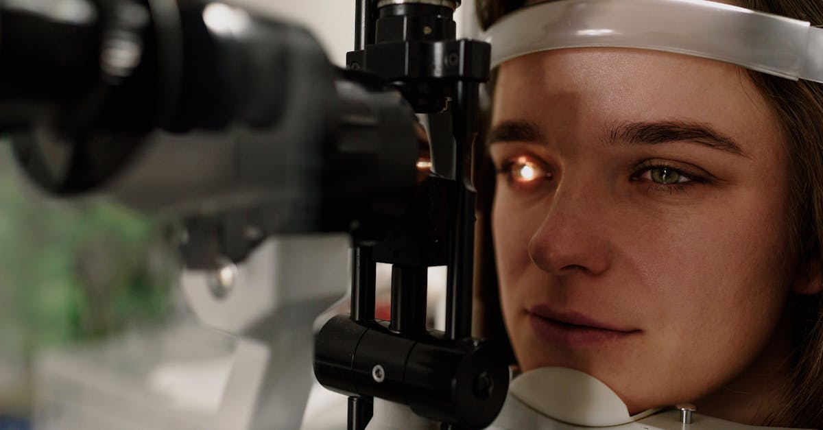
When a diabetic patient presents with unilateral blurry vision, a comprehensive dilated eye examination is mandatory. The ophthalmologist utilizes special dilating drops to widen the pupil, allowing for a complete, unobstructed view of the retina and the optic nerve.
Optical coherence tomography is a critical diagnostic tool. This non-invasive imaging scan uses light waves to take cross-sectional pictures of the retina, mapping its exact thickness and detecting even microscopic amounts of fluid accumulation indicative of macular edema.
Fluorescein angiography may also be employed. A specialized yellow dye is injected into a vein in the arm, which quickly travels to the blood vessels in the eye. A camera captures images as the dye illuminates the retinal vessels, explicitly highlighting any areas of microvascular leakage or abnormal neovascular growth.
13. Treatment Modalities for Retinal Damage
If the blurry vision is caused by macular edema or proliferative retinopathy, advanced therapeutic interventions are required to preserve sight. The standard of care involves the intravitreal injection of anti-VEGF medications.
These medications are carefully injected directly into the vitreous cavity of the eye. They bind to and neutralize the vascular endothelial growth factor, which rapidly halts the leakage from damaged capillaries and commands the abnormal neovascular vessels to regress and shrink.
Focal laser photocoagulation is another intervention. A precise medical laser is used to create microscopic burns around the leaking microaneurysms, effectively cauterizing the vessels and sealing the leaks to reduce macular swelling and restore visual clarity.
14. The Importance of Glycemic Control
While ophthalmic treatments can halt bleeding and reduce swelling, they do not cure the underlying systemic disease. The most critical factor in preventing further visual degradation is the rigorous management of systemic blood glucose levels.
Consistent glycemic control prevents the ongoing destruction of the capillary pericytes and the endothelial lining. By maintaining blood sugar within targeted ranges, the patient eliminates the toxic environment that drives the progression of diabetic eye disease.
Furthermore, managing concurrent systemic factors, specifically hypertension and hyperlipidemia, is essential. High blood pressure compounds the mechanical stress on the already weakened retinal capillaries, accelerating the rate of fluid leakage and increasing the risk of sudden hemorrhage.
15. Preventative Eye Care for Diabetics
Because significant retinal damage can occur before any noticeable blurring of vision presents, proactive and preventative care is the cornerstone of preserving sight in diabetic populations. Early microvascular changes are entirely asymptomatic.
Clinical guidelines mandate that all patients with diabetes undergo a comprehensive dilated eye examination at least once a year, regardless of their current visual clarity. Early detection of microaneurysms allows for closer monitoring and intervention before sight-threatening macular edema develops.
Patients are instructed to immediately report any new visual symptoms, particularly unilateral changes, sudden floaters, or localized blind spots, ensuring rapid access to sight-saving therapies.
16. Frequently Asked Questions FAQ
1. Why is only one eye blurry if high blood sugar affects my whole body?
While diabetes affects all blood vessels, the tiny capillaries in your eyes fail at different rates. A specific blood vessel may rupture or leak fluid near the center of the retina in your left eye, causing immediate blurriness, while the right eye remains temporarily stable.
2. Will my vision clear up once I lower my blood sugar?
If the blurriness is caused by the temporary swelling of the lens inside your eye due to a recent sugar spike, your vision will return to normal after a few days or weeks of stable blood sugar. If the blurriness is caused by retinal damage, it requires specific eye treatments.
3. Can getting new glasses fix the blurry vision from diabetes?
If your blood sugar is currently uncontrolled, getting new glasses is not recommended because the physical shape of your eye’s lens is temporarily swollen. Once your blood sugar stabilizes, the lens shape changes back, and the new glasses will no longer work.
4. Is it normal to see sudden dark floating spots if my blood sugar is high?
No, a sudden shower of dark spots or cobwebs is a medical emergency. It strongly indicates that an abnormal, fragile blood vessel in your retina has ruptured and is actively bleeding into the back of your eye.
5. How often should I see an eye doctor if I have diabetes?
You must have a comprehensive, dilated eye exam at least once a year. Retinal damage often begins silently, and an eye doctor can detect and treat early capillary leakage long before it causes you to experience any blurry vision.
17. Bibliography
Disclaimer: The content is for informational purposes only and does not replace medical advice. Always consult your doctor for personalized treatment.