1. Introduction
Blurry vision during a headache typically indicates a migraine with aura, a phenomenon caused by transient electrical changes in the visual cortex. However, it can also signify increased intracranial pressure or acute glaucoma, requiring immediate medical assessment if the vision loss is sudden, progressive, or entirely unyielding.
Experiencing visual disturbances alongside cranial pain is a highly specific neurological presentation. The visual system is intricately connected to the brain through a massive network of nerves and blood vessels. When an inflammatory, electrical, or pressure-related event occurs within the cranium, the visual pathways are frequently the first structures to exhibit clinical distress.
Differentiating a benign, transient aura from a vision-threatening emergency relies on analyzing the precise characteristics of the blurriness. Clinicians evaluate whether the visual loss occurs in one eye or both, the duration of the symptom, and the specific nature of the accompanying head pain to formulate an accurate diagnosis and prevent permanent neurological damage.
2. The Neurological Connection Between Vision and Pain
The human visual pathway extends from the retina at the front of the eye all the way to the occipital lobe at the very back of the brain. Because this pathway spans the entire length of the intracranial cavity, it is uniquely vulnerable to changes in local blood flow, electrical stability, and physical pressure.
Pain in the head is primarily mediated by the trigeminal nerve. This massive sensory nerve not only provides feeling to the face and scalp but also innervates the meninges, which are the protective membranes surrounding the brain. The trigeminal nerve shares intimate neurological connections with the blood vessels supplying the visual cortex.
When a headache is triggered by a vascular or inflammatory event, the trigeminal nerve releases neuropeptides that cause localized swelling in the cranial blood vessels. This swelling can temporarily compress adjacent visual nerve fibers or alter blood flow to the occipital lobe, resulting in perceived blurriness or visual distortion during the pain episode.
3. Understanding Migraine with Aura
The most prevalent cause of blurry vision during a headache is a migraine with aura. An aura is a transient, reversible neurological deficit that typically precedes or accompanies the headache phase of a migraine attack. Visual auras occur in roughly one-third of all migraine sufferers.
The blurriness experienced during a migraine aura is rarely a simple out-of-focus sensation. Patients frequently describe seeing jagged, shimmering lines, bright flashing lights, or expanding blind spots, known as scotomas. These visual anomalies usually begin in the center of the visual field and slowly drift outward toward the periphery over twenty to thirty minutes.
Importantly, the visual symptoms of a classic migraine aura are homonymous, meaning they affect the visual field of both eyes simultaneously. If a patient covers one eye, the blurry or shimmering spot will still be visible in the corresponding field of the open eye, proving that the phenomenon is occurring in the brain, not the eyeball.
4. Cortical Spreading Depression Mechanisms
The exact biological mechanism driving a migraine aura is a phenomenon called cortical spreading depression. This is a slow, self-propagating wave of electrical depolarization that moves across the surface of the cerebral cortex, followed immediately by a prolonged period of suppressed neurological activity.
When this electrical wave sweeps across the occipital lobe, the region responsible for processing visual information, it triggers erratic firing of the visual neurons. This sudden burst of electrical activity causes the patient to see the bright flashes or shimmering lines characteristic of the aura.
As the wave passes and leaves a wake of suppressed cellular activity, the patient experiences the blind spots or localized blurry vision. Cortical spreading depression also triggers the trigeminal nerve, initiating the neurogenic inflammation that eventually leads to the severe, throbbing pain of the migraine headache itself.
5. Ocular Migraines and Retinal Vasospasm
An ocular migraine, medically termed a retinal migraine, is a rare and distinct clinical entity from a standard migraine with aura. While a standard aura affects the brain and alters the vision in both eyes, an ocular migraine causes temporary visual loss or severe blurriness in only one single eye.
This unilateral visual disturbance is caused by a sudden, transient vasospasm of the blood vessels specifically supplying the retina of the affected eye. The blood flow to the sensitive retinal tissue is momentarily reduced, causing the vision to dim, blur, or completely black out for several minutes.
The visual symptoms in a retinal migraine are typically followed shortly by a throbbing headache on the same side of the head. Because transient monocular vision loss can also be a sign of a stroke or a retinal detachment, a diagnosis of retinal migraine is only made after a physician has rigorously excluded these severe structural emergencies.
6. Intracranial Hypertension and Papilledema
A far more critical cause of blurry vision accompanying a headache is elevated pressure inside the skull, known as intracranial hypertension. The brain, blood, and cerebrospinal fluid are enclosed within a rigid bony vault. If the volume of any of these components increases due to a tumor, bleeding, or poor fluid drainage, the pressure inside the skull rises dangerously.
This high pressure transmits directly down the sheath surrounding the optic nerve. The physical pressure chokes the nerve, causing the optic disc inside the eye to swell massively, a condition called papilledema. As the nerve swells, its ability to transmit clear visual signals is severely compromised.
Headaches caused by elevated intracranial pressure are often worst in the morning or when lying flat, as gravity increases the fluid pooling in the head. The blurriness may initially be transient, occurring only when the patient bends over or coughs, but will become constant as the nerve damage progresses.
7. Acute Angle-Closure Glaucoma
Acute angle-closure glaucoma is an absolute ophthalmic emergency that frequently masquerades as a severe neurological headache. This condition occurs when the drainage angle inside the front of the eye suddenly closes, trapping the aqueous fluid and causing intraocular pressure to spike rapidly to perilous levels.
The patient typically experiences a sudden, agonizing pain in and around one eye, which rapidly radiates outward, presenting as a severe frontal headache. The rapid rise in pressure forces fluid into the cornea, causing it to become waterlogged and cloudy. This corneal edema results in profound, sudden blurry vision.
| Diagnostic Feature | Migraine with Aura | Acute Angle-Closure Glaucoma |
|---|---|---|
| Nature of Visual Blur | Shimmering, zigzag lines, affects both eyes. | Cloudy, hazy vision, halos around lights, affects one eye. |
| Eye Appearance | Eyes appear completely normal. | Eye is very red, pupil is mid-dilated and unreactive. |
| Accompanying Symptoms | Sensitivity to light and sound, nausea. | Severe nausea, vomiting, intense localized eye pain. |
Immediate administration of pressure-lowering eye drops and systemic medications is required to prevent permanent blindness. Clinicians are trained to inspect the pupil in any patient presenting with a severe, unilateral headache and blurry vision to rule out this structural crisis.
8. Temporal Arteritis and Ischemic Optic Neuropathy
Temporal arteritis, or giant cell arteritis, is a serious inflammatory disease of the medium and large blood vessels, predominantly affecting individuals over the age of fifty. The inflammation typically targets the temporal arteries, which supply blood to the scalp and the optic nerve.
When the blood vessels become severely inflamed, their internal diameter narrows, drastically reducing blood flow. Patients typically present with a new, continuous, throbbing headache, often localized to the temple region. The scalp may feel exquisitely tender to the touch, making brushing hair painful.
If the inflammation compromises the blood supply to the optic nerve, it causes anterior ischemic optic neuropathy. This results in a sudden, painless, and severe blurring or complete loss of vision in one eye. This is a medical emergency requiring immediate high-dose intravenous corticosteroids to save the vision in the affected eye and protect the contralateral eye.
9. Tension Headaches and Visual Strain
Not all instances of blurry vision during a headache indicate a severe neurological or vascular crisis. Tension-type headaches, the most common primary headache disorder, are frequently associated with mild, transient visual blurriness that is entirely benign.
This blurriness is usually the direct result of prolonged visual strain and exhaustion of the intraocular ciliary muscles, which are responsible for focusing the lens of the eye. After hours of staring at a computer screen or reading fine print, these muscles become fatigued and spasm, preventing clear focus.
The sustained visual effort also causes the patient to subconsciously tighten their facial and pericranial muscles, triggering the dull, band-like ache of a tension headache. Resting the eyes, applying a warm compress, and utilizing over-the-counter analgesics generally resolves both the muscular ache and the focusing difficulty.
10. Recognizing Red Flag Visual Symptoms
To ensure patient safety, clinicians rely on specific red flag symptoms that separate benign, headache-related visual changes from pathological emergencies. The speed of onset is a critical factor. An aura develops gradually over several minutes, whereas a sudden, instantaneous drop in vision suggests a vascular blockage.
A visual deficit that persists long after the headache has resolved is deeply concerning. Migraine auras typically conclude within sixty minutes. If the vision remains blurry, dark, or distorted for several hours or days, it indicates that physical tissue damage has occurred within the retina or the optic nerve pathways.
Additionally, if the blurry vision is accompanied by new neurological deficits such as slurred speech, profound weakness on one side of the body, or a sudden loss of coordination, the patient is likely experiencing a stroke. These combined symptoms demand immediate activation of emergency medical services.
11. Diagnostic Ophthalmic and Neurological Testing
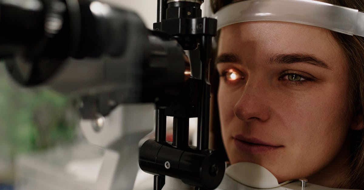
Evaluating a patient who reports blurry vision during headaches requires a multidisciplinary diagnostic approach. A comprehensive ophthalmic examination is the starting point. The physician will measure visual acuity, check intraocular pressure to rule out glaucoma, and perform a dilated fundus exam.
During the dilated exam, the physician visually inspects the optic disc for papilledema, which would confirm elevated intracranial pressure, and examines the retinal blood vessels for signs of vasospasm or inflammatory occlusion.
If the ophthalmic exam is unremarkable but the symptoms are atypical for a simple migraine, neurological imaging is necessary. A magnetic resonance imaging scan of the brain provides detailed views of the visual cortex and cranial nerves, ensuring there are no tumors, aneurysms, or demyelinating lesions interfering with visual processing.
12. Pharmacological Management of Migraine Auras
For patients with confirmed migraine with aura, treatment focuses on aborting the headache and preventing future cortical spreading depression events. Acute treatments, such as triptans, are most effective when taken at the very onset of the aura phase, before the severe pain fully establishes itself.
Triptans function by binding to serotonin receptors, causing the dilated cranial blood vessels to constrict and blocking the transmission of pain signals along the trigeminal nerve. However, because they are potent vasoconstrictors, they are generally contraindicated in patients with a history of retinal migraines due to the risk of worsening the ocular vasospasm.
For patients experiencing frequent auras that disrupt their daily life, preventive medications are initiated. Anti-seizure medications, beta-blockers, and certain antidepressants are very effective at stabilizing the electrical activity of the cerebral cortex, making it significantly harder for the wave of spreading depression to initiate.
13. Lifestyle Modifications for Prevention
Non-pharmacological strategies are essential for managing headaches associated with visual disturbances. Identifying and strictly avoiding personal triggers is the most reliable preventative measure. Common triggers include severe emotional stress, profound sleep deprivation, and drastic changes in barometric pressure.
Dietary factors also play a significant role. Aspartame, monosodium glutamate, aged cheeses containing tyramine, and excessive caffeine intake are notorious for lowering the neurological threshold and triggering migraine events. Maintaining a consistent meal schedule prevents the blood sugar drops that frequently initiate attacks.
Proper ergonomic alignment and limiting continuous screen time are vital for preventing tension headaches and the associated ciliary muscle fatigue. Implementing frequent screen breaks and ensuring adequate ambient lighting reduces the physiological burden on the visual system, lowering the frequency of visually induced cranial aches.
14. When to Seek Emergency Medical Care
While many cases of blurry vision with a headache are related to familiar migraine patterns, patients must err on the side of caution. Any new, severe headache accompanied by visual changes in a patient over the age of fifty requires immediate medical evaluation to definitively rule out temporal arteritis.
If a pregnant woman experiences a sudden, severe headache with blurry vision or flashing lights, she must proceed to an emergency department immediately. This specific symptom cluster is a hallmark sign of preeclampsia, a critical, life-threatening spike in blood pressure that can cause seizures and organ failure.
Finally, a headache described as the sudden, worst pain of one’s life, accompanied by visual changes and a stiff neck, is the classic presentation of a ruptured cerebral aneurysm. Rapid transport to a neurosurgical center is absolutely essential for survival.
15. Frequently Asked Questions FAQ
1. Does needing new glasses cause headaches?
Yes, wearing an outdated prescription forces the intraocular muscles to strain continuously to achieve focus, leading to severe visual fatigue and frequent, dull tension headaches at the end of the day.
2. Can a sinus infection cause my vision to blur?
While a severe sinus infection causes significant pressure around the eyes, it does not typically cause blurry vision; if vision becomes blurry or double during a sinus infection, it suggests the infection has dangerously spread into the orbital cavity.
3. Why do I see zig-zag lines before my headache starts?
These shimmering, jagged lines, known as a fortification spectrum, are the classic visual aura caused by a wave of altered electrical activity moving slowly across the visual processing center in the back of your brain.
4. Is it normal for blurry vision to last after a migraine goes away?
No, the visual aura should resolve completely within an hour; if your vision remains blurry or distorted after the pain is gone, you require a medical evaluation to ensure no vascular damage occurred in the retina.
5. Can high blood pressure cause blurry vision and headaches?
Yes, a severe hypertensive crisis dramatically increases pressure in the cranial blood vessels, causing a throbbing headache and forcing fluid into the retina, which directly results in significant visual blurring.
Bibliography
Disclaimer: The content is for informational purposes only and does not replace medical advice. Always consult your doctor for personalized treatment.