1. Introduction
Sharp pain in the heel of the foot when walking is predominantly caused by plantar fasciitis, a condition involving the structural breakdown and inflammation of the thick band of connective tissue spanning the arch of the foot. Other notable clinical causes include the formation of calcaneal heel spurs, entrapment of the tibial nerve, or localized degradation of the protective fat pad beneath the heel bone. When the foot strikes the ground during the normal gait cycle, the entire weight of the human body is transferred directly through the heel, requiring an intricate network of fascia, ligaments, and fat to absorb and disperse the massive kinetic impact.
The human foot is an engineering marvel designed for dynamic shock absorption and propulsion. However, this biomechanical system is exceptionally vulnerable to repetitive strain and improper structural loading. When factors such as unsupportive footwear, sudden increases in physical activity, or anatomical abnormalities like flat feet place undue tension on the plantar fascia, the collagen fibers sustain microscopic tears. This microtrauma initiates an aggressive localized inflammatory response. Understanding the precise location and timing of the sharp pain is essential for differentiating a fascial strain from a neurological entrapment or a bony stress fracture.
2. Biomechanics of the Foot and Heel
To comprehend the origin of heel pain, one must visualize the architectural mechanics of the foot. The heel bone, or calcaneus, is the largest bone in the foot and serves as the initial point of contact during the heel-strike phase of walking.
Extending from the bottom of the calcaneus, across the arch, and attaching to the base of the toes is the plantar fascia. This thick, inelastic band of connective tissue acts like a bowstring. It holds up the longitudinal arch of the foot and provides critical tension that allows the foot to act as a rigid lever for pushing off the ground.
During the gait cycle, as the body weight rolls forward onto the ball of the foot, the arch flattens slightly, and the plantar fascia stretches tightly. If the foot mechanics are flawed—such as overpronation where the foot rolls too far inward—this bowstring is stretched beyond its biological tolerance, placing severe shear force directly on its attachment point at the heel bone.
3. Pathophysiology of Plantar Fasciitis
Plantar fasciitis is the leading clinical cause of a sharp, stabbing sensation in the heel. Despite the suffix “-itis,” indicating inflammation, advanced medical research demonstrates that plantar fasciitis is primarily a degenerative condition, more accurately termed plantar fasciosis.
The repetitive mechanical overloading of the fascia causes microscopic tears at the precise junction where the tissue anchors into the calcaneus. The body attempts to heal these micro-tears with an inflammatory response, but continuous daily walking disrupts the healing process before it can finalize.
Over time, this chronic cycle of tearing and incomplete healing causes the collagen fibers to degrade and lose their structural organization. The tissue becomes thickened, scarred, and highly irritable. The nociceptors, which are the pain-sensing nerves embedded in the fascia, become hyper-sensitized, generating a sharp, agonizing stab with every step.
4. The Morning Step Phenomenon
The most defining clinical hallmark of plantar fasciitis is the severe, stabbing pain experienced during the very first steps taken in the morning or after a long period of sitting.
During sleep or prolonged rest, the foot naturally rests in a slightly pointed downward position (plantar flexion). In this relaxed position, the plantar fascia shortens and begins to heal and tighten in that shortened state.
When the individual stands up and places their full body weight on the foot, the ankle flexes upward, and the shortened, stiffened fascia is violently ripped and stretched apart. This sudden, forceful tearing of the partially healed tissue produces a breathtakingly sharp pain. As the individual hobbles around for a few minutes, the fascia gradually warms up and elongates, and the sharp pain typically dulls to a persistent ache.
5. Heel Spurs and Bony Exostosis
A heel spur is a physical, bony growth (exostosis) that develops on the underside of the calcaneus bone, extending forward toward the arch. Historically, physicians believed these sharp bony spikes were the direct cause of heel pain, stabbing the tissue like a needle.
However, clinical imaging reveals that many individuals with massive heel spurs experience zero pain, while patients in excruciating agony may have perfectly smooth heel bones. A heel spur is actually a symptom of chronic tension, not the primary cause of the pain.
When the plantar fascia constantly pulls and tears at its attachment point on the bone, the body attempts to secure the connection by depositing calcium and building new bone along the line of tension. The sharp pain is derived from the inflamed, degenerated fascia attached to the bone, rather than the bony spur itself mechanically piercing the flesh.
6. Fat Pad Atrophy
Beneath the calcaneus lies a specialized, thick layer of adipose tissue known as the heel fat pad. This pad serves as a dense biological shock absorber, cushioning the bone against the hard impact of the ground.
As individuals age, or as a consequence of receiving multiple corticosteroid injections into the heel, this fat pad can begin to shrink and degrade, a condition known as fat pad atrophy.
Without this vital cushioning, the hard calcaneus bone strikes the ground almost directly, bruising the bone itself and crushing the delicate capillary beds and nerves in the thin overlying skin. Pain caused by fat pad atrophy is typically described as a deep, severe bruise precisely in the center of the heel, rather than the sharp, tearing pain of plantar fasciitis located closer to the arch.
7. Achilles Tendinopathy
While plantar fasciitis causes pain under the heel, Achilles tendinopathy generates severe pain at the back of the heel. The Achilles tendon is the massive, powerful cord connecting the calf muscles to the posterior aspect of the calcaneus.
Repetitive explosive movements, such as running or jumping, or abrupt increases in training volume, can cause microtrauma to the collagen fibers of this tendon. As the tendon degrades, it becomes thickened, stiff, and exceptionally painful.
When a patient with Achilles tendinopathy walks, the mechanical pull of the calf muscle forcefully stretches the damaged tendon against its bony attachment. This creates a sharp, localized pain at the very back of the heel bone that significantly worsens when attempting to stand on tiptoes or walk up a flight of stairs.
8. Tarsal Tunnel Syndrome
Sharp, shooting, or burning pain in the heel can originate from a neurological entrapment rather than a structural tissue strain. Tarsal tunnel syndrome involves the compression of the posterior tibial nerve.
This large nerve travels down the inside of the ankle and passes through a narrow anatomical space called the tarsal tunnel before branching into the heel and the sole of the foot. If the tunnel is narrowed by severe flat feet, swollen tendons, or varicose veins, the nerve is mechanically crushed.
This nerve compression triggers sharp, electrical, or burning pain that radiates around the inside of the ankle and straight into the heel. Unlike structural fasciitis, nerve pain from tarsal tunnel syndrome often worsens dynamically at night and is accompanied by distinct tingling or numbness in the bottom of the foot.
9. Calcaneal Stress Fractures
In highly active individuals, long-distance runners, or military recruits, sharp heel pain may indicate a calcaneal stress fracture. The repetitive, high-impact striking of the heel on hard surfaces eventually overwhelms the bone’s ability to remodel and repair itself.
Microscopic cracks begin to form within the trabecular structure of the heel bone. The pain of a stress fracture is severe, deep, and occurs relentlessly with every single weight-bearing step.
Unlike plantar fasciitis, which often loosens up after walking a short distance, the pain of a stress fracture progressively worsens the longer the patient is on their feet. Applying physical pressure by squeezing the sides of the heel bone together typically elicits an agonizing pain response, confirming the bony origin of the distress.
10. The Impact of Footwear and Biomechanics
The physical environment supporting the foot heavily dictates the strain placed upon the internal structures. Unsupportive footwear is the leading environmental catalyst for heel pain.
Walking barefoot on hard surfaces, such as hardwood or tile floors, forces the plantar fascia and the fat pad to absorb 100 percent of the kinetic impact without any external shock absorption.
Wearing shoes lacking adequate arch support, such as completely flat sandals or worn-out athletic shoes, allows the foot to overpronate. The arch collapses inward with every step, violently stretching the plantar fascia. Transitioning to structurally rigid, supportive footwear is the foundational requirement for mitigating this mechanical strain.
11. Differential Diagnosis of Heel Pain
Accurately charting the location and characteristics of the pain allows the clinician to differentiate the root cause effectively.
| Pain Characteristic | Anatomical Location | Probable Clinical Cause |
|---|---|---|
| Sharp, tearing pain during the first morning steps | Bottom of heel, slightly toward the arch | Plantar Fasciitis |
| Deep, bruised feeling worsening with standing | Dead center of the heel pad | Fat Pad Atrophy |
| Electric, burning, or tingling pain | Inside of ankle radiating to the heel | Tarsal Tunnel Syndrome |
| Severe pain worsening with every step, side tenderness | Entire heel bone | Calcaneal Stress Fracture |
Identifying these distinct patterns guides the physician toward selecting the appropriate diagnostic imaging.
12. Clinical Diagnostic Imaging
While plantar fasciitis is frequently diagnosed entirely through clinical history and physical examination, prolonged or atypical pain demands diagnostic imaging. A standard weight-bearing X-ray is utilized to rule out acute fractures and visualize the presence of calcaneal heel spurs.
If a stress fracture or severe soft tissue degradation is suspected, a Magnetic Resonance Imaging (MRI) scan is ordered. An MRI vividly displays the thickness of the plantar fascia, highlighting the exact areas of severe inflammation, tears, or surrounding tissue edema.
Diagnostic ultrasound is an increasingly popular, cost-effective tool that allows the physician to dynamically assess the thickness of the fascia and guide targeted therapeutic injections precisely into the most inflamed regions of the tissue without damaging the protective fat pad.
13. Conservative Management and Physical Therapy
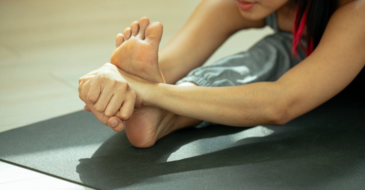
The vast majority of heel pain cases resolve completely with dedicated, conservative biomechanical management. The immediate priority is resting the tissue and aggressively stretching the calf muscles and the Achilles tendon. Tight calf muscles restrict ankle mobility, forcing the foot to compensate by excessively pulling on the plantar fascia.
A physical therapist will implement specific fascial loading exercises and deep tissue massage to break down the disorganized scar tissue and promote the realignment of healthy collagen fibers.
To combat the severe morning pain, patients are frequently prescribed a rigid night splint. This device holds the foot at a strict ninety-degree angle throughout the night, keeping the plantar fascia constantly elongated. This prevents the tissue from healing in a shortened position, completely eliminating the violent, tearing pain of the first morning step.
14. Frequently Asked Questions
1. Why do I feel completely fine walking in high heels but get sharp pain barefoot?
High heels artificially lift the heel bone, which puts your foot in a pointed position (plantar flexion). This position puts slack on the plantar fascia and the Achilles tendon, relieving the mechanical tension. When you walk barefoot, the foot flattens entirely, stretching the inflamed bowstring to its absolute limit and triggering a sharp, tearing pain.
2. Will rolling my foot on a frozen water bottle cure my plantar fasciitis?
Rolling your arch over a frozen water bottle provides excellent temporary relief. The ice numbs the hyperactive nerve endings and constricts the inflamed blood vessels, while the rolling motion provides a gentle physical stretch to the fascia. However, it manages symptoms rather than curing the underlying biomechanical overload.
3. Are steroid injections safe for severe heel pain?
Corticosteroid injections provide profound, rapid anti-inflammatory relief for severe cases. However, they are used sparingly because multiple injections aggressively degrade the collagen fibers of the fascia, significantly increasing the risk of a complete, catastrophic fascial rupture, and they can permanently dissolve the protective heel fat pad.
4. Do custom orthotics actually help, or are store-bought inserts enough?
For mild, acute pain, a firm over-the-counter arch support is often sufficient to unload the fascia. However, if your pain is driven by a severe biomechanical flaw, such as extreme overpronation or drastically unequal leg lengths, a custom-molded podiatric orthotic is necessary to physically realign the bones of your foot and correct the specific gait abnormality.
15. Bibliography
Disclaimer: The content is for informational purposes only and does not replace medical advice. Always consult your doctor for personalized treatment.