1. Introduction
Yes, severe psychological stress can lead to sudden blurry vision. When the body enters a fight-or-flight state, a surge of adrenaline causes the pupils to dilate and the ciliary muscles to spasm, making focusing on near objects difficult. Furthermore, prolonged elevations in cortisol can induce fluid shifts beneath the retina, causing a distinct central blurriness that requires medical evaluation.
The human visual system is intricately connected to the autonomic nervous system. Vision is not merely a mechanical process of light entering the eye; it involves complex neuromuscular coordination and precise fluid balance within the ocular tissues. Any systemic physiological shift, particularly a surge in stress hormones, directly impacts these delicate structures.
Understanding how mental strain translates into physical visual disturbances requires a detailed look at the body’s acute and chronic stress responses. While transient blurriness usually resolves once the nervous system calms down, persistent visual changes during periods of severe anxiety warrant a thorough clinical assessment to rule out structural damage.
2. The Physiology of the Stress Response
The autonomic nervous system regulates involuntary bodily functions and is divided into the sympathetic and parasympathetic branches. During a stressful event, the sympathetic nervous system takes control, initiating the fight-or-flight response. This survival mechanism is designed to prepare the body for immediate physical action.
The adrenal glands release a cascade of hormones, primarily adrenaline and cortisol, into the bloodstream. These chemicals alter blood flow, increase heart rate, and prioritize sensory input that might be crucial for survival. The eyes, being the primary sensory organs for detecting threats, undergo immediate physiological modifications.
While this response is beneficial for escaping physical danger, modern psychological stress triggers the exact same biological cascade. When a person sits at a desk experiencing severe anxiety, their eyes are physically preparing for a physical threat that does not exist, leading to inappropriate visual changes and perceived blurriness.
3. Adrenaline and Pupillary Dilation
Adrenaline has a direct and immediate effect on the iris, the colored part of the eye. It stimulates the radial muscles of the iris, causing the pupils to dilate widely. This dilation is an evolutionary adaptation intended to let more light into the eye, enhancing peripheral vision to spot potential predators.
However, a widely dilated pupil significantly reduces the eye’s depth of field. A narrow pupil acts like a pinhole camera, focusing light sharply on the retina. When the pupil is forced open by stress, excess light floods the eye, causing glare and making it exceptionally difficult to focus on fine details, such as printed text or a computer screen.
Patients experiencing a panic attack frequently report that their vision seems washed out, overly bright, or suddenly out of focus. This is the direct result of adrenaline overriding the normal pupillary light reflex. Once the panic subsides and the adrenaline is metabolized, the pupils constrict, and clear vision typically returns.
4. Ciliary Muscle Spasms and Focusing Issues
The ability to transition focus from distant objects to near objects is called accommodation. This process is controlled by the ciliary muscle, a ring of tissue located behind the iris. To focus on something close, the ciliary muscle must contract, altering the shape of the crystalline lens.
The parasympathetic nervous system governs this resting state of near focus. During periods of severe stress, the sympathetic nervous system dominates and suppresses parasympathetic signals. This neural tug-of-war causes the ciliary muscle to spasm or become temporarily paralyzed.
When the ciliary muscle fails to contract smoothly, the patient experiences accommodative dysfunction. They may look down at a book and find the words completely blurred, unable to force their eyes to focus. This transient loss of near vision is a classic somatic symptom of acute anxiety and mental exhaustion.
5. Cortisol and Systemic Inflammation
While adrenaline handles the immediate acute stress response, cortisol manages sustained, chronic stress. Prolonged elevation of cortisol levels affects almost every organ system, including the eyes. Cortisol alters the permeability of blood vessels and influences fluid retention throughout the body.
In the ocular tissues, elevated cortisol can compromise the blood-retinal barrier, a tight network of cells designed to keep unwanted fluid out of the retina. When this barrier weakens, fluid can leak from the choroidal blood vessels and accumulate in the delicate layers of the macula, the center of the retina.
This fluid pooling disrupts the precise alignment of the photoreceptor cells. Patients may experience a sudden dimming of vision, distorted lines, or a central gray spot. Managing chronic stress is essential to prevent these sustained inflammatory and vascular changes from causing long-term visual impairment.
6. Central Serous Chorioretinopathy
The most significant clinical link between stress and sudden blurry vision is a condition called central serous chorioretinopathy. This disease involves the accumulation of clear fluid beneath the retina, creating a blister-like elevation in the macula.
Ophthalmologists have long recognized the type-A personality connection to this condition. Individuals who are competitive, aggressive, or under profound psychological pressure are statistically much more likely to develop central serous chorioretinopathy. The exact mechanism is linked directly to the systemic circulation of cortisol and epinephrine damaging the retinal pigment epithelium.
Patients with this condition typically report a sudden onset of blurry or distorted central vision in one eye. Straight lines may appear wavy, and objects may look smaller than they actually are. The condition often resolves spontaneously over a few months as stress levels decrease, but recurrent episodes can lead to permanent vision loss.
7. Dry Eye Syndrome and Blink Rate
Psychological stress significantly alters subconscious habits, most notably the blink rate. When individuals are anxious or intensely focused, their blink rate drops dramatically. A normal blink rate is around fifteen times per minute, but under stress, it can drop to three or four.
Blinking is essential for spreading the protective tear film across the cornea. When blinking decreases, the tears evaporate rapidly, leaving the surface of the eye dry and exposed. A dry cornea becomes irregular and rough, scattering incoming light rather than focusing it cleanly on the retina.
This irregular refractive surface causes a smeared, fluctuating blurriness that is often mistaken for a change in optical prescription. The blurriness typically improves momentarily after a forceful blink or the application of artificial tears, confirming that the issue is surface dryness rather than internal structural damage.
8. Extraocular Muscle Tension
The extraocular muscles control the movement and alignment of the eyes. Just as stress causes tension in the neck and shoulders, it also causes involuntary guarding and tension in these small ocular muscles. Both eyes must be perfectly aligned to maintain single, clear binocular vision.
When stress-induced muscle fatigue sets in, the eyes may struggle to maintain proper convergence. The muscles tremble or lose their precise coordination. This subtle misalignment prevents the brain from fusing the images from the right and left eyes effectively.
The result is a subtle doubling or blurring of words, especially during reading or computer work. The patient may feel a deep, aching pressure behind the eyes, a symptom born from the physical exhaustion of muscles trying to perform precise tasks under the influence of systemic anxiety.
9. Stress-Induced Migraines and Visual Auras
Severe mental strain is a primary trigger for migraines. Many individuals experience a visual aura preceding a migraine headache, while others experience the aura without any subsequent pain, a condition known as an ocular migraine or acephalgic migraine.
A visual aura is caused by a wave of altered electrical activity moving across the visual cortex in the brain. It typically presents as a sudden, shimmering blind spot in the center of vision that slowly expands outward over twenty to thirty minutes, leaving a jagged, blurred edge in its wake.
During this episode, the patient’s vision is severely compromised. Reading, driving, and recognizing faces become impossible. While terrifying, visual auras are transient and benign, leaving no permanent damage once the electrical activity in the brain normalizes.
10. Differentiating Stress from Structural Pathologies
It is vital to distinguish functional stress-induced blurriness from sight-threatening medical emergencies.
| Symptom Characteristic | Typical Stress Presentation | Structural Pathology Presentation |
|---|---|---|
| Onset and Duration | Fluctuates with anxiety levels, transient | Sudden onset, persistent, does not improve |
| Visual Field Defect | Overall smearing or transient central gray spot | Dense black curtain falling over peripheral vision |
| Accompanying Symptoms | Rapid heartbeat, dry eyes, muscle tension | Sudden flashing lights, new floaters, eye pain |
| Response to Blinking | Vision clears temporarily after hard blinking | Blinking has absolutely no effect on clarity |
11. When to Seek Emergency Evaluation
While stress frequently causes visual fluctuations, certain symptoms mandate immediate emergency medical care. If sudden blurry vision is accompanied by profound numbness on one side of the body, difficulty speaking, or a sudden severe headache, it could indicate a cerebral stroke or a transient ischemic attack.
Additionally, if the patient perceives a dark shadow advancing across their peripheral vision, a sudden shower of new floaters, or bright flashes of light, emergency ophthalmological evaluation is required to rule out a retinal detachment. One must never assume a sudden, unilateral loss of vision is merely stress without professional confirmation.
12. Clinical Assessment and Ocular Exams
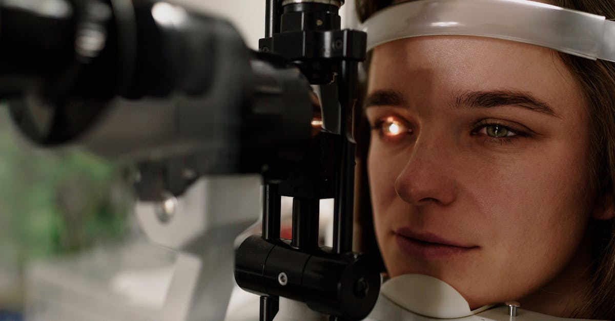
When a patient presents with sudden blurry vision during a stressful period, the ophthalmologist conducts a comprehensive examination to rule out organic disease. A dilated fundus exam allows the physician to inspect the macula for the fluid blisters characteristic of central serous chorioretinopathy.
Optical Coherence Tomography is utilized to capture microscopic, cross-sectional images of the retinal layers. This non-invasive scan definitively identifies any subretinal fluid or swelling that might be compromising visual acuity.
The physician will also perform a thorough refraction and binocular vision assessment to ensure the ciliary and extraocular muscles are functioning synchronously. If the ocular structures are entirely healthy, the diagnosis shifts toward functional accommodative spasm or dry eye syndrome secondary to acute anxiety.
13. Immediate Relaxation Techniques
Addressing stress-induced visual symptoms requires down-regulating the autonomic nervous system. When vision blurs during a panic attack, diaphragmatic breathing is a potent intervention. Slow, deep breaths stimulate the vagus nerve, which activates the parasympathetic nervous system, subsequently constricting the pupils and relaxing the ciliary muscles.
The 20-20-20 rule is essential for relieving ciliary spasm and dry eye. Every twenty minutes, the patient should look at a target twenty feet away for twenty seconds. This completely disengages the near-focusing reflex and encourages normal blinking, quickly restoring the smooth optical surface of the tear film.
Applying a warm compress to the closed eyes for five minutes soothes the tense extraocular muscles and stimulates the meibomian glands to release oils, preventing tear evaporation and alleviating the burning, blurred sensation of stress-related dry eye.
14. Long-Term Stress Management
Preventing recurrent episodes of blurry vision requires systemic stress management. Cognitive behavioral therapy helps patients restructure the thought patterns that trigger physiological panic responses, preventing the massive surges of adrenaline that dilate the pupils and distort vision.
Regular cardiovascular exercise metabolizes excess circulating cortisol, preventing the fluid retention issues linked to central serous chorioretinopathy. A consistent sleep schedule is equally critical, as sleep deprivation leaves the neuromuscular system frayed and prone to focusing spasms.
By recognizing blurry vision as a somatic alarm bell for severe psychological strain, patients can implement the lifestyle changes necessary to protect both their mental health and their long-term visual clarity. For persistent issues, an evaluation by a behavioral optometrist can provide specific exercises to rehabilitate fatigued focusing muscles.
15. Frequently Asked Questions FAQ
1. Can a panic attack cause you to go completely blind?
No, a panic attack cannot cause true structural blindness. However, the severe pupil dilation and hyperventilation can cause temporary tunnel vision or profound blurriness that feels debilitating until the panic subsides.
2. Will my vision return to normal after a period of severe stress?
Yes, functional blurriness caused by dry eyes, ciliary spasms, and dilated pupils will resolve completely once the nervous system calms down and normal physiological balance is restored.
3. Can crying make my vision stay blurry for days?
Crying alters the salt balance of the tear film and causes localized swelling of the conjunctiva and eyelids. While this causes immediate blurriness, it typically resolves within a few hours after washing the face and resting.
4. Is central serous chorioretinopathy permanent?
In most cases, the subretinal fluid resolves spontaneously within three to four months with full visual recovery, but chronic stress can cause recurrent episodes that eventually lead to permanent macular scarring.
5. Does taking deep breaths really help with blurry vision?
Yes, deep breathing directly stimulates the parasympathetic nervous system, which reverses the adrenaline-induced pupil dilation and relaxes the focusing muscles inside the eye, improving visual clarity.
16. Bibliography
Disclaimer: The content is for informational purposes only and does not replace medical advice. Always consult your doctor for personalized treatment.